Complete clinical study: Zuzzi-Krebitz AM, Buchta K, Bergmann M, Krentz D, Zwicklbauer K, Dorsch R, Wess G, Fischer A, Matiasek K, Hönl A, Fiedler S, Kolberg L, Hofmann-Lehmann R, Meli ML, Spiri AM, Helfer-Hungerbuehler AK, Felten S, Zablotski Y, Alberer M, Both UV, Hartmann K. Short Treatment of 42 Days with Oral GS-441524 Results in Equal Efficacy as the Recommended 84-Day Treatment in Cats Suffering from Feline Infectious Peritonitis with Effusion-A Prospective Randomized Controlled Study.Viruses. 2024 Jul 16;16(7):1144. doi: 10.3390/v16071144. PMID: 39066306; PMCID: PMC11281457.
The discovery of GS-441524 as an effective antiviral drug for cats with feline infectious peritonitis (FIP) has enabled feline patients to survive this once incurable, fatal disease. In the UK and Australia, GS-411524 is already legally available, while in the US the drug has only recently been available through selected compounding pharmacies. An 84-day treatment cycle has been shown to be successful in various clinical studies and has become an unofficial standard protocol. From a practical point of view, the daily administration of the drug for 12 weeks, as well as the cost of such treatment, can make it difficult or even impossible for cat owners to complete the entire prescribed treatment.
The aim of the researchers in Germany and Switzerland was to evaluate whether a 42-day treatment with GS-441524 is as effective as the currently recommended 84-day protocol. In a prospective randomized controlled treatment study, 40 cats were randomized to receive 15 mg/kg GS-441524 orally once daily for 42 or 84 days. Patients were diagnosed with FIP based on either FCoV RNA detected by RT-qPCR or RT-PCR in effusion in at least one body cavity with altered laboratory parameters typical of FIP. In addition to the diagnosis of FIP, other inclusion criteria included the presence of abdominal and/or pleural effusion, negative FeLV and FIV status, a body weight of at least 2 kg, and the absence of other serious diseases. The age of the cats ranged from 5.1 to 116.3 months, with 17 of the 40 cats being less than 1 year old. Breed distribution was as follows: 40 % Domestic Shorthairs (DSH), 20 % British Shorthairs (BSH) and 40 % other breeds. At the start of the study, 63 % cats had abdominal effusion, 12 % pleural effusion, and 25 % effusion in both cavities.
Each patient was treated for the first 7 days at the Center for Clinical Veterinary Medicine at the LMU in Munich. Treatment groups were blinded until day 7 of the study. The cats remained in their owners' homes for the remaining days of the study and returned every 2 weeks for follow-up examinations and diagnostic tests at the clinic. Tests included abdominal and thoracic ultrasonography, blood chemistry, hematology, urinalysis, measurement of viral RNA in effusion, blood, and feces, and anti-FCoV antibodies. Detailed cardiological and neurological examinations were performed at study entry. The final re-examination was performed 168 days after the start of treatment.
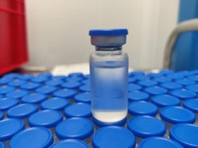
GS-441524 was supplied as 50mg tablets and was legally imported from the UK. Owners kept diaries documenting items such as activity, stool consistency, food intake and body weight. 19 cats (of 20) in each treatment group completed treatment. Two cats were euthanized during treatment (days 3 and 31) due to secondary complications.
Clinical remission was observed between days 14 and 84 with a median of 28 days, and within the first 42 days 37/40 cats went into complete clinical remission. Every cat that completed treatment showed significant improvement in hematological and clinical chemistry parameters. At the beginning of the study, viral RNA was detected in the blood of 35/40 cats, and by day 28 no more viremia was noted in any cat. During the second phase (days 42 to 84) of the study, in which only the long-term treatment group received the drugs, no significant differences were found in viral load in blood, effusion and feces or anti-FCOV antibodies. By 168 days, all 38 cats remaining in the study were in complete remission. Two cats with neurological or ocular signs also fully recovered during treatment.
The most frequently observed adverse events were diarrhea in 25/40 cats (20 % of which were diagnosed as severe based on stool evaluation), elevation of liver enzymes (mild to moderate) in 24/40 cats between days 1 and 84, lymphocytosis in 27/40 cats and a slight increase in SDMA above the reference interval in 25/40 cats. None of the patients experienced adverse effects related to the administration of GS-441524.
This study demonstrated that a shorter treatment of 42 days with oral GS-441524 was as effective as the currently recommended 84-day treatment. GS-441524 was generally well tolerated, with no significant adverse reactions noted. Limitations include that all patients received continuous professional veterinary care during the first 7 days of treatment, which is not common in most clinical practice. In addition, only patients with wet FIP were included and only the oral form of GS-441524 was used. The preparation used in the study was legally manufactured in a strictly controlled manner by BOVA Specials in London, UK. Many cat owners around the world still purchase oral and/or injectable GS-441524 from "black market" sources, so it is unknown whether the 42-day treatment is equally effective in these patients. -BJP
More details:
Pedersen NC, Perron M, Bannasch M, Montgomery E, Murakami E, Liepnieks M, Liu H. Efficacy and safety of the nucleoside analog GS-441524 for treatment of cats with naturally occurring feline infectious peritonitis.J Feline Med Surg. 2019 Apr;21(4):271-281. doi: 10.1177/1098612X19825701. Epub 2019 Feb 13. PMID: 30755068; PMCID: PMC6435921.
Murphy BG, Perron M, Murakami E, Bauer K, Park Y, Eckstrand C, Liepnieks M, Pedersen NC. The nucleoside analog GS-441524 strongly inhibits feline infectious peritonitis (FIP) virus in tissue culture and experimental cat infection studies.Vet Microbiol. 2018 Jun; 219:226-233.doi: 10.1016/j.vetmic.2018.04.026. Epub 2018 Apr 22. PMID: 29778200; PMCID: PMC7117434.
We thank Richard Malik and Sally Coggins for their advice and assistance in the preparation of this paper.
FIP treatment protocols - what's new?
Antivirals currently legally available in the UK and other countries through importation include remdesivir (injection), GS-441524 (oral suspension and oral tablets) and EIDD-1931 (oral tablets). The following recommendations are based on published and unpublished data and experience. The treatment of individual cases remains within the competence of the attending veterinarian. The dosage below is based on experience with the use of reputable preparations with known antiviral content. The extrapolation does not apply to other oral preparations for which the active ingredient and/or its content is unknown or not provided by the manufacturer.
Use of oral GS-441524 throughout treatment, including initiation of treatment
Oral GS-441524 (available as a 50 mg/ml suspension and 50 mg tablets) can be used from the start of FIP treatment for a full (eg, 12-week/84-day) cycle. It is important to support owners in their cats' medication, which can be difficult. GS-441524 oral suspension or tablets can be given with a small treat (tablets can be crushed for this) or directly into the cat's mouth. Further study is needed to examine the effect of food on absorption, but it is recommended that it be given in a small treat or on an empty stomach, with an hour or more gap before a larger meal.
Fasting cats at night can increase their hunger to facilitate the administration of the medicine in the morning, and similarly for the evening dose. However, starving kittens is never recommended as they will not be able to handle it. Any withholding of food must be adapted to the cat's age.
Injectable remdesivir is intended for cats that cannot be treated orally
Injectable remdesivir (10 mg/ml) is effective in the treatment of FIP but is associated with some side effects (see below), particularly pain on subcutaneous injection occurring in 50 % cats. Previous FIP treatment protocols suggested that this drug be used initially before switching to oral GS-441254. However, we now know that cats with FIP can be successfully treated with oral GS-441524 from the first day of treatment. This avoids injection pain and reduces treatment costs (the dose per cat weight using GS-441524 is cheaper than remdesivir). The use of injectable remdesivir should be reserved for the following situations:
Severe neurological symptoms and inability to swallow or tolerate oral medications;
Extremely dehydrated/unruly cats;
Cats that cannot be treated orally for other reasons.
In certain circumstances, if the cat is hospitalized and has decreased appetite, which affects the ability to administer medication, 48 hours of remdesivir (given intravenously, not subcutaneously) may result in significant clinical improvement that may facilitate subsequent oral treatment with GS-441524. The rest of the treatment cycle can then be administered in the form of oral GS-441524.
The switch between remdesivir and oral GS-441524 can be immediate, ie from one treatment to the other.
The current recommendation is to treat for at least 84 days. Some cats have been successfully treated with shorter courses, but large-scale case studies have not yet been published. If cost constraints require a shorter duration of treatment, the dose used should not be reduced and treatment should last as long as possible.
What dosage of GS-441524 and remdesivir should I use to treat FIP?
With experience and as yet unpublished data on therapeutic drug level monitoring (TDM), dosing recommendations have increased over previous FIP treatment protocols. However, evidence shows that more than 85 % cats respond to previously recommended drug dosing, which is still a high rate. However, based on TDM studies, we now know that individual cats vary in their absorption of oral GS-441524, with those that absorb it poorly requiring higher doses to achieve clinical and biochemical remission. Ideally, dosage should be adjusted based on TDM, if available (see below), or based on response to treatment.
Compared to previous FIP treatment protocols, the following changes in dosing recommendations are important:
GS-441524 is administered orally in divided doses twice daily (every 12 hours) to optimize serum levels of GS-441524;
Higher dosages may overcome malabsorption problems in some cats and have a better chance of crossing the blood-brain barrier and the blood-ocular barrier;
Dosage should be adjusted according to response and TDM if available.
Clinical presentation
GS-441524 PO dosing
Remdesivir IV or SC injection
Effusion present No ocular or neurological symptoms
6-7.5 mg/kg q 12h ie 12-15 mg/kg divided into 2 doses per day
10 mg/kg q 24h
Absence of effusion No ocular or neurological symptoms
6-7.5 mg/kg q 12h
12 mg/kg q 24h
Ocular symptoms
7.5-10 mg/kg q 12h
15 mg/kg q 24h
Neurological symptoms
10 mg/kg q 12h
20 mg/kg q 24h
PO, per os – orally; IV-intravenous; SC – subcutaneously; q – every x hours
Cats should be re-examined after 1-2 weeks (sooner if not improving or worsening) and dosage adjusted depending on monitoring at this point.
NOTE ON WEIGHING CATS: During treatment, it is very important to weigh the cats once a week, using accurate scales, e.g. on cat or baby scales. With successful treatment, the kittens will gain weight and/or grow, necessitating an increase in the dose to ensure that the dose of antiviral given is still adequate for the type of FIP being treated according to Table 1. Failure to increase the dose as the kitten grows appears to be one of the most common causes poor response to treatment and treatment failure.
What should I do if FIP relapses?
e.g. recurrence or insufficient resolution of effusion, pyrexia, development of new ocular or neurological symptoms, or persistent clinicopathological abnormalities:
Make sure you are still sure the cat has FIP; reassess the diagnosis, seek further pathology and consider repeat sampling (eg, external laboratory analysis and culture of any fluid; cytology or lymph node biopsy ± detection of feline coronavirus antigen or RNA, but remember that with treatment the virus is more difficult to find), AGP;
Consider TDM, if available, to monitor GS-441524 serum levels to inform dosing;
If relapse occurs during treatment, increase the dose of GS-441524 (or remdesivir) by 2-3 mg/kg per dose and monitor as above, ensuring that treatment is not stopped before the cat is in normal clinical condition and based on clinical pathology results for at least 2 weeks. Dosage increases depend on the dosage the cat is receiving at the time of relapse, the nature of the relapse and financial resources, but can be up to the recommended dosage for neurological FIP (see dosage chart above) or even higher (seek advice when considering this option );
If relapse occurs after stopping treatment, restart GS-441524 (or remdesivir) at a higher dose (at least 2-3 mg/kg higher than previously used) and ideally continue treatment for an additional 12 weeks. The increased dosage used will depend on the dosage the cat was receiving prior to the relapse and the nature of the relapse, but may be up to the dosage recommended for neurological FIP;
If the cat is already on a high dose of GS-441524 and/or serum TDM levels are adequate, consider switching to EIDD-1931 (see below) and seek advice (email advice for FIP or specialists) as adjunctive therapy such as mefloquine feline interferon or a polyprenyl immunostimulant may be an option (see below).
Treatment with EIDD-1931
This drug is another antiviral effective in the treatment of FIP in cats, although there is much less evidence of its use than GS-441524. The recommended dosage is 15 mg/kg every 12 hours and is available as 60 mg tablets for oral use. Potential adverse effects include cytopenia, especially neutropenia, rarely pancytopenia, decreased appetite/nausea, increased ALT enzyme activity, and potential renal compromise. The use of EIDD-1931 should be reserved for:
cats that do not respond to treatment with GS441524 or remdesivir despite adequate dosing (ideally assessed by TDM);
cats that relapse after treatment with GS-441524 or remdesivir at an appropriate dose.
Treatment with feline interferon (IFN), polyprenyl immunostimulant or mefloquine
In some cats, combinations of IFN omega, the immunostimulant polyprenyl, and mefloquine were used in the post-treatment period with GS-441524 (or remdesivir); however, there is currently no evidence to suggest that they are necessary, as even without this adjunctive treatment, a high success rate of over 85 % has been reported;
Mefloquine is also used to treat cats with FIP when financial constraints make it absolutely impossible to use a full course or increased dosage of more potent antivirals such as GS-441524. Studies are needed to evaluate its effectiveness, but it should only be used when absolutely no alternatives are available, as GS-441524 is known for its high potency.
A cat with Feline Infectious Peritonitis (FIP) in Cyprus shows several signs of the disease, including a swollen abdomen due to fluid build-up in the abdominal lining, unkempt fur and poor muscle condition.
The first signs of problems appeared at the end of last year (2022) in Cyprus, an island state in the eastern Mediterranean, known for its abundance of free-roaming cats. Some veterinarians there have begun to see an increase in cases of Feline Infectious Peritonitis (FIP), a fatal disease of cats.
The cats had a fever, were lethargic, losing weight and did not want to eat. Some had swollen abdomens, others had tumor-like lesions. Some were staggering, uncoordinated. Some had inflamed, cloudy or discolored eyes.
FIP usually occurs in cats as a rare reaction to infection with a common pathogen, feline enteric coronavirus (FECV). The virus is shed in the feces of infected cats, from where it can be spread to other cats. FECV is a subtype of feline coronavirus (FCoV), which is one of hundreds of known coronaviruses and does not infect humans. However, this virus is very common among stray cats and cats that live with several other cats. Cats infected with FECV are generally asymptomatic and remain healthy. However, sometimes the virus mutates and causes FIP.
In Cyprus, thousands of cats were diagnosed in the first months of this year. The disease spread rapidly, contradicting common ideas about how FIP develops.
“It’s just not right,” said Dr. Danielle Gunn-Moore, a professor of feline medicine at the Royal (Dick) School of Veterinary Medicine at the University of Edinburgh, when she found out this summer that the number of diagnoses on the island had increased 40-fold compared to the previous year.
So far unpublished work, which was published on the bioRxiv portal in November before being published in a peer-reviewed journal, offers preliminary answers to the question of why so many cats fell ill in Cyprus. Based on RNA sequencing of samples from dozens of cats with FIP, the authors of the paper argue that a strain of coronavirus that arose from separate feline and canine coronaviruses may have combined, linking the fecal excretion and infectivity of the common FECV virus with the virulence of the mutated FIP virus to one pathogen.
"In normal FIP, the FIP virus is rarely spread," said Gunn-Moore, the author of the paper. "That's a huge difference in the new outbreak. Everything suggests it's directly transmitted."
Veterinary researchers asked by VIN to comment on the paper called the evidence “interesting” and “highly suggestive” but not definitive. They say further research is needed, which the authors say is underway.
Concerns are growing on "Cat Island"
In short
Thousands of cats on the Mediterranean island of Cyprus have been diagnosed this year with feline infectious peritonitis, a fatal and usually rare feline disease.
The disease spread quickly among the many free-ranging cats on the island, upsetting the conventional wisdom about how FIP develops.
The authors of the new paper, which has not yet been peer-reviewed, say the outbreak was caused by a new strain of the pathogen that evolved from separate cat and dog coronaviruses, a recombination that increases its ability to spread.
Veterinarians in Cyprus are treating many sick cats with antivirals, some of which have been developed to treat humans with Covid-19.
A cat imported to the UK from Cyprus in August has been confirmed to be infected with the new strain.
Dr. Demetris Epaminondas, vice president of the Pancyprian Veterinary Association (PVA), first learned of the “alarming increase in FIP cases” in December last year from his wife, a practicing veterinarian. He soon began receiving similar reports from other doctors.
The potential rise in the deadly feline disease is of particular concern in an area sometimes called “cat island,” where cats roam, scurry, hunt and sleep everywhere. Large colonies of the cats, revered as holy, live in and around monasteries, where they are cared for by monks. Elsewhere, residents feed and care for neighborhood felines. There is no official estimate of the number of cats on the island.
PVA sent a questionnaire to 150 veterinary clinics in Cyprus to try to find out what was going on. Twenty-four clinics reported a total of approximately 500 cases of FIP in the first three months of 2023, a tenfold increase over the first three months of the previous year. In April, the number of reports peaked at around 2,000 cases.
These numbers only include cats on about half the island, an area of more than 2,000 square miles. Cyprus has been divided since the 1970s: The area to the south and west is under the control of the Republic of Cyprus, which has a Greek Cypriot majority and is a member of the European Union. The area to the north is occupied by Turkish military forces and is not under the effective control of the Cypriot government. There have been no official FIP announcements from the north.
dr. Charalampos Attipa, a veterinary pathologist from Cyprus, noticed the increase in cases in January while reviewing test results for Vet Dia Gnosis Ltd., a veterinary diagnostic laboratory he helped found in 2021.
There is no single test that can diagnose every case of FIP. Therefore, the diagnosis is often made based on a summary of clues from the patient's history, physical examination, and various diagnostic tests, including PCR tests. PCR stands for polymerase chain reaction, a technique that allows users to rapidly amplify a small sample of genetic material for study.
In the diagnosis of FIP, small segments of the genetic material of the coronavirus are identified by PCR in a sample of fluid from the lining of the abdomen (peritoneum) or lungs (pleura) or from the spine, or from a biopsy of tumor-like lesions.
In 2021 and 2022, Vet Dia Gnosis recorded three and four positive PCR tests in cats with FIP in Cyprus. From January to August 2023, 165 positive FIP-related PCR tests were recorded.
"These are just cats whose owners paid to have PCR done," Attipa said. "This is most likely the tip of the iceberg. But we don't really know the size of the iceberg. That's the problem."
Attipa, who started at the University of Edinburgh in April, is the lead author of the preprint paper. He is also a key member of an international collaboration investigating the virology, epidemiology and therapy of the current epidemic.
At the beginning of the year, Attipa, Epaminondas and others focused on raising awareness among veterinarians and the public. The effort led to several missteps, including a report picked up by multiple news outlets that 300,000 cats had died from the disease. Epaminondas said the figure was an unofficial and inaccurate estimate by animal welfare organizations. Additionally, the PVA estimate of 8,000 infected cats by mid-July was incorrectly reported by the Associated Press as 8,000 dead. There is no official estimate of the number of dead.
The number of PCR-confirmed cases began to decline in April, but this may not be cause for celebration just yet.
“Initially, people didn’t know what the disease was, so they took their animals to the vet to get a diagnosis,” Epaminondas said. But as awareness grew, cat owners and caretakers became aware of the rise in FIP and the clinical signs, such as a swollen abdomen, present in one form of the disease. “Because they can find treatment on the black market, they don’t want to spend money to get the disease properly diagnosed,” he said.
In recent years, antiviral compounds have shown remarkable promise in reversing the course of FIP. However, these compounds are not approved for veterinary use in many countries. As a result, a black market for antiviral drugs, mostly made in China, is flourishing, fueled by desperate cat owners who treat their animals on their own. A 2021 study of cats given unlicensed antivirals in the US found a survival rate of 80 % and above.
The number of PCR tests increased again in August, which may be due to the Cypriot government approving the veterinary use of molnupiravir, an antiviral drug used to treat Covid-19 in humans. Cats must be PCR positive for their owners or caretakers to receive a prescription.
According to Epaminondas, caseload data for most of the second half of the year, based on clinic surveys, should be available soon.
Dr. J. Scott Weese, an infectious disease veterinarian at the Ontario Veterinary College at the University of Guelph, said in an email to VIN News that the epidemiology of the outbreak is not well described, adding that it is "a common problem in field studies where information is piecemeal and often anecdotal."
According to Weese, the number of cases confirmed by PCR - 165 - is very small, especially for a country with a lot of feral cats. He also said that the rate of diagnosis based on questionnaires begs the question: Are more diagnoses due to a significant increase in the disease, a significant increase in testing, or both?
"There appears to be an increase in FIP in Cyprus. It's hard to say by how much," he said. "As there is more discussion and awareness, there are also more diagnoses of an endemic disease that may have been there all along, just ignored. Often times these are mixed situations where there is a real increase or a small local cluster, but the increased discussion and testing leads to an overestimation of the rate of change."
He added: "I'm not ruling out that this was a real epidemic or that this is a worrying new strain. We just don't know (or at least I don't know). This work shows that we need to look more closely at this issue."
Introducing FCoV-23
Basic information of FIP
If you find the specifics of feline infectious peritonitis difficult to understand, take a cue from Dr. Brian Murphy: It is. “FIP is probably the most complicated virus in veterinary medicine,” said Murphy, a veterinary pathologist and FIP researcher at the University of California, Davis, School of Veterinary Medicine. Much of what science knows about FIP was pioneered by Murphy’s now-retired mentor, Dr. Niels Pedersen.
FIP is caused by mutations in a ubiquitous and otherwise insignificant pathogen called feline enteric coronavirus. These mutations allow the virus to infect immune system cells called macrophages, which multiply and cause deadly inflammation. FIP is estimated to affect 1.3 % cats, most often kittens in catteries and shelters.
There are two forms of FIP: wet and dry. Cats may initially have one form and later develop another. In wet FIP, the fluid created as a result of inflammation accumulates most often in the abdomen, less often in other parts of the body. In dry FIP, the patient develops tumor-like lesions in the abdomen, chest, eyes, and/or brain. Early symptoms of FIP include fever, loss of appetite, weight loss, and depression. Cats with neurological FIP may develop lack of coordination, seizures, and dementia. Eye disease can cause inflammation, discoloration, or clouding of a cat's eyes, which impairs vision.
No test can diagnose every case of FIP with absolute sensitivity and specificity. Therefore, the diagnosis is often established based on a summary of clues from the patient's history, physical examination, and various diagnostic tests.
First identified in the 1950s, FIP was considered a death sentence for decades. However, in recent years, antivirals – including those used against the coronavirus that causes Covid-19 in humans, such as remdesivir, molnupiravir and Paxlovid – have been shown to reverse the course of FIP in cats. None of these antivirals are approved for veterinary use in the United States (except at universities that are researching these drugs in cats with FIP). These medicines are variously available for veterinary use in several European countries, including Cyprus, Finland, Norway, Sweden and the United Kingdom, as well as Australia and New Zealand.
In countries where the treatment is not approved by regulators, pet owners have resorted to buying unlicensed antivirals, mostly made in China, to treat sick cats themselves.
Understanding why the new strain behaves differently than what some researchers call “traditional FIP” requires a closer look at how FECV leads to FIP.
For reasons that are not fully understood, FECV sometimes mutates inside a cat. These changes allow it to escape from intestinal cells and infect a key cell of the immune system, the macrophage. This macrophage-infecting virus is known as the FIP virus or FIPV. It can now travel throughout the body, “ravaging the environment,” Gunn-Moore says, causing potentially fatal inflammation.
Once established, the FIPV coronavirus has two important characteristics: First, it is no longer an enteric virus, so it can only very rarely return to the gut to be excreted as FIPV in the feces. Second, FIPV has a gene sequence that is unique to a given cat.
FCoV-23, as the new strain has been named, appears to violate both of these schemes. Gunn-Moore said it thrives in the intestines of Cypriot cats. Furthermore, based on RNA sequencing from PCR samples, the virus in many cats had the same genetic sequence. The genetic sequencing was carried out by researchers at the Roslin Institute, an animal science research center at the University of Edinburgh.
Gunn-Moore had some early hypotheses about what might have caused the outbreak, but genetic analysis points to her main theory — that a pantropical canine coronavirus combined with a feline enteric coronavirus. Pantropic viruses can spread to different tissues in the body, a property that would allow FCoV-23 to enter other organs and nerves, as well as continue to multiply in the intestinal tract.
"I think a dog came to Cyprus and defecated on the floor. The cat, who already had FECV, then got dog feces on its feet, licked its paws and got both viruses," she said. "And that's what these viruses do, they recombine, they're party animals. They get together and go, 'Hey, do you want some of this? I'll give you some of that?'"
Gunn-Moore added that further work is planned to confirm the case for their direct transmission.
"We are currently conducting experiments to sequence the virus from feces because we need to sequence the virus and prove that it is the same sequence as in the blood," she explained.
dr. Brian Murphy, a veterinary pathologist and FIP researcher at the University of California, Davis, School of Veterinary Medicine, said the recombinant feline and canine coronavirus described in the Cyprus paper had been identified before, including by a team at Washington State University's School of Veterinary Medicine. Medicine in the 1970s.
"This is a replication of this virus, but it's a highly virulent form of the virus," Murphy said of the evidence so far. He welcomes the scientists' plans for further genetic analysis and suggests further investigation.
“Sequencing an enteric corona or a virus that comes from the gut is compelling, but it’s not proof,” he said. “It wouldn’t be a bad experiment to take fecal material containing the virus that comes from the gut, infect cats with it, and then when they get sick, save those cats with antiviral treatment. That would be good evidence of transmission of a virulent form of the virus.”
Murphy acknowledged that this kind of testing on companion animals is essentially banned in Europe and would likely be very controversial. “I think most people probably wouldn’t agree with me,” he said. “But I think it can be done ethically because we have high-quality, highly effective drugs.”
Dr. Maria Lyraki, an internal medicine specialist in Athens, Greece, who coordinates treatment protocols with veterinarians in Cyprus and is an author of the paper, said Murphy is right that the experiment would be proof, although some of the infected cats may not get sick if they develop an adequate immune response.
"However, it is something that we would not be able to implement from an ethical point of view," she said.
Thousands of sick cats
If the Cypriot epidemic had occurred 10 years ago, countless cats would probably have died because until recently there was no known way to stop the disease.
Antivirals have changed this situation.
Two promising products are remdesivir, made by pharmaceutical company Gilead to treat Covid-19 in humans, and the related compound GS-441524. GS, as it's called for short, was shown to be effective in reversing FIP in cats in an infectious disease study at the University of California, Davis in 2018.
Gilead has not granted a license to develop GS-441524 as a veterinary medicinal product. Remdesivir is not approved as a veterinary medicine in the United States, Canada, and some European countries.
However, since August, Cypriot vets have been able to import compounded versions of GS and remdesivir, manufactured by Bova, a veterinary pharmaceutical company based in the UK and Australia, under a special permit, to based on instructions UK Veterinary Medicines Directorate.
GS and remdesivir “are the first line of drugs that we use because we have the most literature on them,” Lyraki said. “But they are really expensive … which is really challenging, especially for such a large number of stray cats.”
According to Epaminondas, treating one cat with these drugs can cost from €3,000 to €7,000 ($3,250 to $7,580).
“We contacted people around the world who treat FIP cases,” Lyraki said. “And the specialists advised us that they were using molnupiravir. There is published literature on this drug and we have a lot of anecdotal discussion among FIP specialists around the world that it is indeed effective.”
The government's decision to allow veterinary use of molnupivirus has made a huge difference.
“It actually works quite well,” Lyraki said. Initially, boxes of molnupiravir were donated to the PVA to help fight the epidemic. They were available at an exceptionally low price of around 100 euros ($108) per cat, a significant saving. The actual price of the drug is much higher, but still significantly cheaper than GS.
However, the replacement of molnupivir will not last long. Merck Sharp and Dohme BV, which sells the antiviral under the name Lagevrio, will stop making it for Europe next year after the European Medicines Agency decided earlier this year not to support the registration of molnupiravir based on its finding that the antiviral's benefit in treating Covid in adults has not been proven.
"We are now working to find alternatives," Lyraki said.
Limitation of dissemination
A healthy free-roaming cat lies outside a bakery in Paphos, Cyprus. Stray cats are everywhere on the island - although no one knows how many there are. In response to the FIP outbreak, the Cyprus Veterinary Association, together with ThePetzApp, is launching an initiative to engage cat owners and carers in Cyprus to count the cats in their homes or in the stray cat colonies they care for.
In addition to the immediate situation in Cyprus, there are fears that the strain could spread to other countries or that it is already in other countries from which it has reached Cyprus, as yet unrecognized.
The new strain was confirmed in a cat imported to the UK from Cyprus in August. This cat has been quarantined and is responding to treatment.
Keeping the cats on the island under control can be a challenge. Cyprus is home to many cat rescue activities. Stray cats are regularly collected and taken to other parts of Europe where they are given a new home. According to Epaminondas, there are no regulations regarding the export of cats from Cyprus.
But veterinarians like Lyraki are urging rescue groups to be cautious. “Our team of experts has issued a recommendation that cats be tested before traveling outside Cyprus and that only cats with negative antibodies to FCoV be exported,” said Dr. Lyraki. Once the cats arrive at their destination, they should be quarantined for three weeks and then retested for antibodies “until the outbreak is under control and the number of affected cats has significantly decreased.”
Meanwhile, Greece, which has its own large stray cat population, has also seen an increase in FIP cases, according to Lyraki.
"We believe it's a matter of time before this outbreak spreads to Greece because Greece and Cyprus are very, very close culturally, geopolitically. There is a lot of exchange and travel between them," she said. "So it's something we're actively monitoring."
We had hoped that in 2023 one or more antivirals for cats would be legalized. With the exception of a few countries outside the US, this has not happened. Still, there is hope that studies being conducted at the University of California, Davis and elsewhere around the world will help advance conditionally and/or fully approved human drugs such as Remdesivir, Molnupiravir and Paxlovid for use by veterinarians. Even if drugs are approved for use in animals, drugs marketed for human use are not ideal because they must be purchased at the price set for humans. Therefore, the unapproved market will remain the main source of cheaper antivirals for many years to come. However, SOCK FIP appreciates the efforts of countless cat owners and lobbying industry and government agencies to allow the use of effective antivirals for cats. These efforts have had varying degrees of success in many countries outside the US.
FIP research at UC Davis in 2023 supported by SOCK FIP contributions
2023 continues to support SOCK FIP and feline coronavirus research at UC Davis, and we couldn't do it without the help of many donors. Two ongoing research projects receiving SOCK FIP funding are of particular interest. The first project involves testing antiviral drugs and is led by Drs Krystle Regan and Brian Murphy. Patients and owners were drawn from across the US. The first study compared two antiviral drugs in cats with wet FIP to test cure rates with either oral GS-441524 or Remdesivir (Gilead). This study, which was published, showed that oral Remdesivir worked as well as oral GS-441524. So if Remdesivir gets full approval in the United States, veterinarians can safely prescribe it to cats with wet FIP. Other studies comparing GS-441524 and Remdesivir in cats with dry FIP and Molnupiravir (Merck) in cats with wet FIP have also been completed. The results of these studies should be published in early 2024. The latest study involving Paxlovid (Pfizer) was recently fully approved and widely available in the United States, and if it proves to be a safe and effective treatment for cats with FIP, it will a third human antiviral drug to treat FIP that may one day be used by veterinarians. Drs Regan and Murphy also used their field test cases to study the causes of death during the first two weeks of treatment. This population represents up to 10 % treated cases worldwide. Necropsies showed the existence of serious complicating diseases, which often included bacterial sepsis, often with highly resistant organisms to antibiotics, as well as serious heart disease. More work is needed to determine the nature of the heart disease and how much of it may be pre-existing disease and how much is caused by the FIP virus.
The second major research project focused on the prevention of FIP is implemented by Dr. Patricia Pesavento, one of our veterinary pathologists, and her research team, which includes veterinary microbiologist Terza Brostoff, biomedical engineer Randy Carney, immunologist Dennis Hartigan O'Connor, and lab technician Ken Jackson. Their study involves the development of an mRNA vaccine against a portion of the nucleocapsid protein that is common to virtually all known feline coronavirus isolates. The theory is that an immune response to this protein, compared to the spike protein commonly used in COVID-19 vaccines, will protect cats exposed to the common enteric form of feline coronavirus from developing FIP. This would be analogous to the protection against severe and chronic forms of COVID-19 reported with mRNA vaccines. Team Dr. Pesaventa developed a vaccine based on ideal manufacturing parameters and tested it for safety and efficacy in a rodent model. The development of this mRNA vaccine will only be a first step as it will need to be further tested in a limited number of cats as a prelude to much more extensive field testing in larger populations of cats such as breeding stations or temporary/rescue stations experiencing ongoing cases of FIP.
Areas of future FIP research
The discovery of a cure for FIP does not end the need for further FIP research. We hope that veterinary scientists from around the world who are still active in academia and industry will consider some of the other promising areas of research. Such studies cover all aspects of FIP pathogenesis, from the basic enteric coronavirus, which is enzootic in virtually all healthy cat populations and exists in the lower intestinal tract, to mutant forms that have acquired the ability to infect monocytes/macrophages in and outside the abdominal cavity. The exact nature of immunity to feline coronaviruses, both the minimally pathogenic enteric form and the highly lethal form causing FIP, needs to be clarified. We know that immunity to both intestinal and extraintestinal forms of the virus is weak, short-lived and susceptible to weakening by internal and external stressors. Immunity to enteric coronavirus appears to involve locally produced antibodies, whereas immunity to FIP-causing mutant viruses involves more systemic lymphocyte-mediated (cellular) immune responses. Accurate knowledge of the strengths and weaknesses of both types of immunity will be essential for all future vaccine development efforts. Will you prevent FIP by attacking underlying enteric coronavirus infections or by attacking FIP-causing mutants when they emerge?
There is a great need to develop tests that can accurately determine when a cat has been cured by antiviral therapy. We know that some cats can heal in as little as 4-6 weeks, while others need up to 12 weeks. We suggest 12 weeks of treatment because this gives the maximum cure rate, but we know that some cats will be treated for an unnecessarily long time. The only current way to tell when a cat is cured is to stop the treatment and see if the disease returns. Regular complete blood counts and basic serum biochemistry are useful in conjunction with physical health indicators in monitoring and managing treatment, but return to normal test values and general health do not guarantee that treatment will not relapse. On the contrary, the persistence of minor abnormalities in the blood and health status is not always a sign that there has been no cure and that it is necessary to increase the dosage or prolong the treatment. This is especially true for cats with neurological FIP, where blood test results and the state of neurological deficits do not always indicate a cure.
Although there is hope that even more effective antiviral drugs will be found in the future, the well-documented safety and efficacy profiles of current drugs leave little room for further improvement. However, drug resistance is currently being observed in some cats. What is known about how drug resistance develops in chronic infections such as HIV/AIDS should be applied to FIP. The most effective way to combat drug resistance in HIV/AIDS is to combine two or more antivirals with different mechanisms of action before resistance develops.
It appears that some strains of feline coronavirus may be more neurotropic than others. A penchant for infecting the central nervous system can be developed by specific mutations in enteric strains of the coronavirus that are enzootic in the environment or by mutations that occur as part of the FIP biotype. The role of the blood-brain barrier and the apparent compartmentalization of immunity between the central nervous system and the rest of the organism are other areas that require study.
Most cat owners are currently aware of the large outbreak of FIP occurring on the island of Cyprus. It is still uncertain whether this outbreak qualifies as epizootic (epidemic) or enzootic (endemic). Preliminary research suggests that the outbreak is linked to closely related isolates of FIP virus serotype 2 (similar to canine coronavirus). It is clear that for cats in all parts of the world, whether this outbreak is related to the spread of the virus from cat to cat (ie, an epizootic disease) or to factors promoting the disease in the environment (ie, an enzootic disease) is important. The worst possible scenario is a panepizootic disease like COVID-19. Hopefully, researchers in Cyprus, the UK and elsewhere will be able to resolve the nature of this outbreak as quickly as possible.
Between January and April 2023, the Urolite Center in Minnesota received three shipments of atypical stones (Figure 1). All three samples were obtained from cats. All three cats were under 1 year old. Cats came from North and South America. In each case, the infrared spectrographic pattern of the stones was identical. Urinary stones usually contain large amounts of phosphorus, calcium and magnesium. In these cases, electron dispersion spectroscopy revealed a high proportion of nitrogen, carbon and oxygen.
Mystery solved. When asked about their medical history, all three cats were diagnosed with feline infectious peritonitis. All three were treated with either Remdesivir or its metabolite GS-441524. We requested samples of their antiviral drugs for analysis. The antiviral drugs were spectrographically identical (Figure 2). The stones were composed of GS-441524.
Figure 2: FT-IR spectroscopy of urinary stone of patient and reference sample GS-441524
After administration, GS-441524 is excreted primarily in the urine. Although GS-441524 is very soluble in organic solvents such as DMSO (10-59 mg/ml), it is poorly soluble in aqueous solutions such as water (0.0004 to 0.1 mg/ml). Its limited solubility makes GS-441524 a prime candidate for stone formation. Observation of urinary symptoms in cats receiving Remdesivir or GS-441524 is an indication to look for stones. Observation of atypical crystalluria or uroliths may be an indication to limit the dose of the drug (if possible) and increase water consumption to minimize stone formation.
Basic information Feline infectious peritonitis (FIP) is a viral disease of cats caused by certain strains of the coronavirus that has a high mortality rate.
The goal This case series reports the results of treatment of cats with FIP with molnupiravir.
The animals Eighteen cats diagnosed with FIP at You-Me Animal Clinic, Sakura-shi, Japan between January and August 2022 and whose owners gave informed consent for this experimental treatment.
Methods In this prospective observational study, molnupiravir tablets were prepared directly at the You-Me Animal Clinic. Owners administered 10-20 mg/kg PO twice daily. The standard duration of treatment was 84 days.
The results Of the 18 cats, 13 cats had effusive FIP and 5 cats had non-effusive FIP. Three cats had neurological or ocular signs of FIP before treatment. Four cats, all with effusive FIP, died or were euthanized within 7 days of starting treatment. The remaining 14 cats completed treatment and were in remission at the time of writing (139-206 days after initiation of treatment). Elevated serum alanine transaminase (ALT) activity was found in 3 cats, all on days 7–9, and all recovered without intervention. Two cats with jaundice were hospitalized, 1 during treatment (day 37) and 1 with severe anemia at the start of treatment.
Conclusions and clinical significance This case series suggests that molnupiravir may be an effective and safe treatment for domestic cats with FIP at a dose of 10–20 mg/kg twice daily.
1. Introduction
Feline infectious peritonitis (FIP) is a viral infectious disease occurring mainly in domesticated cats.1, 2 FIP is an aberrant immune response to infection with feline coronavirus (FCoV), which is ubiquitous, especially in breeding and rescue breeds, usually with no or mild clinical signs. Fecal-oral transmission of FCoV often occurs, especially in environments with multiple cats3 and the incidence of FIP in cats exposed to FCoV is up to 14 %.4, 5
Feline infectious peritonitis is usually classified as either effusive or non-effusive based on clinical presentation.1, 6, 7 Until the development of specific antiviral therapy, the case fatality rate associated with FIP was high, and most affected cats died within weeks to months of the onset of clinical signs.
Some nucleoside analogs, including remdesivir (GS-5734) and its active metabolite GS-4415248, inhibit viral RNA synthesis and have high antiviral activity against FCoV causing FIP in cats.9, 10 Despite the expectations of veterinarians and cat owners, the developer decided not to apply for approval of GS-441524 for the treatment of FIP. As a result, many cats with FIP are being treated with the unapproved drug GS-441524, and concerns have been raised about the quality, purity, and efficacy of the unapproved products available on the world market. Mutian has excellent efficacy and safety.11-14 Although the manufacturer did not disclose the chemical structure of the active substance and its exact concentration, its active substance is GS-441524.12
Molnupiravir is an oral nucleoside antiviral prodrug with activity against severe acute respiratory syndrome coronavirus 2 (SARS-CoV-2) and coronavirus disease (COVID-19). As of 2021, molnupiravir is approved in Japan to treat people with COVID-19. There are published reports on the efficacy and safety of molnupiravir in cats15 , but sufficient data on the use of molnupiravir in cats with FIP are lacking. Due to the lack of treatment options for FIP, we began offering molnupiravir to clients at our clinic, using small tablets prepared in-house for easy administration to cats. Here we present the results of the first 18 cats that underwent this FIP treatment in our clinic.
2 MATERIALS AND METHODS
2.1 Cats
All cats attending the You-Me Animal Clinic in Sakura-shi, Japan since January 2022 diagnosed with FIP and whose owners provided informed consent were included in this case series. Feline infectious peritonitis was diagnosed based on a combination of clinical signs (decreased appetite, enlarged abdominal lymph nodes, weight loss, fever, effusions, or uveitis) and laboratory test results for anemia and hyperglobulinemia, including the albumin-to-globulin (A/G) ratio and values of α1-acid glycoprotein (AGP). The presumptive diagnosis of FIP was based on the identification of FCoV RNA in samples from abdominal or pleural effusion (effusive FIP) or whole blood (non-effusive FIP), or from fine needle aspiration (FNA) of pyogranulomatous lesions. Virus detection was performed by reverse transcription polymerase chain reaction (RT-PCR) in the following testing laboratories: abdominal effusion and FNA samples at IDEXX, Japan (using a LightCycler 480 System II, Roche Diagnostics KK, Basel, Switzerland) and whole blood at Canine Lab., Japan (using CFX Connect, Bio-Rad Laboratories, Inc, Irvine, CA, USA). Abdominal or pleural effusion samples (1 ml each) were taken by ultrasound-guided abdominocentesis, respectively. by thoracentesis and were evaluated for the total number of nucleated cells, protein content, A/G ratio and cytology. Whole blood samples (1 ml) were collected and sent in ethylenediaminetetraacetic acid (EDTA) tubes.
2.2 Preparation of the medicine
Tablets containing molnupiravir 20 mg were prepared at the You-Me Animal Clinic. Briefly, molnupiravir powder was extracted from 20 commercially produced molnupiravir 200 mg capsules (MOVFORE, Lot No. HH2201001 [HETERO HEALTHCARE, Hyderabad, India]) and mixed with powdered cellulose (microcrystalline cellulose powder, NICHIGA, Takasaki, Japan) using a mortar and pestle (Matsuyoshi Medical Instruments Co, Ltd, Tokyo, Japan) to make a total of 12 g of powder mixture. The powder was shaped into approximately 200 6 mm wide sectional scored tablets using a generic tablet press made in China.
2.3 Treatment
Treatment with molnupiravir was initiated when FIP was highly suspected on the basis of clinical presentation or when FCoV RNA was detected by PCR; this date was marked as the first check. The following dosages were chosen: 20 mg/kg/d (10 mg/kg twice daily) for cats with effusive type, 30 mg/kg/d (15 mg/kg twice daily) for cats with non-effusive type and cats with pyogranulomatous lesions, and 40 mg/kg/d (20 mg/kg twice daily) for cats with neurological or ocular signs of FIP. Dosage may be increased or decreased in animals showing clinical deterioration or adverse reactions. Dosage was chosen based on estimated animal dosages listed on the Internet16, 17 and based on the adult human COVID-19 dose of molnupiravir, which is 800 mg every 12 hours.18 This would correspond to a dose per kilogram of 10 to 13.3 mg/kg twice daily for adults weighing 60 to 80 kg. As no pharmacokinetic information is available in relation to cats, we have chosen the dosage for cats based on the assumption that metabolism of drugs in cats is equivalent to metabolism in humans.
Owners were instructed to administer the tablets twice a day with 12 hours between doses. The expected standard duration of treatment was 84 days according to study GS-441524.10
2.4 Measurements
Owners were instructed to record body weight, body temperature, physical activity, appetite, and voiding/urinating daily and were asked to visit the clinic at 1, 2, 6, and 10 weeks. The following laboratory tests were required at each visit: red and white blood cell count, hemoglobin, hematocrit (HCT), AGP, total protein, albumin, aspartate transaminase (AST), alanine transaminase (ALT), total bilirubin, creatinine, blood urea nitrogen (BUN ) and the A/G ratio. Samples were analyzed in the clinic using a Catalyst One chemistry analyzer and a ProCyte One hematology analyzer (both IDEXX Laboratories, Westbrook, Maine). The A/G ratio was determined either from a whole blood plasma sample or from a fractionated protein sample. We performed abdominal and thoracic ultrasound evaluation of each cat at baseline and after 2, 6, and 10 weeks of treatment using a Prosound α7 (Aloka, Japan), including assessment of cardiac function (fractional shortening, left atrial to aortic diameter ratio, and valvular regurgitation).
2.5 Adverse Events
Any abnormal laboratory test values or medical events that occurred during treatment were considered adverse events and decisions were made to continue/discontinue treatment.
2.6 Statistical analysis
As this is a case series, no statistical calculations were performed other than descriptive statistics.
2.7 Ethics
All owners provided written informed consent prior to initiation of treatment. The experimental use of molnupiravir was approved by our Institutional Animal Study Review Committee.
3 RESULTS
3.1 Characteristics of the disease and treatment
Eighteen cats completed treatment by August 4, 2022 and are included in this summary report.
The presentation of the 18 cats is summarized in Table 1 and Table S1. The median age was 6.5 (range: 3-93) months. All 18 cats had a low serum A/G ratio, 16 cats had a loss of appetite, and 14 cats had mild to severe anemia by hemoglobin level and HCT. Thirteen cats had effusive FIP and 5 cats had non-effusive FIP. Neurological or ocular signs suggestive of FIP were present in 3 cats before treatment, including epileptic seizures/neurological signs (No. 8), blunted postural reflexes (No. 10), and slow pupillary reflex (No. 18) (Table S1). All but 2 cats (#8 and #15) were treated exclusively on an outpatient basis. Cat no. 8 was hospitalized from day 37 for 3 days due to jaundice. Cat no. 15 required hospitalization for 5 days after starting treatment due to anemia and jaundice accompanied by increased bilirubin concentration and ALT activity. During hospitalization, the cat received molnupiravir as scheduled, was hydrated with Ringer's solution, and treated with oral ursodeoxycholic acid (Towa, Japan) 10–15 mg twice daily to reduce bilirubin. There was no evidence of intravascular hemolysis (HCT remained stable), and microscopic examination of blood smears was negative for hemotropic mycoplasma infection.
TABLE 1. Basic characteristics of the cats in the described case series.
Age at disease onset, months, median (range)
6.5 (3-93)
Breed, n (%)
Housecat
9 (50.0)
Exotic shorthair
2 (11.1)
British Shorthair
2 (11.1)
Other
5 (27.8)
Sex, n (%)
Male not neutered/male neutered
4 (22.2)/7 (38.9)
Female not spayed/female spayed
2 (11.1)/2 (11.1)
Weight in kg, mean (SD)
2.72 (0.77)
Duration from onset of illness to initiation of treatment, days, median (range)
16.5 (2-49)
Effusive type, n (%)
13 (72.2)
Pyogranulomatous lesions in the abdominal cavity, n (%)
5 (27.8)
Neurological manifestations of FIP, n (%)
2 (11.1)
Ocular symptoms of FIP, n (%)
1 (5.6)
Temperature, °C, mean (SD)
39.3 (0.9)
Hematocrit, %, mean (SD)
27.3 (8.1)
Albumin/globulin ratio, mean (SD)
0.35 (0.10)
Sample type, n (%)
Abdominal effusion
11 (61.1)
Pleural effusion
1 (5.6)
FNA of a pyogranulomatous lesion
2 (11.1)
Full blood
3 (16.7)
None
1 (5.6)
The attending physician decided to extend the treatment to 99 days for cat no. 1. Consciousness disturbances appeared on day 8 in this cat and the dose was subsequently increased to 40 mg/kg. This symptom disappeared on the 15th day; however, the A/G ratio with the fractionated protein sample did not return to normal. On day 99, although the A/G ratio was still below the reference range (0.6), the clinician decided to stop treatment because the cat showed no clinical progression or deterioration.
3.2 Outputs
Clinical response in 14 cats was rapid. Dosing, findings during treatment, and outcomes in these animals are summarized in Tables S2 and S3. The fever subsided and the appetite returned within 2-3 days after the start of treatment. Remissions were also achieved in cats with severe clinical signs. Among them were cats no. 8, 17 and 18, which had pyogranulomatous lesions ≥ 2 cm in size, cat no. 4, which had severe anemia and a low A/G ratio, cat no. 14, which had a pleural effusion and difficulty breathing, and cat no. 15, who had an enlarged kidney. The pyogranulomatous lesions shrank or were undetectable on ultrasound in all 5 cases, and laboratory values returned to normal in all cats. Three cats had neurological signs of FIP before treatment. Cat no. 12 had no neurological symptoms of FIP before treatment, but had an epileptic seizure on day 7. The dosage was subsequently increased to 40 mg/kg. On the 2nd day, on the slit lamp at cat no. 7 found anisocoria that was probably related to uveitis. The dosage of molnupiravir was increased to 40 mg/kg from day 15, and all neurological or ocular signs of FIP resolved within 15 days.
Of the 14 cats that achieved remission, no relapses occurred through August 3, 2022, during 55 to 107 days of post-treatment follow-up. Three cats died (No. 2, No. 11 and No. 16) and one (No. 13) was euthanized; all these cats had the effusive form of FIP but had no neurological or ocular signs of the disease. All died within one week of starting treatment.
3.3 Security
Alanine transaminase activity higher than the reference value was found in 4 cats; the value of each of them was 286 U/l (cat #8 on day 37), 283 U/l (cat #9 on day 9), 154 U/l (cat #10 on day 7), and 117 U/l (cat No. 17 on the 9th day). Three cats with early ALT elevations on days 7 to 9 recovered without the need for intervention. In cat no. 8 developed jaundice on day 37 and was hospitalized for 3 days.
No abnormalities in BUN or creatinine concentrations were noted during treatment with molnupiravir.
4 DISCUSSION
In our series of cats with presumptive FIP treated off-label with a molnupivir compound, 14 of 18 cats achieved remission and remained in remission for up to 107 days of observation at the time of writing. Four cats showed signs of potential hepatic adverse effects; 3 cats developed ALT activity above the reference range during the first 7 to 9 days of treatment, all of which resolved without treatment, and 1 cat developed jaundice requiring hospitalization on day 37 of treatment.
The approved human formulation of molnupiravir is in 200 mg capsule form, but for animals it must be divided into smaller doses to facilitate administration of an appropriate dose based on body weight. We decided to prepare molnupiravir in the form of small tablets to simplify administration. We hypothesized that cats might refuse to swallow the drug in powder form or in aqueous solution, and it might be difficult for owners to administer the entire powder dose each time.
The minimum effective dose of molnupiravir in FIP is recommended to be 4.5 mg/kg PO every 12 hours for cats without neurological/ocular signs of disease, increasing to 12 mg/kg PO every 12 hours for cats developing ocular or neurological signs. symptoms of FIP.17 Others recommend a dose of 25 mg/kg every 24 hours for dry/wet FIP, 37.5 mg/kg every 24 hours for ocular FIP, and 50 mg/kg every 24 hours for neurological FIP.16 Since none of these recommended dosages have been established in prospective controlled studies, the dosage in this case series was determined by the author based on these estimates, adult dosages, and his experience. Nevertheless, the dosage used in our case series (10 mg/kg twice daily for cats with effusive FIP, 15 mg/kg twice daily for cats with non-effusive FIP or pyogranulomatous lesions, and 20 mg/kg twice daily for cats with neurological or ocular signs FIP) appears to be effective and safe and may help guide dosing in future clinical trials.
Four cats died during this study. Each of these cats had the effusive type of FIP; however, considering that some cats that survived had symptoms as severe or even more severe than those that died, no signs were found to predict early death. Unfortunately, the attending veterinarian was given little information about the deaths of the 3 cats that died at home, and no post-mortem examinations were performed. One cat (#2) died after vomiting the drug on day 6, so it is possible that this animal had swallowing problems.
The use of GS-441524 in 31 cats with FIP, of which 26 cats completed at least 12 weeks of treatment, resulted in remission in 25. 10 Eight of the 26 cats relapsed or became reinfected within 3 to 84 days after this period. . The length of observation in our case series is shorter than in study GS-44152410; however, observation of cats in our series is currently ongoing and more cats with FIP are being treated with molnupiravir. Further observation will provide data on longer-term efficacy. The major adverse events reported with injection in study GS-441524 were injection site reactions in 16 of 26 cats.10 Because the treatment in our study was administered orally, no injection site reactions occurred in any of the cats in our series. In our series, the most frequent adverse event during treatment was an increase in ALT activity. However, longer-term follow-up is necessary to more adequately assess liver-related reactions, and a larger group of animals is needed to more fully assess the adverse effects of molnupiravir.
Molnupiravir is active against SARS-CoV-2 and other RNA viruses19 and in cell culture it creates only low resistance.20 – 22 The efficacy of oral molnupiravir was evaluated in a phase 3 randomized control trial in 1,433 people with COVID-19, with a lower percentage of hospitalizations or deaths by day 29 in the molnupiravir group compared to placebo.18 Another important clinical question is whether molnupiravir-resistant viruses can develop and how many cats relapse or reinfection after treatment.
All owners who provided informed consent to participate were included in this study, so the risk of any bias should be minimal. Nevertheless, selection bias should be considered as this case series was enrolled in a single center in Chiba Prefecture, Japan. Another potential limitation of our case series is that the diagnosis of FIP was probable in all cases. Cats can have FCoV viremia without FIP, so RT-PCR detection of FCoV RNA is not specific for FIP.7 although this technique has a high sensitivity (90 %) and specificity (96 %) for FIP when applied to FNA specimens.23 In our series, the combination of RT-PCR with clinical signs and other serum biochemical tests, including a low A/G ratio, was highly suggestive of FIP.7 This case series suggests that molnupiravir may be an effective and well-tolerated treatment for FIP.
Table S1. Feline demographics and disease status before treatment. Table S2. Overview of the course of treatment. Table S3. Test values obtained at the first visit and the last test, based on which the decision was made to stop treatment.
References
Addie D, Belák S, Boucraut-Baralon C, et al. Feline infectious peritonitis. ABCD guidelines on prevention and management. J Feline Med Surg. 2009; 11: 594-604.
Wang YT, Su BL, Hsieh LE, Chueh LL. An outbreak of feline infectious peritonitis in a Taiwanese shelter: epidemiologic and molecular evidence for horizontal transmission of a novel type II feline coronavirus. Vet Res. 2013; 44: 57.
Addie DD, Toth S, Murray GD, Jarrett O. Risk of feline infectious peritonitis in cats naturally infected with feline coronavirus. Am J Vet Res. 1995; 56: 429-434.
Foley JE, Poland A, Carlson J, Pedersen NC. Risk factors for feline infectious peritonitis among cats in multiple-cat environments with endemic feline enteric coronavirus. J Am Vet Med Assoc. 1997; 210: 1313-1318.
Murphy BG, Perron M, Murakami E, et al. The nucleoside analog GS-441524 strongly inhibits feline infectious peritonitis (FIP) virus in tissue culture and experimental cat infection studies. Vet Microbiol. 2018; 219: 226-233.
Pedersen NC, Perron M, Bannasch M, et al. Efficacy and safety of the nucleoside analog GS-441524 for treatment of cats with naturally occurring feline infectious peritonitis. J Feline Med Surg. 2019; 21: 271-281.
Jones S, Novicoff W, Nadeau J, Evans S. Unlicensed GS-441524-like antiviral therapy can be effective for at-home treatment of feline infectious peritonitis. Animals (Basel). 2021; 11: 2257.
Krentz D, Zenger K, Alberer M, et al. Curing cats with feline infectious peritonitis with an oral multi-component drug containing GS-441524. Viruses. 2021; 13: 2228.
Katayama M, Uemura Y. Therapeutic effects of Mutian([R]) Xraphconn on 141 client-owned cats with feline infectious peritonitis predicted by total bilirubin levels. Vet Sci. 2021; 8: 328.
Katayama M, Uemura Y. Prognostic prediction for therapeutic effects of Mutian on 324 client-owned cats with feline infectious peritonitis based on clinical laboratory indicators and physical signs. Vet Sci. 2023; 10: 136.
Roy M, Jacque N, Novicoff W, Li E, Negash R, Evans SJM. Unlicensed molnupiravir is an effective rescue treatment following failure of unlicensed GS-441524-like therapy for cats with suspected feline infectious peritonitis. Pathogens. 2022; 11: 1209.
Jayk Bernal A, Gomes da Silva MM, Musungaie DB, et al. Molnupiravir for oral treatment of Covid-19 in non-hospitalized patients. N Engl J Med. 2022; 386: 509-520.
Agostini ML, Pruijssers AJ, Chappell JD, et al. Small-molecule antiviral β-dN (4)-hydroxycytidine inhibits a proofreading-intact coronavirus with a high genetic barrier to resistance. J Virol. 2019; 93:e01348.
Dunbar D, Kwok W, Graham E, et al. Diagnosis of non-effusive feline infectious peritonitis by reverse transcriptase quantitative PCR from mesenteric lymph node fine-needle aspirates. J Feline Med Surg. 2019; 21: 910-921.
What is FIP? – FIP is caused by a common and mostly harmless enteric coronavirus, similar to those that cause the common cold in humans and diarrhea in foals, calves and poultry. Most cats are infected with feline enteric coronavirus (FECV) at around 9 weeks of age and may be reinfected before 3 years of age, when cycles of infection become less frequent. Specific mutations that allow FECV to escape from the cells lining the lower intestine and infect the most basic cell of the immune system, the macrophage, occur in about 10 % infections. However, this macrophage infection is eliminated in all but 0.3–1.4 % cats. Predisposing conditions that lead to disease in this small proportion of cats include young age, genetic susceptibility, sex, overcrowding, poor nutrition, and a number of stressful events in the environment. The site of initial onset of the disease is in the lymphoid tissue in the lower small intestine, cecum, and proximal colon. Infected macrophages leave these initial sites of disease and migrate locally and in the bloodstream to small veins in the lining of the peritoneal cavity, the uveal tract of the eye, the ependyma, and the meninges and spine. Symptoms of the disease appear within days, weeks, sometimes months, and rarely a year or longer. The form of the disease that manifests itself is simply referred to as wet (effusive) or dry (non-effusive). The two forms are easily distinguishable, although there may be intermediate forms between them. Some cats may have symptoms of dry FIP but later develop wet FIP, or vice versa. Overall, about two-thirds of cats have wet FIP and one-third have dry FIP. The duration of illness until death, usually by euthanasia, used to be only a matter of days or weeks. Fewer than 5 % diseased cats, especially those with milder forms of dry FIP, survive longer than one year with the best symptomatic care.
Manifestations and forms of FIP
Clinical manifestations of FIP – The clinical manifestations of wet (Table 1) and dry (Table 2) FIP differ depending on the site(s) in the body where the infected macrophages end up causing inflammation. The intensity and nature of the inflammation are responsible for the form of the disease. Wet FIP is a more acute and severe form of FIP and is characterized by the accumulation of inflammatory fluid in either the abdominal cavity and/or the chest cavity. Involvement of the central nervous system (CNS) and eyes is relatively rare in the wet form of FIP (Table 1). The dry form of FIP is not characterized by diffuse inflammation and fluid discharge, but rather by fewer and more tumor-like lesions (ie, granulomas) in organs (e.g., kidneys, cecum, colon, liver, lungs, lymph nodes) in the abdomen or chest cavity or in the eyes and brain (Table 2). While the brain and/or eyes are involved in only 9 % cases of the wet form, neurological and/or ocular disease is the main clinical sign in 70 % cats with the dry form of FIP.
TABLE 1. VARIABILITY OF CLINICAL SYMPTOMS OF THE EFFECTIVE (WET) FIP IN CATS AVOIDED AT UC DAVIS
Symptoms associated with:
occurrence (%)
Peritoneal cavity
58%
Peritoneal and pleural cavities
22%
Pleural cavity
11%
Peritoneal cavity, eyes
2,8%
Peritoneal cavity, CNS *
1,9%
Peritoneal and pleural cavity, CNS
0,9%
Peritoneal and pleural cavity, eyes
0,9%
Pleural cavity, CNS, eyes
0,9%
Peritoneal cavity, CNS, eyes
0,9%
* CNS - Central nervous system (brain, spine)
TABLE 2. VARIABILITY OF CLINICAL SYMPTOMS OF NON-FUSION (DRY) FIP IN CATS AVOIDED AT UC DAVIS
Symptoms associated with:
occurrence (%)
Peritoneal cavity
30%
CNS
22%
Eyes
14%
CNS and eyes
8%
Peritoneal cavity, eyes
7%
Peritoneal and pleural cavities
4%
Peritoneal and pleural cavity, CNS
3%
Peritoneal and pleural cavity, eyes
2%
Peritoneal cavity, CNS, eyes
2%
Pleural cavity
1%
Blood-brain and blood-eye barrier
Basic facts - The eye and central nervous system (CNS) are protected from harmful substances by blood-eye barriers (blood-eye barrier) and blood-brain (blood-brain barrier). These barriers are of great evolutionary importance because they protect brain and eye functions from the effects of systemic toxins and infectious agents. Such barriers have been developed over millions of years by positive selection of the most capable individuals. The blood-brain barrier in cats does not pass about 80% most drugs, while the blood-eye barrier about 70%. Therefore, if a given dose of a drug such as GS-441524 reaches an effective blood level (plasma) of 10 μM, the levels in the brain (cerebrospinal fluid) will be only 2 μM and the level in the eye (ventricular water) will only be 3 μM. However, higher levels are likely to be reached in inflamed tissues and will decrease as inflammation subsides. This may be one of the explanations for the rapid improvement that is often observed in the first days of treatment.
Several other aspects of these two blood barriers need to be considered. First, their impermeability of undesirable substances varies from individual to individual. Second, the effectiveness of this barrier decreases in inflamed tissues and increases as inflammation subsides. This is good for treatment in the early stages of the disease, but bad for treatment in the final stages when the inflammation disappears and only the virus remains. Third, there are no simple, safe or effective means of weakening these barriers, and the only way to increase the level of the drug in the brain or eyes is to increase their level in the blood plasma by administering a higher dose, either orally or parenterally.
How these barriers affect forms of FIP - Paradoxically, ocular and neurological forms of FIP are also a consequence of the same barriers, but in this case in neurological and / or ocular FIP, the main problem is the entry of antibodies and immune lymphocytes. The phenomenon of neurological disease after a common systemic viral infection is well known in humans and animals. A typical example is polio-encephalomyelitis in humans and canine distemper in dogs. Poliomyelitis virus (polio) is a common intestinal pathogen and usually causes a mild or mild intestinal infection. However, in some people, the virus also penetrates the brain and spinal cord. Humans develop a strong systemic immune response to the polio virus, which is highly effective in eliminating the virus in all parts of the body, except the nervous system, where the limits of the blood-brain barrier are an obstacle to immunity. These unfortunates develop a classic neurological form of infection. A similar phenomenon occurs in canine distemper. Canine distemper virus, which is closely related to the human measles virus, causes an acute respiratory infection in young dogs, which manifests 7-14 days after exposure and lasts one to two weeks. Most of these dogs recover completely, but some develop neurological disease in three or more weeks. This highly lethal secondary form of canine distemper is caused by a virus that has escaped from the body to the brain and spinal cord during the respiratory phase of the infection and is protected from the host's immune system by the blood-brain barrier.
The distribution of the disease between the CNS and other parts of the body may also explain why blood tests are rarely abnormal in cats with primary neurological disease or in those who have relapsed to these forms during or after treatment with non-neurological forms of FIP. It appears that inflammation at privileged sites such as the CNS may not elicit a systemic inflammatory response and may not cause significant changes in hematology, nor an increase in total protein and globulin, and a decrease in albumin to globulin A: G ratio.
Preliminary diagnosis of ocular and neurological FIP
Preliminary diagnosis – Eye and neurological diseases are much less common in cats with wet than with dry FIP (Tables 1, 2). They also occur in primary and secondary forms. Primary disease accounts for approximately one-third of cases of dry FIP (Table 2), and lesions outside the eyes and central nervous system (CNS) are either absent or not readily discernible. Secondary neurological and ocular forms of FIP become much more common as a result of antiviral therapy and occur either during the initial treatment of the common extra-ocular/CNS forms or as a relapse during the 12-week post-treatment observation period.
The initial suspicion of neurologic and/or ocular FIP is based on age, origin, and presenting clinical signs. FIP occurs mainly in cats under 7 years of age, three-quarters of them under 3 years of age and with the highest incidence between 16 weeks and 1.5 years. Common symptoms in both ocular and neurological FIP were stunted growth in kittens and adolescent cats, weight loss in adults, and vague signs of ill health often associated with fever.
It is believed that the diagnosis of FIP, especially the dry form, is difficult. However, a preliminary diagnosis is relatively easy to establish due to stereotypic signaling, clinical history and physical findings, and the rarity of disease confusion in the group with the highest risk of FIP. Neurological and/or ocular forms of FIP can be confused with systemic feline toxoplasmosis, so many cats with these forms of FIP are tested for toxoplasmosis and treated with clindamycin. However, systemic toxoplasmosis is an extremely rare disease in cats, especially compared to FIP. FIP is easily distinguished by the cat's origin (breeding station, foster/rescue station, shelter), signaling (age, sex, breed) and basic blood test results. Deep fungal infections (coccidioidomycosis, blastomycosis, histoplasmosis) can cause ocular and sometimes neurological symptoms similar to FIP, but are still rare even in their endemic areas. Lymphoma can also be a differential diagnosis of dry FIP, but this disease is usually sporadic and occurs in older cats. A number of congenital disorders can also present with progressive neurological signs, but these occur mainly in younger cats and are not associated with the inflammatory manifestations of infectious diseases such as FIP, toxoplasmosis or deep mycoses.
Symptoms of ocular FIP - Ocular disease occurs as the sole or primary symptom in about one-third of cats with dry FIP and in two-thirds of cases associated with extra ocular lesions (Table 2). Eye disease is an unusual manifestation in cats that initially had wet FIP (Table 1). The initial clinical manifestation is unilateral or bilateral anterior uveitis, manifested by a change in iris color, turbidity and remnants of flocculant in the anterior chamber, keratic clots on the back of the cornea, and anisocoria (unequal pupil size). In some cats, retinitis (inflammation of the retina) is an accompanying feature, and is manifested by focal wallpaper hyporeflectivity associated with local inflammation and microhemorrhage (minor bleeding) of the retinal vessels. Less than one-third of cats with ocular FIP also show indeterminate or overt neurological symptoms (Table 2). In some cases, glaucoma, usually unilateral, and panopthalmlitis (inflammation of all layers of the eye) occur, which can lead to enucleation (removal of the eye).
Symptoms of neurological FIP - the same prodromal signs have often been observed in cats with neurological signs, but include vague signs of dementia, aggressive behavior, compulsive licking of inanimate objects and other cats, reluctance to jump to high places, spontaneous muscle twitching, abnormal swallowing movements and occasional seizures. Later symptoms include posterior ataxia, inability to jump to high places, physical and auditory hyperesthesia, hyperreflexia, and cerebellar-vestibular signs (cruciate extensor reflex, loss of conscious proprioception), seizures, and increasing incoordination and dementia. Symptoms of spinal involvement often include fecal and/or urinary incontinence, paralysis of the tail and hind limbs, pain in the lower back. Catastrophic decerebral symptoms are also associated with sudden and severe herniation of the brain into the spinal cord.
Confirmatory tests of ocular and neurological FIP
Basic facts - The definitive diagnosis of FIP is based on the identification of the presence of viral antigen or RNA in macrophages in typical effusions or lesions by PCR or immunohistochemistry (IHC). Definitive diagnosis can be a difficult and expensive process in many cats, and PCR / IHC can be false negative in up to 30% samples. In most cases, however, it is not necessary to go that far because of the diagnosis. A comprehensive set of historical, physical, and less direct laboratory abnormalities may be sufficient to make a diagnosis.
Laboratory symptoms - The diagnosis of ocular and neurological FIP can usually be made by combining characteristic changes in cerebrospinal fluid (CSF) and aqueous humor (high protein, high cell counts, neutrophils, lymphocytes, macrophages) with significant abnormalities in history and history, physical examination, CBC ), biochemistry, or MRI. Total protein concentration is often increased (mean, 9.4 g / l; median 3.6 g / l; range 0.85-28.8 g / l) as well as increased erythroblast (NRBC) count (mean 196 / μL median 171 / μL; range 15–479 / μL). Neutrophils are the dominant inflammatory cell in most cats, while lymphocytes and a mixture of neutrophils and lymphocytes are observed in a smaller proportion.
MRI is a useful tool for diagnosing neurological FIP, especially in combination with routine signaling / history, typical clinical signs, and CSF analysis. MRI identified three different clinical syndromes in 24 cats with an autopsy confirmed by neurological FIP (Rissi DR, JVDI, 2018.30: 392–399): 1) T3-L3 myelopathy, 2) central vestibular syndrome, and 3) multifocal CNS disease. In all cases, MRI abnormalities were found, including increased meningeal contrast, increased ependymal contrast, ventriculomegaly, syringomyelia, and foramen magnum herniation. 15 cases showed hydrocephalus (10 cases), cerebellar herniation through the foramen magnum (6 cases), swelling of the brain with flattened gyri (2 cases) and fibrin accumulation in the ventricles (2 cases) or leptomening (1 case). Histologically, 3 main different distributions of neuropathological changes were observed, namely periventricular encephalitis (12 cases), rombencephalitis (8 cases) and diffuse leptomeningitis with superficial encephalitis (6 cases).
In one study, the most useful anti-mortem indicator of neurological FIP was the positive titer of IgG anti-coronavirus antibodies in the CSF. Antibody titers in CSF 1: 640 or higher were found only in cats with FIP and RT-PCR was always positive. Initial studies indicated that the antibody present in the CSF was produced, at least in part, in the CNS. However, in another study, the antibody was detected only in cats with serum titers of 1: 4096 to 1: 16384, and the researchers concluded that the antibodies in the CSF were obtained passively. In another attempt to measure local CNS antibody production in cats with FIP, the albumin quotient and IgG index were measured to determine if the proteins in the CSF were of blood origin or of local origin. Neither the albumin quotient nor the IgG index identified a pattern consistent with intrathecal IgG synthesis in cats with the CNS form of FIP. In conclusion, anti-coronavirus antibodies appear to enter the CSF at high levels, when they are also at high serum levels. Indeed, IFA serum coronavirus antibody titers in cats with ocular and neurological FIP are among the highest in any form of FIP.
PCR test performed from a sample of CSF and aqueous humor with a higher number of proteins and cells is highly sensitive and specific for ocular and neurological FIP. However, it is recommended that only a PCR test targeting the FCoV 7b gene be used, and no less sensitive PCR to FIPV specific mutations in the S gene. This FCoV gene is often used for PCR because it is the most abundant viral transcript and is therefore likely to that it will be detected. In some PCR assays, the FCoV M gene was targeted because it is highly conserved in all isolates, but transcripts are less numerous than in the 7b gene.
Immunohistochemistry on cells collected from spinal fluid is as sensitive and specific as PCR on samples with higher protein and cell counts. The antigen is localized specifically to macrophage-like cells.
The rapid FIP response to GS-441524 is being used as a confirmatory test increasingly. However, it should only be used in cases where there is other supporting evidence for a diagnosis of FIP. However, the truth is that there are probably no other simpler or cheaper means available at the moment to facilitate the diagnosis.
Treatment of neurological and ocular FIP
Difficulties in obtaining authorization for veterinary use of medicinal products for human use – Pharmaceutical companies such as Gilead Sciences and Merck have refused to compromise the development and approval processes of their promising anti-coronavirus drugs such as GS-5734 (Veklury®/Remdesivir) and EIDD-2801 (Molnupiravir®) or their respective biologically active forms GS-441524 and EIDD -1931. Out of desperation, cat owners around the world have turned to the Chinese black market for drugs like GS-441524. This black market was not entirely motivated by profit – China's FIP problem also grew at the same time as the domestic cat population. Moreover, even if Gilead Sciences had approved the use of GS-441524 in animals, the immediate need for an effective treatment for FIP has overtaken the official approval and commercialization process, which takes many years. Chemical companies and a dozen or more vendors of injectable and oral products have been able to satisfy the demand for GS from tens of thousands of desperate cat owners around the world. Veterinarians have been reluctant to pressure human pharmaceutical companies like Gilead to license their promising antiviral drugs for use in animals, but they are increasingly involved in helping owners with treatments. It therefore appears that the unapproved use of human drugs such as GS-441524, which are also desperately needed in veterinary species, will be the norm for many years to come.
(This paragraph comes from the original article from 1/4/2021.)
Virus-specific inhibitors – Inhibition of viral genes regulating specific stages of infection and replication has become the mainstay of treatment for chronic RNA virus infections in humans, such as HIV and hepatitis C virus. Currently, two classes of antiviral drugs have been shown to be effective against FIP. The first class consists of RNA synthesis inhibitors and includes the nucleoside analogs GS-441524 (the active ingredient in Remdesvir) and EIDD-2801 (molnupiravir). The second class of drugs consists of viral protease inhibitors, such as GC376 (prodrug of GC373) and Nirmatrelvir (prodrug of nitrile modification of GC373). Protease inhibitors are much less effective at crossing the blood-brain and blood-ocular barriers than nucleoside analogues and are not recommended for the treatment of neurological or ocular FIP.
Treatment with GS-441524 – GS-441524 has become the drug of first choice for the treatment of cats with all forms of FIP, and both injectable (SC) and oral forms are available in the off-label Chinese market. However, oral absorption is less than 50 % effective compared to injection, thus requiring twice the dosage of oral GS-441524. Suppliers of oral GS-441524 almost never disclose the actual concentration of GS-441524 in tablets or capsules, but rather label them as an equivalent injection dose. There is also an upper limit to the absorption efficiency of oral GS, making it difficult to achieve the higher blood levels needed to reach sufficient amounts of the drug in the brain and eyes. Therefore, if cats with ocular and neurological disease fail despite high equivalent doses of oral GS-441524, a switch to injectable GS-441524 should be considered before switching to a drug such as molnupiravir is considered.
The starting dose for cats with wet or dry FIP and no signs of ocular or neurological disease is 4-6 mg/kg daily for 12 weeks, with younger and wet cases tending towards the lower end and dry cases towards the higher end. Cats with eye lesions and no neurological signs are started at 8 mg/kg daily for 12 weeks. Cats with neurological signs are started at 10 mg/kg daily for 12 weeks. If cats with wet or dry FIP initially develop ocular or neurological signs, they are switched to the appropriate ocular or neurological doses. The dose of GS is adjusted weekly to account for weight gain. Weight gain can be huge in many of these cats, either because they are in poor condition to begin with or because their growth has been stunted. If the cat does not gain weight during treatment, this is considered a bad sign. The initial dosage is not changed unless there are serious reasons for this, such as ineffectiveness of treatment or improvement in blood test values, improvement is very slow, low activity level, initial clinical symptoms have not resolved, or the disease form has changed with the appearance of ocular or neurological symptoms. If there are good reasons to increase the dosage, it should always be from +2 to +5 mg/kg per day and for at least 4 weeks. If these 4 weeks exceed the original 12-week treatment time, the treatment time is extended. A positive response to any increase in dosage can be expected, and if you don't see an improvement, it means that the dosage is still not high enough, drug resistance is emerging, the GS mark is not what it should be, the cat does not have FIP, or there are other diseases that confuse the treatment.
One of the most difficult decisions is determining when to stop treatment. Although some cats, often younger with wet FIP, can be cured as early as 8 weeks and possibly earlier, the usual treatment period is 12 weeks. Some cats may even require dose adjustments and even longer treatment periods. Critical blood levels such as hematocrit, total protein, albumin and globulin levels and absolute lymphocyte counts usually return to normal in curing cats after 8 to 10 weeks, when there is often an unexpected increase in activity levels. We believe, but there is no evidence yet, that after 8-10 weeks, the cat will have its own immune response against the infection. This is a situation that occurs in the treatment of people with hepatitis C, which is also a chronic RNA virus infection that often requires antiviral treatment for up to 12 weeks or more.
Cats with ocular disease and no neurological impairment show a rapid response to GS, and complete recovery of vision with minimal or no residual damage is expected in as little as two weeks. Cats that develop neurological abnormalities, develop neurological disease during the treatment of other forms of FIP, or develop neurological symptoms during the 12-week post-treatment observation period also improve rapidly, but the dose is much higher, the duration of treatment often longer and the cure rate slightly lower. Treatment failures in cats with neurological FIP are due to either insufficient dose or the development of drug resistance.
Unfortunately, there is no simple blood test that can determine when a cat with neurological impairment has fully recovered. Many cats with neurologic FIP show minimal blood abnormalities, especially those with primary neurologic FIP, and the abnormalities often disappear by the end of treatment, even though residual sites of inflammation remain in the brain or spinal cord. In addition, some cats that recover from the infection will have mild to moderate neurological deficits that are residual effects of the previous illness. These facts make it difficult to use blood test results or residual neurological deficits as indicators of cure or undertreatment. Although a thorough eye examination can clearly rule out active signs of disease, the true state of the disease in the brain and spinal cord can only be determined by an MRI, ideally together with an analysis of the cerebrospinal fluid. These procedures are expensive, not available to everyone, and may not provide definitive proof that the infection in the CNS has been cleared.
Fear of relapses means that many people involved in GS treatment are too cautious about a single blood parameter that is slightly abnormal (eg, slightly high globulin or slightly low A: G ratio), or final ultrasound results suggesting suspiciously enlarged abdominals. lymph nodes, small amounts of abdominal fluid or blurred irregularities in organs such as the kidneys, spleen, pancreas or intestines. It should be borne in mind that the normal range of blood values applies to most animals, but it is a bell-shaped curve, and that there are a few non-standard patients who will have values at the edge of these curves. Ultrasonographers must consider the degree of pathology that can occur in the FIP of the affected abdomen and how scars and other permanent consequences can change the normal appearance of successfully treated cats. In situations where such questions arise, it is better to focus in more detail on the overall picture and not just on one small part. The most important outcome of treatment is a return to normal health, which has two components - external signs of health and internal signs of health. External signs of health include a return to normal activity levels, an appetite, adequate weight gain or growth, and coat quality. The latter is one of the best criteria for cat health. Internal health symptoms include the return of certain critical values to normal based on periodic complete blood count (CBC) monitoring and serum chemical profiles. The most important values in CBC are hematocrit and relative and absolute total white blood cell, neutrophil and lymphocyte counts. The most important serum values for chemical analysis (or serum electrophoresis) are total protein, globulin, albumin and A: G ratio levels. Bilirubin is often elevated in cats with effusive FIP and may be useful in monitoring the severity and duration of inflammation. There are many other values in the CBC panels and serum, and it is not uncommon for some of them to be slightly higher or lower than normal, and it is better to ignore these values unless they are significantly elevated and associated with clinical signs. For example, high BUN and creatinine, which is also associated with increased water consumption, excessive urination, and urinary abnormalities. The number of machine-counted platelets in cats is notoriously low due to the trauma of blood collection and platelet aggregation and should always be verified by manual examination of blood smears. The final decision to discontinue or extend treatment when faced with unclear doubts about different testing procedures should always be based on external manifestations of health than on any single test result.
(This paragraph comes from the original article from 1/4/2021.)
Relapses usually refer to infections that have escaped into the central nervous system (brain, spine, eyes) during treatment for wet or dry FIP that are not accompanied by neurological or ocular symptoms. Doses of GS-441524 used to treat these forms of FIP are often insufficient to effectively cross the blood-brain or blood-ocular barrier. The blood-brain barrier is even more efficient than the blood-ocular barrier, which explains why eye lesions are easier to heal than brain and/or spinal cord infections. Post-treatment relapses involving the eyes, brain, or spine are usually treated for at least 8 weeks at an initial daily dose at least 5 mg/kg higher than the dose used during primary treatment (eg, 10, 12, 15 mg/kg per day). Cats that fail to clear the infection at doses up to 15 mg/kg per day are likely to have developed varying degrees of resistance to GS-441524. Partial resistance may allow suppression of disease symptoms but not cure, while complete resistance is manifested by varying severity of clinical symptoms during treatment.
Different groups focused on the treatment of FIP have made various modifications in the treatment protocols. Some groups will treat with an extremely high dose of GS from the beginning and not increase the dose when indicated, or will recommend discontinuing or extending the high dose for the last two weeks in the hope that this will reduce the risk of relapse. In addition to GS, systemic prednisolone is often prescribed, but should only be used temporarily to stabilize serious illness. Systemic steroids reduce inflammation but tend to mask the beneficial effects of GS, and if used for an unreasonably long time and in high doses, can interfere with the development of immunity to FIP. Restoration of immunity to FIP is thought to be an important part of successful GS treatment. Therefore, some people advocate the use of interferon omega or non-specific immunostimulants to further stimulate the immune system, and some come up with other modifications. There is no evidence that using an extremely high dose will improve the cure rate. Also, interferon omega and non-specific immunostimulants have not been shown to have beneficial effects on FIP, whether given as a single treatment or as an adjunct to GS. The practice of adding another antiviral drug, the viral protease inhibitor GC376, to the treatment of GS in cats that develop resistance to GS is also emerging, but this still requires further research. Finally, it is common for owners, treatment groups and veterinarians to add many supplements, tonics or injections (eg B12) to increase hematopoiesis or to prevent liver or kidney disease. However, such supplements are rarely necessary in cats with pure FIP.
Molnupiravir (EIDD-2801) – Molnupiravir is very similar to GS-441524, but is a cytidine rather than an adenine nucleoside analog. It is widely used as an oral treatment for early cases of COVID-19 in humans, but in the last 1-2 years it has been increasingly used to treat cats with FIP. Due to the toxicity observed in cats at higher doses and as yet unknown chronic side effects, it is most often recommended for cats that developed resistance to GS-441524 during primary treatment or relapsed with neurological/ocular signs after treatment with high doses of GS- 441524. Fortunately, molnupiravir has a different resistance profile than GS-441524.
The safe and effective dosing of molnupiravir in cats with FIP has not been established in properly controlled and monitored field studies such as those performed for GC376 and GS-441524. However, the estimated starting dose of molnupiravir in cats with FIP was derived from published EIDD-1931 and EIDD-2801 in vitro cell culture studies and other laboratory and experimental animal studies. Molnupiravir (EIDD-2801) has an EC50 of 0.4 µM/µL against FIPV in cell culture, while the EC50 of GS-441524 is approximately 1.0 µM/µL. Molnupiravir begins to show cellular cytotoxicity at concentrations of 400 µM or higher, while GS-441524 is non-toxic at 400 µM. Both have similar oral absorption of around 40-50 %. The current recommended starting dose of molnupiravir for neurologic and ocular FIP is 8–10 mg/kg orally every 12 hours for 84 days. Depending on the response to treatment, it may be necessary to increase it to a maximum of 15 mg/kg orally every 12 hours. At higher doses, molnupiravir toxicity is likely to occur as indicated by changes in the complete blood count.
Causes of treatment failure
Incorrect dosage adjustments - It is important to start treatment with the appropriate dosage and to monitor it closely with regular checks on temperature, weight and external signs of improvement. The CBC and serum chemical analysis panel, which contains baseline protein values (total protein, albumin, globulin (TP - albumin = globulin) and A: G), should be performed at least once a month. with GS-441524 Expensive serum protein electrophoresis does not provide much more valuable information.
Low quality GS-441524 - GS-441524 is not approved for marketing in any country and is sourced from a small number of Chinese chemical companies which sell it to distributors as pure powder. Vendors dilute it into injectable solutions or prepare oral forms for sale under their trade names. There is no independent mechanism to ensure the quality of the final product sold to cat owners. Nevertheless, the main providers of dilute forms for injectable solutions and / or oral preparations are surprisingly honest, and some even offer limited guarantees if treatment with some of their products does not cure the disease. However, the batches sold by some providers appear to be counterfeit and some are not in the specified concentration. There may also be differences between batches, probably due to occasional problems with the supply of raw GS by retailers or problems with meeting the needs and expectations of the cat owner. Various groups of FIP Warriors have good information about the most reliable brands.
Drug resistance - resistance to GS-441524 may already exist at the time of diagnosis, but this is unusual. It occurs more frequently during treatment and is initially only partial and requires only higher doses. In some cats, it may become complete. Resistance is the biggest problem in cats with neurological disease, or they develop brain infections during treatment or within a few days or weeks after stopping treatment. Many cats with partial drug resistance may be "treated" for their symptoms, but they relapse as soon as treatment is stopped, as is the case with HIV treatment, for example. There are cats that have been able to partially or completely treat the symptoms of FIP for more than a year, but without a cure. Resistance eventually worsens and the symptoms of the disease worsen, treatment difficulties become unbearable for the owner or the owner's financial resources run out.
GS side effects
GS-441524 treatment is incredibly free of systemic side effects. It can cause mild kidney damage in cats without significant kidney damage, but does not lead to latent disease or kidney failure. Systemic drug reactions such as vasculitis have been observed in several cats and can be confused with injection site reactions. However, these drug reactions are in places where injections are not given, and often stop on their own or respond well to short-term low-dose steroids. The main side effect of GS treatment is pain at the injection sites, which varies from cat to cat and according to the abilities of the person giving the injections (usually the owner). Swelling or ulcers at the injection site sometimes occur in owners who do not change the application site often enough (do not stay between the shoulder blades) and do not inject into the muscular and nervous layers under the skin. I recommend choosing places starting one inch behind the shoulder blades, down from the back to 1 to 2 inches in front of the root of the tail and one third to half way down to the chest and abdomen. Many people use gabapentin to relieve pain before injections. Swollen spots and ulcers at the injection site should be stripped of surrounding hair and gently cleaned 4 or more times a day with sterile cotton swabs soaked in homemade hydrogen peroxide diluted 1: 5. They usually do not require any more complicated treatment and will heal in about 2 weeks.
Prognosis of treatment with GS441524
Exact cure rate data with GS-441524 are not yet available, but it seems possible to cure more than 80% cats with confirmed FIP. Treatment failure is due to misdiagnosis of FIP, inadequate treatment monitoring and dose adjustment, complicating diseases, poor GS, resistance to GS, or economic difficulties. The cure rate is slightly lower in cats with neurological forms of FIP and in older cats. Older cats are more susceptible to other chronic diseases, which either predispose cats to FIP or complicate overall health.
Cats with neurological FIP may suffer permanent residual symptoms of the disease. This is especially true for cats with spinal involvement and urinary and/or fecal incontinence or hind paralysis. Hydrocephalus and syringomyelia are common complications of neurological FIP and often persist to some extent after the infection has cleared. Fortunately, most cats with neurologic FIP recover normal or near-normal function despite persistent traces of hydrocephalus and syringomyelia.
Legal treatment for FIP?
We hope that the legal form GS-441524 will be available soon. The drug, called Remdesivir, is the greatest hope of the present because Remdsivir breaks down into GS when given intravenously to humans, mice, primates and cats. Remdesivir (Veklury®) has been fully approved by the US FDA and similar approval is likely to follow in other countries. If so, it can be prescribed by any licensed human physician as well as veterinarians. However, the use of Remdesivir in the United States was initially limited to a specific subset of patients with Covid-19 and only under controlled conditions and with ongoing data collection. Until all restrictions are lifted, it will not be easily accessible for human use. We have no experience in treating cats with Remdesivir instead of GS-441524. The molar basis of Remdesivir is theoretically the same as GS-441524. GS-441524 has a molecular weight of 291.3 g / M, while Remdesivir is 442.3 g / M. Therefore, 442.3 / 291.3 = 1.5 mg of Remdesivir would be required to obtain 1 mg of GS-441524. The diluent for Remdesivir is significantly different from the diluent used for GS-441524 and intended for intravenous use in humans. How diluted Remdesivir will behave when given by subcutaneous injection over 12 weeks or more is not known. Mild signs of hepatic and renal toxicity were observed in humans. GS-441524 causes mild and progressive renal toxicity in cats, but without apparent hepatic toxicity. It is uncertain whether renal toxicity observed in humans receiving Remdesivir is due to its active substance (ie GS-441524) or to chemical agents designed to increase antiviral activity.
The GC376 approval process for cats (and humans) is ongoing at Anivive, but will take two or more years. GC376 is a viral protease inhibitor and, unlike GS-441524, which inhibits the initial stage of viral RNA replication, GC376 prevents viral replication in the final stage of its replication process. Therefore, it is unlikely to have a significant synergistic viral inhibitory effect and its use in combination will be much more important in inhibiting drug resistance (e.g. in combination antiviral therapy for HIV / AIDS).
Improper use of GS-441524
Some veterinarians, in collaboration with major Chinese supplier GS-441524, have advocated its use to eliminate feline enteric coronavirus (FECV) infection. The reason is to prevent the occurrence of a mutant virus causing FIP (FIPV) and thus prevent FIP. This approach was supported by limited and highly controversial studies with shelter cats, which were naturally exposed to the FECV. Although this approach is attractive at first glance, it is a very incorrect use of GS-441524 in cats. FECV infection originally occurs in kittens and is not associated with any significant symptoms of the disease. Elimination lasts for weeks, months, and in some cases indefinitely, but in most cats, it eventually stops when immunity develops. Most cats over the age of three will no longer shed the virus. GS-441524 treatment is highly unlikely to result in more permanent immunity than is observed in nature and to eliminate cycles of infection and reinfection in younger cats.
Although our current knowledge of FECV infection seriously challenges this approach, there are even more compelling reasons why we will not treat healthy cats GS-441525 or other antiviral agents in the future. We already know from published studies that some primary strains of FIPV are resistant to GS-441524 (and GC376). We also know that drug resistance has become a long-term problem in cats with long-term treatment for GS-441524, especially in neurological forms of FIP. Therefore, the use of drugs such as GS-441524 in a large population of healthy cats will undoubtedly lead to widespread resistance to enzootic FECV. This resistance will also manifest itself in FIP-causing FECV (FIPV) mutations from these populations, making it impossible to use GS-441524 in more and more FIP cats. Unfortunately, veterinary medicine does not have the means of human medicine, it is not stimulated by potential benefits, which would lead to the discovery, testing and approval of more and more antiviral drugs to circumvent either natural or acquired drug resistance, which is already the case in HIV / AIDS treatment. achieved (at least on time).
(This part comes from the original article from 1/4/2021.)
Niels C. Pedersen, DVM PhD Distinguished Professor Emeritus UC Davis, Center for Animal Health Companion January 4, 2021, updated February 10, 2023
dr. Sam Taylor BVetMed(Hons) CertSAM DipECVIM-CA MANZCVS FRCVS Prof. Séverine Tasker BVSc BSc DSAM PhD DipECVIM-CA FHEA FRCVS, Prof. Danielle Gunn-Moore BSc(Hon), BVM&S, PhD, MANZCVS, FHEA, FRSB, FRCVS Dr. Emi Barker BSc BVSc PhD PGCertTLHE DipECVIM-CA MRCVS, Dr. Stephanie Sorrell BVetMed(Hons) MANZCVS DipECVIM-CA MRCVS
Given the current situation, Sam Taylor, Séverine Tasker, Danièlle Gunn-Moore, Emi Barker and Stephanie Sorrell discuss treatment protocols to help doctors manage this viral disease.
Introduction
Figure 1: Remdesivir intended for intravenous or subcutaneous administration
In August 2021, remdesivir (Figure 1) became legally available to UK vets to treat FIP in cats. Since then, many cats and kittens have been and are still being successfully treated. As with any new product, protocol modifications are adopted with experience, and in light of the recent release (November 2021) of oral GS-441524 (50 mg tablets) from a specialist UK manufacturer (Figure 2), this article has been drafted to support general practitioners in the use of remdesivir and GS-441524 in the treatment of FIP. It should be kept in mind that treatment may need to be tailored to the individual cat based on the client's response, compatibility and financial capabilities. The specific protocols below may help veterinarians and their clients, but will not be appropriate for all cases.
Treatment protocols (updated November 2021)
The dosage of the drugs has been increased compared to previous recommendations based on the experience of our Australian colleagues, who have treated more than 600 cats so far. Although some cats responded to previously recommended lower doses, they found that relapse was possible at or near the end of the 84-day (12-week) treatment period, leading to the need to extend treatment with a higher daily dosage. This was ultimately more expensive than starting treatment at a higher dosage.
Figure 2: GS-441524 oral tablets
With the use of remdesivir and/or GS-441524, treatment options are now available including a 12-week course of injectable remdesivir, switching from injectable remdesivir to oral GS-441524, or an exclusively oral GS-441524 protocol.
Suggested dosing, benefits, and limitations of each protocol are listed below. Remdesivir cannot be taken orally. The recommended dosage of drugs (Table 1) depends on the clinical picture - ie whether there is an effusion or not and whether there is eye and/or neurological involvement - this is due to differences in drug penetration into tissues. In case of doubt, it is more appropriate to use a higher dosage.
Please note that these dosages of oral GS-441524 are higher than reported in some publications - this is because these publications used so-called black market preparations of GS-441524 in which the amount of active ingredient administered to cats was not confirmed. The dosages given in this article are based on experience using an oral formulation of known GS-441524 that is legally available in the UK and Australia. Therefore, extrapolation cannot be used for other oral preparations for which the active ingredient and/or its concentration is not known or is not indicated by the manufacturer.
Combined injection and oral treatment protocols
The decision when to switch from injectable remdesivir to oral GS-441524 may depend on tolerability of injections (or oral tablets), differences in product cost (including cost of needles, syringes, sharps disposal, losses), owner preference, and finances.
Experience suggests that this transition may occur between days 7 and 14 after initiation of intravenous or subcutaneous remdesivir therapy. The change can be made directly; remdesivir is given for one day and GS tablets are started the next day.
The protocol chosen depends on the severity of the FIP disease in the cat. Dosage is shown in Table 1.
Serious condition
If the condition is severe (anorexia, dehydration, the cat is usually hospitalized):
Initial treatment with remdesivir given once daily intravenously (Table 1) for three to four days – ie days 1, 2, 3 and/or 4. This will achieve the loading dose of the drug. Each day, dilute the required dose of remdesivir to a total volume of 10 mL with saline and administer slowly over 20 to 30 minutes by hand or pump.
Subsequently, administer SC remdesivir once daily at the same dose (Table 1) until days 7 to 14.
On days 8-15, switch to oral GS-441524 once (or twice) daily (Table 1) and continue until at least day 84.
Table 1: Overview of dosing recommendations for remdesivir and GS-441524
Clinical presentation
Remdesivir - by injection
GS-441524 – oral
Cats with effusion and no ocular or neurological signs
10 mg/kg once a day
10-12 mg/kg once a day
No effusion and no ocular or neurological symptoms
12 mg/kg once a day
10-12 mg/kg once a day
Ocular symptoms present (effusive and non-effusive FIP)
15 mg/kg once a day
15 mg/kg once a day
Neurological symptoms (effusive and non-effusive FIP)
20 mg/kg once a day
10 mg / kg twice daily (ie 20 mg/kg in divided doses)
Translator's Note: the injectable form of GS-441524 is not used for legal treatment in the UK. Given that the molecular weight of remdesivir is approximately 2x higher than the molecular weight of GS-441524, the recommended dosage of remdesivir is approximately 2x higher than that of GS-441524. Coincidentally, the bioavailability of GS-441524 when administered orally is about 50%, so the dosage for tablets with the stated real GS content from the manufacturer BOVA is practically identical to the dosage for remdesivir injection.
Less serious condition
Regarding a less severe condition (normal hydration, food intake):
Initial treatment with remdesivir SC 1x a day (Table 1) until the 7th to the 14th day.
Change to 1x (or 2x if a very high neurological dose is required) daily oral administration of GS-441524 (Table 1) on days 8-15 and continue until at least day 84.
An exclusively oral protocol
In the event that injectable treatment is not tolerated/financially feasible, only the oral GS-441524 treatment protocol is recommended:
1x (or 2x if a very high neurological dose is required) daily oral GS-441524 (Table 1) for at least 84 days.
Possible side effects of remdesivir:
Remdesivir appears to be well tolerated. However, the following side effects have been reported:
Transient local discomfort/stinging on injection (see prevention later).
Development/worsening of a pleural effusion (not always proteinaceous) during the first 48 hours of treatment, sometimes requiring drainage.
Cats may be depressed or nauseous for several hours after intravenous administration.
An increase in the activity of the enzyme alanine aminotransferase has been reported (whether due to the underlying disease of FIP or an adverse effect of the drug is unclear).
Mild peripheral eosinophilia has been reported.
A note on weighing cats
During treatment, it is very important to weigh cats weekly using an accurate scale - with successful treatment, kittens will gain weight and/or grow, which will require a dose increase to ensure that the dose of antiviral given is still appropriate for the type of FIP being treated.
Options for clients with a limited budget
Please note that ideally, treatment should be administered using the recommended preparations and dosage for as long as possible (up to 84 days) to increase the likelihood of a cure.
Use the options below only when absolutely necessary, as a relapse may occur, which then requires longer treatment, leading to increased costs:
Administer oral treatment with GS-441524 only for 84 days as above.
Administer injectable remdesivir or oral GS-441524 for as many days as the owner can tolerate, then switch to oral mefloquine 62.5 mg two to three times weekly (in large cats three times weekly) or 20 mg to 25 mg orally once daily (if possible to change the composition of the tablets - for example, Novalabs) to complete the 84-day treatment protocol; mefloquine is less expensive than remdesivir and GS-441524, but further research is needed to assess its effectiveness in this setting.
If it is necessary to increase the dose of remdesivir (for example, due to a neurological disease that appears during treatment), but it is not possible to afford it, mefloquine treatment can be added as an adjunctive treatment, because it is cheaper than remdesivir, although it is necessary to assess the effect of this combination further research.
Feline interferon omega has also been used in the post-remdesivir/GS-441524 treatment period, but further research is needed to assess whether this combination is necessary.
Is the oral treatment given with or without food?
GS-441524 is administered on an empty stomach (with some water) - food may be administered 30 minutes after administration of the drug.
Mefloquine is given with food, otherwise vomiting often occurs.
Remember to support clients when giving oral medications, as this can also be challenging for them. Direct clients to the website iCatCare, where you can find information and videos.
How can I help owners with remdesivir SC application?
Remdesivir injection may cause temporary local discomfort. The following measures can help reduce discomfort and improve cooperation:
Make sure owners use a new needle each time to withdraw medication from the vial (this will reduce the risk of bacterial contamination of the vial, as well as rubbing the top of the reusable seal vial with alcohol before inserting the needle).
Make sure owners change the needle after removing the medicine from the bottle and before giving the injection (puncturing the reusable seal will blunt the needle).
Needle size preferences vary; some prefer a 21G needle to make the injection faster; others find the finer 23G needle better tolerated, so it may be worth trying both if you have problems.
Alternate injection sites.
Allow remdesivir to warm to room temperature before administration.
Oral gabapentin (50 mg to 100 mg per cat) and/or intramuscular or SC buprenorphine given at least 30 to 60 minutes before remdesivir injection may be useful to induce mild sedation/analgesia.
The area to be injected may also be shaved to help owners locate a suitable injection site and to allow topical EMLA cream to be applied 40 minutes prior to injection, although superficial desensitization may not help as discomfort is usually caused by remdesivir under the skin.
Ensure that the full injection dose is always administered and encourage owners to report any failures as this may influence decisions in case of relapse.
Cats will need several weeks of treatment. Encourage owners to make the injection more enjoyable by using treats at the time of the injection or by petting, combing or playing with the cat if it is less motivated to eat. Suggest that owners spend time with their cat in a positive way each day to avoid any damage to the cat-owner relationship that may reduce cooperation.
What can I expect during treatment?
During the first two to five days, you should see an improvement in behavior, appetite, resolution of pyrexia, and a decrease in abdominal (Figure 3) or pleural fluid if an effusion is present (please note that in some cases pleural fluid may be transient in the first few days worsen - if the cat is at home, advise the owner to measure the resting respiratory rate and respiratory effort) - the effusion usually subsides within two weeks.
If discharge is still present after two weeks, consider increasing the dosage.
Serum albumin increases and globulin decreases (that is, normalizes) within one to three weeks, but note that globulins may initially increase when a large volume of effusion is absorbed.
The resolution of lymphopenia and anemia may take longer, up to 10 weeks.
Mild peripheral eosinophilia is a common finding and may be a favorable marker for disease resolution, similar to that seen in patients with COVID-19.
The size of the lymph nodes will decrease within a few weeks.
If progress is not as expected, consider reassessing the diagnosis (see below) and/or increasing the dosage.
Figure 2. Cat with FIP and ascites. Effusions should begin to subside within three to five days of starting treatment.
What should be observed during treatment?
Ideally, serum biochemistry and hematology after two weeks and monthly thereafter.
For clients on a limited budget, monitor only weight/behavior/effusion/neurological signs/key biochemical abnormalities (for example, measuring only globulin and bilirubin).
Note that the activity of the enzyme alanine transaminase (ALT) may increase - it is not clear whether this is due to the pathology of FIP or a reaction to the drug, and it is not usually a reason to stop treatment. It is not known whether the addition of hepatoprotective treatment (eg S-adenosyl-L-methionine) helps in these cases.
Ultrasonography in the outpatient clinic to monitor the resolution of the effusion and/or the size of the lymph nodes.
If I observe a positive response to the treatment, when should I stop the treatment?
Not earlier than after 84 days (12 weeks).
Verify the disappearance of previous abnormalities (clinical, sono, biochemical and hematological examination).
Discontinue treatment only after the cat has been normal (clinically, biochemically and hematologically) for at least two weeks (ideally four weeks).
What should I do if I have no or only a partial response to treatment?
Make sure the cat actually has FIP - reevaluate the diagnosis, look for other pathologies, consider repeated sampling (eg, external laboratory analysis of any fluid; cytology or lymph node biopsy).
If biochemical abnormalities (especially hyperglobulinemia and albumin to globulin ratio) remain present after 6 to 8 weeks, increase the dosage as for relapse (see below) by 3 mg/kg to 5 mg/kg daily and continue treatment, not stopping until parameters have normalized for at least 2 weeks, as indicated above under “when to stop treatment?” – This may mean extending treatment beyond 12 weeks.
What should I monitor after treatment?
Advise the owner to monitor the cat closely for recurrence of the clinical condition - this monitoring should continue for 12 weeks after the end of treatment.
Ideally, repeat serum biochemistry and hematology two weeks and one month after stopping treatment (to detect any changes that might indicate an early relapse).
Note that relapse may occur with clinical symptoms but without any significant biochemical/hematological abnormalities.
Relapse
In case of relapse – e.g. recurrence of effusion, pyrexia, development of ocular or neurological symptoms, or return of hyperglobulinemia:
Make sure the cat has FIP - reassess the diagnosis, consider other pathologies, consider repeat sampling (for example, external laboratory analysis of any fluid, cytology or lymph node biopsy).
If relapse occurs during treatment, increase the dose of remdesivir or GS-441524 and monitor treatment as before, making sure that treatment is not stopped before the cat has been normal for at least two weeks. The increased dosage depends on the dosage the cat is receiving at the time of the relapse, the nature of the relapse and the financial possibilities, but can be up to the recommended dosage for neurological FIP (see above).
If relapse occurs after stopping treatment, restart remdesivir or GS-441524 at a higher dose (usually 3 mg/kg to 5 mg/kg daily higher than previously used doses) and continue treatment for an additional 12 weeks. The increased dosage used depends on the dosage the cat was receiving at the time of the relapse and the nature (eg severity and/or development of neurological signs) of the relapse, but may be up to the dosage recommended for neurological FIP (20 mg/kg - see Table 1). It is possible that some cats will respond to a shorter treatment, but ideally relapse treatment is continued for the full 12 weeks after treatment has been completed to prevent relapse.
If the dose of remdesivir or GS-441524 cannot be increased (for example, the highest neurologic dose of 20 mg/kg is already being used), consider mefloquine as adjunctive therapy (see above) while continuing remdesivir or GS-441524 at the same dose.
Castration and routine measures during the treatment of FIP
If the cat responds to treatment, neutering should ideally be done one month after the treatment has been completed. However, if leaving the cat unneutered causes significant stress – for example, escape attempts or stress when the mother is in heat – it is advisable to neuter during treatment. If the latter option is required, neutering should ideally be done when the cat is responding well to treatment and has at least two weeks of treatment remaining after neutering (so that the antiviral treatment is given during the potential “stress” of post-neutering).
There is no contraindication for routine deworming and flea treatment in cats treated with remdesivir or GS-441524.
No information is available on vaccination of cats treated for FIP. If the cat is well during treatment, it should be vaccinated as usual, as it is still likely that the vaccination will have a protective effect. For cats that have completed the initial round, consider giving a third dose of vaccine after completing FIP treatment (see WSAVA Vaccination Guidelines).
If veterinary procedures are required, the clinic stay should be minimized and protocols and handling should be implemented according to Cat Friendly Clinicto avoid stressing the cat.
Complementary treatment
If a cat is receiving prednisolone, it should be discontinued during administration of remdesivir or GS-441524, and then discontinued completely, unless needed for short-term treatment of a specific immune-mediated disease resulting from FIP—for example, hemolytic anemia.
Supportive therapy such as antiemetics, appetite stimulants, fluid therapy, and analgesics may be given along with remdesivir or GS-442415 as needed.
Possible future updates
We are constantly learning during treatment with these drugs, and recommendations may change over time. Other substances have been tested in cats, such as protease inhibitors (such as GC376) and other nucleoside analogues (such as molpurinavir), but these are not currently commercially available. How these agents and other immunomodulatory agents (such as polyprenyl immunostimulant) will fit into future protocols is currently unknown.
Translator's Note: The original article was published and updated in February 2022, since molnupiravir officially became available for the treatment of COVID-19 in humans, and there is also the possibility of its use in the treatment of FIP.
Acknowledgement
We thank Richard Malik and Sally Coggins for their advice in the preparation of this article.
dr. Richard Malik DVSc MVetClinStud PhD FASM graduated from the University of Sydney in 1981. He is a specialist in small animal internal medicine with a special interest in infectious diseases of dogs and cats. She works at the Center for Veterinary Education and helps organize CPD.
dr. Sally Coggins BVSc (hons I) MANZCVS (Feline Medicine) she graduated from the University of Sydney in 2007 with first class honours. Sally is currently investigating novel antiviral therapeutics for the treatment of feline infectious peritonitis and is conducting clinical trials open to national recruitment.
FIP advisory line
The above experts have come together to launch an email address ‘FIP advice’ (fipadvice@gmail.com) where they volunteer to answer questions about the new treatment and spread the word to vets and veterinary nurses in the UK. They have so far answered over 150 emails on the advice line.
Meagan Roy 1, Nicole Jacque 2, Wendy Novicoff 3, Emma Li 1,Rosa Negash 1 , Samantha JM Evans 1 *
Department of Veterinary Biosciences, College of Veterinary Medicine, The Ohio State University, Columbus, OH 43210, USA
Independent Researcher, San Jose, CA 95123, USA
Departments of Orthopedic Surgery and Public Health Sciences, School of Medicine, University of Virginia, Charlottesville, VA 22903, USA
* Author to whom correspondence should be addressed.
Academic editors: Alessia Giordano and Stefania Lauzi Pathogens 2022, 11(10), 1209; https://doi.org/10.3390/pathogens11101209 Received: 19/09/2022 / Revised: 9/10/2022 / Received: 19/10/2022 / Published: 20/10/2022 (This article is part of a special issue of Advances on Feline Coronavirus Infection)
Feline infectious peritonitis (FIP) is a complex and historically fatal disease, although recent advances in antiviral therapy have revealed treatment options. A newer therapeutic option, unlicensed molnupiravir, is used as first-line therapy for suspected FIP and as salvage therapy for cats that have persistent or recurrent clinical signs of FIP after treatment with GS-441524 and/or GC376. Treatment protocols for 30 cats were documented based on owner-reported data. 26 cats treated with unlicensed molnupiravir as rescue therapy were treated with a mean starting dose of 12.8 mg/kg and a mean final dose of 14.7 mg/kg twice daily for a median period of 12 weeks (IQR = 10-15). A total of 24 of the 26 cats were still living without signs of disease at the time of writing this report. One cat was euthanized after treatment due to persistent seizures and the other cat underwent retreatment due to relapse of clinical signs. Few adverse effects have been reported, with the most prominent - drooping ears (1), broken whiskers (1) and severe leukopenia (1) - occurring at doses above 23 mg/kg twice daily. This study provides proof of principle for the use of molnupiravir in cats and supports the need for future studies to further evaluate molnupiravir as a potentially safe and effective therapy for FIP.
Keywords: FIP; coronavirus; antiviral drug; EIDD-2801; black market
1. Introduction
Feline infectious peritonitis (FIP) is a complex and historically fatal disease caused by mutation of the ubiquitous feline enteric coronavirus (FECV) [1]. Recent advances in feline and antiviral medicine have revealed potential treatment options for FIP. The 3C-like protease inhibitor GC376 was the first targeted antiviral therapy used against this disease [2]. GC376 was highly effective in improving clinical signs of FIP in 19 of 20 naturally infected cats, but showed limited ability to manage long-term disease [2]. Pedersen et al. continued to investigate the antiviral compound GS-441524, a nucleoside analog and active metabolite of remdesivir (GS-5734). GS-441524 demonstrated superior ability to treat and control disease in naturally infected cats compared to GC-376, with 25 of 31 cats disease-free at the time of writing [3]. Since these discoveries, cat owners worldwide have obtained these mostly unlicensed drugs to treat their FIP cats with remarkably high success rates [4]. Legal FIP treatment is in high demand in the United States due to ethical and legal concerns regarding the unlicensed drugs GC376 and GS-441524. In addition, some cats with FIP have exhausted all current treatment options due to disease relapse and/or treatment failure after GS-441524, GC376 and/or combination therapy. Therefore, an effective and legal treatment option for FIP is urgently needed. In connection with the recent outbreak of SARS-CoV-2, a number of new antivirals have entered the market. Molnupiravir (EIDD-2801), manufactured by Merck, is currently available under an emergency use authorization (EUA) from the FDA for the treatment of COVID-19 in adults [5]. It is an oral prodrug of the nucleoside analog BD-N4-hydroxycytidine, which increases guanine to adenine and cytosine to uracil nucleotide transition mutations in coronaviruses [6]. This mechanism increases the rate of mutations above the accepted limit, which in turn inactivates the virus [7]. Molnupiravir has been found to be safe and well tolerated at doses up to 800 mg twice daily in patients with COVID-19 [8]. Some studies have reported significant reductions in hospitalizations and deaths in mild-to-moderate COVID-19 patients, although efficacy appears to be lacking in severe COVID-19 patients [7].
Because of molnupiravir's strong potential to treat other coronavirus infections, cat owners have begun using unlicensed molnupiravir (or its active metabolite EIDD-1931) purchased over the Internet to treat FIP. However, the use of molnupiravir for the treatment of FIP is currently not documented in any scientific literature. Unlicensed molnupiravir can be used as first-line therapy for suspected FIP, but also as rescue therapy to treat cats that have persistent or recurrent clinical signs of FIP after GS-441524 and/or GC376 therapy. The aim of this study is to document this use and provide proof of principle for molnupiravir as a potential treatment for FIP based on owner-reported data.
2. Materials and methods
The survey was conducted using the Qualtrics XM program (Qualtrics Version May-August 2022, Provo, UT, USA) under license from Ohio State University. The survey (Supplementary Data S1) was written in English and consisted of 94 multiple-choice and free-response questions asking about FIP diagnosis, clinical signs, initial therapy (used before molnupiravir), molnupiravir treatment, adverse events, duration of treatment, and remission time. The number of free-response questions was limited to limit recall bias. The survey also allowed owners to upload relevant documents (eg veterinary medical records and laboratory results). The survey was formatted using questions from previous studies to maintain consistency of language and style, as well as newly developed questions specific to the experience of molnupivir treatment. The logic of the survey dictated that some questions appeared only after a particular answer was selected, while others were skipped when a particular answer was selected. This conditional logic was used to reduce questionnaire completion bias and questionnaire fatigue. The survey took approximately 20-30 minutes to complete and could be saved and completed later if needed. This study was approved by the Ohio State University Institutional Review Board (Protocol No. 2021E0162).
The survey was distributed to participants individually by email and data were collected from June to August 2022. Participants were selected from a subset of owners seeking molnupiravir therapy for their cat with suspected FIP through popular FIP therapy and social media support groups. Inclusion criteria were surveys of cats suspected of having FIP based on veterinary diagnosis, failure to respond to initial therapy, or recurrence of clinical signs after completion of initial therapy other than molnupiravir (eg, GS-441524 or GC376) and completion of 8–10 weeks of oral molnupiravir therapy (or those who subsequently died or were euthanized during therapy). This study also included a small group of cats that received molnupiravir for 8-10 weeks as initial and sole therapy, which will be referred to as first-line therapy in the rest of this paper, when FIP is suspected. Exclusion criteria were surveys with incomplete data or cats not diagnosed with FIP by a veterinarian.
3. Results
3.1 Demographic data
A total of 80 potential participants were identified through a social media FIP support group and 37 invitations to complete the questionnaire were sent to those participants with available contact details. A total of 33 questionnaires were mailed and 21 participants were sent follow-up emails to obtain complete questionnaire data. Seventeen owners attached relevant documents to the submitted questionnaires and two additional owners sent relevant documents containing veterinary medical records, laboratory results and diagnostic images to the study email address. These submitted documents were used to document adverse reactions reported by one participant. One response was a refusal to participate. Two cases were excluded because the cats did not have a diagnosis of FIP from a veterinarian (one of them was reported to have been diagnosed based on the loss of a sibling due to FIP and the other was examined by a veterinarian who concluded that blood tests were not consistent with FIP). A total of 30 cats with suspected FIP were therefore included in this study, 4 of which had not received any treatment prior to molnupiravir administration. These four cats were included as a separate small cohort of first-line molnupiravir treatment. A flow chart of these cases is shown in Figure 1. The countries of origin represented were the United States (25), Germany (2), Poland (2), and Sweden (1). The sex/neuter status of the cats at the time of diagnosis was 40 % neutered males, 40 % spayed females, and 20 % intact males. The mean age at diagnosis was 9.7 months, with a range of 1 month to 6 years. The majority of cats were of mixed or unknown breed (70 %); These included seven purebred cats and two special crossbreeds (e.g., a Balinese-Ragdoll-Siamese cross). Responses that referred to the cat as "American Shorthair" or "American Longhair" were instead categorized as mixed breed, given the commonly reported confusion among American owners regarding the nomenclature of this breed.
Figure 1. This flowchart represents the number of cases in each treatment block.
Regarding comorbidities, feline leukemia virus was reported in only one cat and calicivirus was reported in one cat. Several cats also had a history of external and/or internal parasitic infections (3), conjunctivitis/ocular infections (2), and bacterial skin infections (pyoderma) (1). A total of 16 cats had neurological signs of FIP. Three cats had both neurological and ocular manifestations of FIP, and two cats had only ocular manifestations of FIP. Of the remaining cases, seven were effusive, while five cases were non-effusive. The full breakdown of FIP types is shown in Table 1.
Cat
Age at diagnosis (months)
Sex/castration status at diagnosis
Tribe
Previous medical conditions
Country of origin
FIP form
Duration of initial treatment (weeks)
Disease-free period
Second therapy
Duration of the second therapy (weeks)
Disease-free period
The third therapy
Duration of the third therapy (weeks)
Disease-free period
1
4
Male
European shorthair
parasitic infections, URI at an early age
Germany
neurological
injectable oral GS-441524
8
none
injectable and oral GS-441524
15
none
2
15
neutered cat
Burmese
none
Sweden
effusive, non-effusive, neurological
injectable GS-441524
12
less than 4 weeks
injectable GS-441524
14
17 days
oral GS-441524
5 weeks
none
3
9
neutered cat
British Shorthair
none
Poland
effusive, neurological, ocular
injectable GS-441524
13
less than 2 weeks
injectable GS-441524
12
more than 6 months, less than 1 year
4
5
neutered cat
Abyssinia
none
USA
effusive
injectable GS-441524
12
less than 2 weeks
injectable GS-441524
14
less than 4 weeks
5
4
neutered cat
Balinese/Ragdol mix
calicivirus, conjunctivitis, giardiasis, tapeworm, URI
USA
non-effusive
injectable GS-441524
13
less than 8 weeks
6
7
neutered cat
Siamese
none
USA
neurological
injectable and oral GS-441524, injectable GC, injectable and oral molnupiravir
12
none
7
7
neutered cat
American Shorthair
none
USA
non-effusive
injectable and oral GS-441524
5
none
8
6
neutered cat
American Shorthair/Siamese mix
tapeworm, FCoV
USA
effusive, neurological
injectable and oral GS-441524
5
none
9
4
neutered cat
Homemade mixed
broken pelvis
USA
effusive
injectable and oral GS-441524
14
less than 6 months
oral GS-441524
13
less than 4 weeks
oral GS-441524/injectable GC
6 weeks in combination then 6 weeks of oral GS
none
10
4
neutered cat
Homemade mixed
none
USA
effusive
injectable GS-441524
23
less than 4 weeks
11
72
neutered cat
Homemade mixed
FeLV
USA
non-effusive
oral GS-441524
12
less than 6 months
12
5
Male
Homemade mixed
none
USA
non-effusive, neurological, ocular
injectable and oral GS-441524
17
none
13
01.V
Male
Savannah
none
USA
effusive, neurological
injectable and oral GS-441524
24
less than 6 months
injectable and oral GS-441524
12
less than 4 weeks
14
4
neutered cat
Homemade mixed
Skin and eye infections, fleas
Poland
non-effusive, neurological
injectable GS-441524
12
less than 2 weeks
injectable GS-441524
17
less than 4 weeks
15
12
neutered cat
American Shorthair
none
USA
effusive
injectable GS-441524/GC
01.V
none
16
5
neutered cat
Homemade mixed
none
USA
effusive, neurological
injectable GS-441524
12
less than 4 weeks
17
4
Male
American longhair
none
USA
ocular
injectable and oral GS-441524, GC376
13
none
18
6
neutered cat
Homemade mixed
none
USA
effusive
injectable GS-441524
12
none
19
12
neutered cat
Homemade mixed
none
USA
non-effusive
injectable and oral GS-441524
12
less than 2 weeks
injectable GS-441524
12
none
20
6
neutered cat
Unknown
none
USA
non-effusive, neurological
injectable GS-441524
4
none
oral GS-441524
3
none
21
4
neutered cat
Norwegian forest
none
USA
neurological
injectable GS-441524
12
less than 6 months
injectable GS-441524
01.V
none
Molnupiravir, GS-441524, GC
12 weeks
none
22
6
neutered cat
Homemade mixed
none
USA
neurological, ocular
oral GS-441524
3
none
23
12
neutered cat
Unknown
none
Germany
neurological
injectable GS-441524
16
less than 6 months
24
3
Male
Homemade mixed
none
USA
neurological
injectable GS-441524
12
less than 6 months
25
6
neutered cat
American Shorthair
none
USA
effusive
oral GS-441524
13
less than 1 week
26
1
Male
Unknown
none
USA
non-effusive
injectable GS-441524
12
less than 1 week
27
7
neutered cat
Homemade mixed
none
USA
non-effusive, neurological
Molnupiravir
12
less than 1 week
*Molnupiravir
28
24
neutered cat
Homemade mixed
none
USA
effusive
Molnupiravir
29
12
neutered cat
Homemade mixed
none
USA
non-effusive, ocular
Molnupiravir
30
24
neutered cat
Homemade mixed
none
USA
neurological
Molnupiravir
Table 1. Signaling and initial therapy characteristics of all 30 cats treated with unlicensed molnupiravir for suspected FIP.
3.2. Initial treatment before initiation of molnupiravir
A total of 26 of 30 cats received initial treatment for suspected FIP with unlicensed GS-441524 or a drug combination containing unlicensed GS-441524 as the main base drug (GS-441524-based). Half (13) of the cats were treated with injectable GS-441524. Only three cats were treated with oral GS-441524, while the other seven cats were treated with a combination of injectable and oral GS-441524 throughout the treatment period. Two cats were treated with a combination of the unlicensed drug GS-441524 and the unlicensed drug GC376. Cube no. 6 was treated with all previously mentioned drugs along with molnupiravir for 12 weeks of a very complicated regimen (Supplementary Data S2). Dosing of combination drugs used as part of primary therapy (eg, GC376 and molnupiravir) was not determined. Reported initial doses of the unlicensed GS-441524 ranged from 2 mg/kg to 10 mg/kg; the most frequently reported dosages were 5-6 mg/kg (eight cats) and 10 mg/kg (seven cats). Most (21) cats received a dose once a day. Only four were dosed twice daily, and one cat was dosed twice daily for one week at first, then switched to once daily dosing. The median duration of treatment based on GS-441524 was 12 weeks (IQR = 12-13). In fifteen cats, a change in daily doses was reported during treatment. For several cats, the daily dose was increased by body weight to maintain the same dosage in mg/kg. Others increased the mg/kg dosage because of insufficient clinical response or a change in route of administration (eg, from injectable to oral GS-441524). No participant reported dose reduction during treatment.
A total of 6 of 26 cats completed a shorter than average 12-week treatment with GS-441524 due to insufficient clinical response and were immediately started on another treatment. Two of the six cats initiated a different route or dose of unlicensed GS-441524 treatment as shown in Table 1. One cat switched from injectable to oral GS-441524 treatment on the second treatment. In the second cat, the dose of GS-441524 was simply increased during the second treatment. The remaining four cats started treatment with unlicensed molnupiravir at this time, as shown in Table 2. Of the 20 cats that completed at least 12 weeks of treatment with GS-441524, 16 experienced clinical remission. All 16 were in remission for less than 6 months, with 2 cats in remission for less than a week before clinical signs returned. All 16 started a second round of treatment, with 10 receiving a second round of GS-441524-based treatment and 6 starting molnupiravir at this time. Four cats that completed treatment with GS-441524 but did not achieve clinical remission were immediately started on molnupivir. A total of 26 cats received primary treatment with GS-441524 and all 26 relapsed or did not respond adequately. A total of 10 of 26 completed a second round of GS-441524-based treatment and 16 started molnupivir treatment.
Cat
Clinical symptoms at the beginning of treatment
Brand name
Initial dosage and frequency
Final dosage and frequency
Duration of treatment (weeks)
Time to improve
Persistent clinical symptoms
The result
Adverse effects
1
diarrhea, vomiting
Aura Plus
11 mg/kg twice daily
11 mg/kg twice daily
12
less than 1 week
none
clinical remission
none
2
none reported
Aura
12 mg/kg twice a day
12 mg/kg twice a day
12
uncertain
none
clinical remission
none
3
anisocoria, colored spots in the eye, polydipsia, pica, weight loss
Aura 2801
28 mg/kg twice daily
14 mg/kg twice a day
12
within 2 weeks
none
clinical remission
none
4
anorexia, lethargy, weight loss
EIDD
7 mg/kg twice a day
7 mg/kg twice a day
12
less than 1 week
none
clinical remission
none
5
colored spots in the eye, diarrhea, hiding and lack of socialization
Aura 2801
6 mg/kg once daily
13 mg/kg once daily
10
within 2 weeks
none
clinical remission
none
6
anisocoria, constipation, anorexia, fecal and urinary incontinence, lethargy, paralysis, seizures, pale gums, weight loss
Aura 2801
20 mg/kg twice a day
20 mg/kg twice a day
11
less than 1 week
none
clinical remission
none
7
anorexia, difficulty walking, hiding, lack of socialization, jaundice, lethargy
anorexia, heavy walking, hiding, lack of socialization, lethargy, unusual timidity
Aura 2801
11 mg/kg twice daily
16 mg/kg twice a day
18
more than 4 weeks
nothing physical but the MRI is still not normal
clinical remission
none
15
blindness, constipation, anorexia, diarrhea, enlarged abdomen, hiding, lack of socialization, lethargy, pale gums, weight loss
Aura 2801
16 mg/kg twice a day
16 mg/kg twice a day
12
less than 1 week
none
clinical remission
none
16
anorexia, difficulty walking, lethargy, seizures, tremors, weight loss
Aura 2801
14 mg/kg twice a day
14 mg/kg twice a day
12
less than 1 week
none
clinical remission
none
17
cough, anorexia, difficulty breathing, hiding, lack of socialization, lethargy, vomiting, weight loss
Aura 2801 and Aura 1931
12 mg/kg twice a day
17 mg/kg twice a day
20
within 3 weeks
anorexia
clinical remission
nausea/vomiting, anorexia
18
constipation, anorexia, difficulty walking, hiding, lack of socialization, weight loss
Aura 2801
12 mg/kg twice a day
12 mg/kg twice a day
8
within 2 weeks
none
clinical remission
none
19
lethargy, anorexia
Aura 2801
12 mg/kg twice a day
12 mg/kg twice a day
7
within 2 weeks
none
clinical remission
none
20
trembling/shaking
Aura 2801
10 mg/kg twice a day
23 mg/kg two to three times a day
10
less than 1 week
in remission about 1 1 weeks before the onset of seizures
euthanasia
decreased appetite when dosed three times a day, severe leukopenia, loss of beard, scaly skin on ears
21
difficulty walking, fecal incontinence
Aura 2801 and Aura 1931
13 mg/kg twice a day
30 mg/kg twice a day
14
less than 1 week
difficult walking, difficult jumping, fecal incontinence persisted during the study (1 week post treatment)
relapse and euthanasia
drooping ear tips, muscle weakness
22
colored spots in the eye, anorexia, difficulty walking, hiding, lack of socialization, lethargy
Aura 2801
16 mg/kg twice a day
19 mg/kg twice a day
9
within 2 weeks
none
clinical remission
none
23
difficulty walking, anorexia, loss of balance
EIDD aura
12 mg/kg twice a day
15 mg/kg three times a day
10
within 2 weeks
heavy walking
clinical remission
none
24
blindness, colored spots in the mouth, anorexia, difficulty breathing, difficulty walking, enlarged abdomen, urinary incontinence, jaundice, lethargy, paralysis, tremors
Aura 2801
15 mg/kg twice a day
15 mg/kg twice a day
16
less than 1 week
none
clinical remission
none
25
difficulty breathing, difficulty walking, hiding, lack of socialization, lethargy, URI
Aura 2801
7 mg/kg twice a day
7 mg/kg twice a day
16
within 2 weeks
none
clinical remission
none
26
lethargy, anorexia
Aura 2801
14 mg/kg twice a day
14 mg/kg twice a day
15
less than 1 week
neurological twitches, elevated liver enzymes
clinical remission
none
Table 2. Treatment and outcome characteristics of 26 cats receiving unlicensed molnupiravir as rescue therapy.
3.3. The second round of treatment before the initiation of molnupiravir
Overall, 10 of 26 cats that received initial GS-441524 treatment and subsequently relapsed were reported to have received a second round of unlicensed GS-441524 treatment prior to initiation of molnupiravir. Again, most cats received injectable GS-441524 (6), with two receiving oral GS-441524 and two receiving both injectable and oral GS-441524. Reported dosages ranged from 4-5 mg/kg to 15 mg/kg; the most frequently used dosages were 7-8 mg/kg (two cats) and 15 mg/kg (two cats). Most cats were dosed once daily (seven cats), one cat was dosed twice daily and one cat was dosed three times daily. In most cats, the dose was varied during treatment. The two doses were weight-adjusted to maintain the same dosage in mg/kg. Dosing in mg/kg was increased in five cats that did not respond adequately or developed new clinical signs (eg, neurological signs). The median duration of treatment was 12.5 weeks (IQR 9.75–14.25). Only two cats did not undergo at least 12 weeks of therapy. One of the two added GC376 and molnupiravir to current GS-441524 therapy, and the other started molnupiravir as sole therapy. Of the eight cats that completed at least 12 weeks of GS-441524 therapy, two did not achieve clinical remission. Both cats started treatment with unlicensed molnupiravir at that time. The remaining six cats were reported to achieve clinical remission after a second round of treatment with GS-441524. Five of the six cats were in remission for less than 4 weeks, with the exception of one cat that was in remission for more than 6 months but less than a year. Seven out of ten cats started taking unlicensed molnupiravir at this time.
3.4. The third round of treatment before starting molnupiravir
The remaining three cats received a final round of GS-441524-based treatment before switching to molnupiravir. Cat no. 2 received oral GS-441524 for 5 weeks prior to initiation of molnupiravir. Cat no. 9 was treated for 6 weeks with oral and injectable GS-441524 and then continued for 6 weeks with oral GS-442524 alone. Dosing and frequency in both cats are unknown, as the survey collected data on only two therapies prior to molnupivir. Cat no. 21 received a combination of GS-441524, GC376 and molnupiravir for 12 weeks. The dosage, frequency and duration of each varied radically over the course of 12 weeks (Supplementary Data S3). All three cats started treatment with molnupiravir without clinical remission from this third round of treatment.
3.5. Molnupiravir as rescue therapy
Of the 26 cats receiving unlicensed molnupiravir as rescue therapy, most were using the Aura brand, with only 2 cats using a different brand of molnupiravir. More than 81 % cats (18) were treated with Aura 2801, 1 cat was treated with Aura 1931, and another 2 cats were treated with both Aura preparations. The mean initial dosage was 12.8 mg/kg twice daily. One cat was dosed only once a day and two cats were dosed 2 to 3 times a day. The most commonly used initial dosage was 12 mg/kg twice daily. Dosage ranged from 6 to 28 mg/kg twice daily. 11 dosage changes were reported, all but one being an increase in dosage. Reduction of dosage in cat no. 3 was not explained in any way. The mean final dosage was 14.7 mg/kg twice daily, with the same three cats differing in dosing frequency. The most common final dosage was also 12 mg/kg twice daily. The dosage range was 7 to 30 mg/kg twice daily.
Median duration of treatment was 12 weeks (IQR 10-15). Overall, a wide range of 7-20 weeks was reported. Only eight cats were treated for less than 12 weeks. A cat that completed only 7 weeks of treatment was reported to have discontinued treatment due to achieving clinical remission. All 26 cats completed treatment at 7 weeks or longer and all 26 cats survived. No cases of missed doses of molnupiravir have been reported.
Owners reported improvement in clinical signs in more than 92 % cats within three weeks of initiation of molnupiravir treatment, with 84.6 % cats showing improvement within two weeks and nearly half (46.2 %) within one week. Only two cases were reported differently, with one cat showing no signs of improvement for up to 1.5 months, and the owner of the other cat being unsure of the timescale and degree of improvement in clinical signs. A total of seven cats with persistent clinical signs of FIP were reported. In one of them, the disappearance of clinical symptoms was reported after one week of the observation period. Others are thought to have had residual symptoms such as difficulty walking or jumping, tremors, MRI changes and fecal incontinence. The full range of persistent clinical signs is shown in Table 2. Only three cats reported adverse reactions in response to molnupiravir, including nausea/vomiting, anorexia, drooping ear tips (Figure 2), brittle whiskers, leukopenia, scaly skin and muscle wasting. At the time of publication, 24 of 26 cats are living in clinical remission of FIP after oral molnupiravir treatment. One cat reportedly died 1 week after discontinuation of molnupiravir due to a prolonged seizure, and the other cat (No. 21) was disease-free 4 weeks before relapse. Cat no. 21 then started a second round of molnupiravir at the same dose, but was subsequently euthanized due to insufficient response to treatment.
Figure 2. Dropped ear tips were reported as an adverse effect of unlicensed molnupiravir treatment in cat no. 21.
In cat no. Severe leukopenia was reported in 22 cases. Through veterinary records, it was found that cat no. 22 has moderate panleukopenia with lymphopenia, neutropenia, and normal hem and thrombograms on 4 of 5 sequential complete blood counts, which were confirmed through veterinary records of sequential complete blood counts. The initial white blood cell count recorded was 10,700 cells per microliter (reference range 3,500–16,000 cells per microliter). Four more complete blood tests showed white blood cell counts ranging from 1,200 to 1,900 cells per microliter. The initial neutrophil count was 8560 cells per microliter (reference range 2500-8500 cells per microliter). The other four neutrophil counts ranged from 696 to 1292 cells per microliter. The initial lymphocyte count was 1177 cells per microliter (reference range 1200-8000 cells per microliter). The other four lymphocyte counts ranged from 330 to 532 cells per microliter.
3.6. Molnupiravir as primary therapy
A small group of four cats were treated with unlicensed molnupiravir as sole therapy for suspected FIP, as shown in Table 3. Three of them reportedly chose molnupiravir over the unlicensed counterpart GS-441524 due to financial constraints. Cat no. 29 received 12 weeks of oral molnupiravir 12 mg/kg twice daily prior to the treatment shown in Table 3. This cat was disease-free for less than one week prior to restarting oral molnupiravir 19 mg/kg twice daily for 10 weeks.
Cat
Clinical symptoms at the beginning of treatment
Brand name
Initial dosage and frequency
Final dosage and frequency
Duration of treatment (weeks)
Time to improve
Persistent clinical symptoms
Conclusion
Adverse effects
* 27
Hiding, lack of socialization, lethargy, anorexia, URI, vomiting, weight loss
Aura 2801
19 mg/kg twice a day
19 mg/kg twice a day
10
less than 1 week
none
clinical remission
none
28
Anorexia, difficulty walking, distended abdomen, hiding, lack of socialization, lethargy
Aura 2801
8 mg/kg twice a day
8 mg/kg twice a day
13
in two weeks
none
clinical remission
none
29
Anisocoria, blindness, eye color changes, anorexia, hiding, lack of socialization, urinary incontinence, lethargy,
Aura 2801
10 mg/kg twice a day
10 mg/kg twice a day
13
in two weeks
none
clinical remission
none
30
Hiding, lack of socialization, lethargy, pale gums, weight loss
Aura 2801
10 mg/kg twice a day
12 mg/kg twice a day
10
in two weeks
none
clinical remission
none
Table 3. Treatment and outcome characteristics of 4 cats receiving unlicensed molnupiravir as primary therapy.* They received two rounds of molnupiravir treatment; the first round is documented in Table 1.
All four cats were treated with oral molnupiravir Aura 2801 at a mean starting dose of 11.75 mg/kg twice daily (range 8-19 mg/kg) and a mean final dose of 12.25 mg/kg twice daily (range 8-19 mg/kg ). The median duration of treatment was 11.5 weeks (IQR 10-13), with two cats treated for 10 weeks and two cats treated for 13 weeks. A Mann-Whitney test was performed and no significant difference was found between the median duration of molnupivir as rescue therapy (12) and the duration of molnupivir as initial therapy (11.5) (p = 0.692). All owners reported seeing clinical improvement within two weeks and one cat showed improvement within one week. All cats survived the treatment, were disease-free at the time of publication, and no adverse effects of the treatment were reported.
3.7. Molnupiravir by type of FIP
The above information was collected for all 30 cats and then further divided according to the clinical forms of FIP. First, 16 cats with a reported neurological form of FIP were evaluated. Subsequently, the other cats were divided according to ocular (2), effusive (7) and non-effusive (5) forms. The mean starting dose of molnupiravir in the neurological form of FIP was 14.4 mg/kg twice daily, with two cats treated 2-3 times daily. The mean final dosage was 16.4 mg/kg twice daily, with two cats treated 2-3 times daily. The most commonly used initial and final dosage was 12 mg/kg twice daily. Median duration of treatment for neurological FIP was 12 weeks (IQR 10-12,641).
In the two remaining cases of ocular FIP, the mean initial dose was 11 mg/kg twice daily and the mean final dose was 13.5 mg/kg twice daily. The treatment lasted an average of 16.5 weeks. Seven cases of effusive disease were treated with a mean initial dose of 10.5 mg/kg twice daily and a mean final dose of 11.1 mg/kg twice daily. Treatment lasted an average of 13 weeks (IQR 12–16). Five non-effusive cases were treated with a mean initial dose of 10.6 mg/kg twice daily and a mean final dose of 12.8 mg/kg twice daily. One cat was treated once a day. The average duration of treatment was 10 weeks (IQR 8.5-13.5).
3.8. Costs and owner satisfaction
The majority of cats in this study were switched to unlicensed molnupiravir due to treatment failure/relapse or insufficient response. In addition to cats that relapsed or did not respond to unlicensed GS-441524-based therapy, one cat did not tolerate the injectable form of GS, and three owners were restricted due to the cost of treatment. Owners were not required to disclose the financial cost of treatment; this information was provided on a voluntary basis only. In addition, “0” responses reported in the report were not included in the calculation of the following averages because it was not possible to distinguish whether “0” represented no cost or unknown cost. The average reported cost for the first round of GS-441524-based therapy was $3448.83, and similarly, the average reported cost for the second round of GS-441524-based therapy was $3509.09. Only 4 owners reported paying for molnupiravir treatment, while 16 others reported “0” (or no cost/cost unknown). The overall average for the 20 owners who responded to the survey question regarding financial costs (including “0” responses) for molnupiravir was $209. The average cost for the four owners who did not respond with a “0” response was $1045. While 90 % owners reported being “very” or “somewhat” satisfied with their experience of their cat’s molnupiravir treatment, three were “very dissatisfied” with their experience. Unfortunately, no explanation was provided for the reported dissatisfaction.
4. Discussion
In this work, we describe the first known use of unlicensed molnupiravir for the treatment of suspected FIP in cats based on owner-reported data. For the treatment of cats using unlicensed molnupiravir as primary therapy for suspected FIP, the combined data from this study suggests that dosing at 12 mg/kg twice daily for approximately 12 weeks is effective in achieving clinical remission. For the treatment of cats receiving molnupiravir as rescue therapy when failing or relapsing after GS-441524-based therapy, the combined data from this study suggests that dosing at 12-15 mg/kg twice daily for 12-13 weeks is effective in achieving clinical remission. However, when broken down by clinical form of FIP, it was found that neurological cases of FIP were generally treated with a higher dosage than the average for all types of FIP. Ocular, effusive and non-effusive cases were treated with a dosage of around 12 mg/kg twice daily, with some variations. Therefore, dosing of 15 mg/kg molnupiravir twice daily for 12 weeks appears to be effective for neurological cases of FIP. For ocular, effusive, and non-effusive cases, 12 mg/kg molnupiravir twice daily for 12–13 weeks appears to be effective.
These data are somewhat inconsistent with the proposed treatment protocol of the company manufacturing the unlicensed molnupiravir under the trade name HERO Plus 2801. The recommended dosage in the package insert is 25 mg/kg once daily for effusive and non-effusive FIP, 37.5 mg/kg once daily for ocular FIP, and 50 mg/kg once daily for neurological FIP [9]. The package insert for HERO Plus 2801 also reports preliminary results from a study entitled “Effect of Oral Nutrition Therapy on Survival and Quality of Life in Feline Infectious Peritonitis,” which included 286 cats diagnosed with FIP. According to this package insert, 28 cats were cured after 4 weeks of treatment and 258 cats were cured after 8 weeks of treatment, with no deaths at the time of writing [9]. Data from this study have not yet been published in the scientific literature.
However, the cats in this study were using molnupiravir from a different supplier, Aura, which did not provide specific treatment recommendations. The treatment protocols used were therefore based on advice and information shared in groups on social networks, worksheets published on the Internet [10,11] and information on possible adverse effects contained in information published as part of human drug approval applications [12].
The molnupivir treatment protocol derived from this study more closely matches an independently designed protocol [10] published on the Internet. Based on data from in vitro cell cultures of EIDD-1931 and EIDD-2801, laboratory and field studies of GS-441524, and human pharmacokinetic studies, these authors extrapolated the effective dosage of oral molnupiravir [10]. Their calculations suggested a dosage of 4.5 mg/kg every 12 h for effusive and non-effusive FIP, 8 mg/kg every 12 h for ocular FIP, and 10 mg/kg every 12 h for neurological FIP [10]. Although the dosage in this study was generally higher than the dosage suggested by the cited authors, the high survival rate and low relapse rate at the time of the study termination suggest that the manufacturer's unlicensed recommendations may not represent the lowest effective dosage. Ultimately, controlled scientific experiments are greatly needed to evaluate the lowest effective dosage of molnupiravir in cats with suspected FIP.
Several cats were treated with Aura 1931, which is the active metabolite of molnupiravir, EIDD-1931. The reported dosages used were in a similar range to those reported for molnupiravir. Nominally, because the molecular weight of EIDD-1931 is lower than that of EIDD-2801, these cats received more active drug than cats using molnupiravir. However, a previous study showed decreasing oral bioavailability with increasing doses in mice. Therefore, the difference in bioavailability may not be proportional [13]. Pharmacokinetic studies of both molnupiravir and EIDD-1931 in cats are unfortunately unknown.
No adverse effects were reported in the package insert for HERO Plus 2801, which is contrary to what was reported in this study. Among the reported side effects of molnupiravir, the most prominent were drooping ears, hair loss, and severe leukopenia. No skin or follicular lesions have been reported in the human medical literature to match the whisker shedding or ear folding reported here. However, it should be noted that the cats that experienced these side effects received the two highest doses of molnupiravir shown in this study: 23 mg/kg three times daily and 30 mg/kg twice daily.
Severe bone marrow toxicity was reported in dogs during a 28-day study that was discontinued due to severe drug effects [12]. At the dosage of 17 mg/kg/day and 50 mg/kg/day, all hematopoietic cell lines were affected [12]. Cat no. 22 received a maximum dosage of 23 mg/kg three times daily, which was much higher than the toxic dosage in dogs of 17 mg/kg once daily. In the study group with a dose of 17 mg/kg, the possibility of reversibility was noted when the treatment was stopped [12].
There are concerns about the content of unlicensed brands of molnuviravir, as these brands are not currently regulated and often do not list the actual ingredients. The Hero brand (same manufacturer as HERO Plus 2801) shown in Figure 3 was analyzed by our group in December 2021 through Toxicology Associates Inc. (Columbus, OH). It was found to contain 97.3 % of molnupivirus, with no other contaminants detected. The Aura 2801 product used by the majority of participants in this study was analyzed in September 2022 by the same laboratory. It was found to contain 96.8 % of pure molnupivirus. A more controlled assessment of the actual content and purity of the unlicensed preparations of both GS-441524 and molnupiravir is of great interest to the veterinary community and is an active research topic in our group.
Figure 3. Images of Hero brand unlicensed molnupiravir packaging.
Some limitations of this study result from the retrospective nature and legality of the therapies used. First, all data used in this study were obtained based on owner reports. Working closely with the owners and administrators of the social media websites that supported this group enabled a better understanding and interpretation of many of the survey responses. Due to the lack of a definitive ante-mortem diagnosis of FIP available for practical use, it was also not possible to confirm that the cats included in this study had FIP. In addition, the data are likely to be biased toward positive outcomes and may be burdened by recall error. During the distribution phase, a potential study participant responded by requesting to be removed from our email list and stating that he did not wish to participate in the study. Their cat did not respond to molnupiravir treatment and was eventually euthanized. We assume that others may have had the same feeling, since three other potential participants did not respond to the invitation to the study. This may have narrowed the number of participants with an adverse outcome and falsely inflated apparent survival rates. Therefore, the data presented here are intended to serve as evidence of the feasibility of using molnupiravir as primary or rescue treatment for FIP, not as an indication of the true rate of efficacy.
In cats using unlicensed molnupiravir as rescue therapy, the cause of failure to respond or relapse after GS-441524-based therapy was not determined. It could be related to the quality of the drug, the resistance of the virus or another factor. As there is currently no testing or regulation in the US, unlicensed versions of GS-441524 or GC376 may be of insufficient purity or concentration, leading to treatment failure. Another possible cause is natural or acquired resistance to GS-441524. These two causes may also be linked, as acquired resistance may be promoted when an insufficient amount of antiviral is used in treatment, for example with low-quality drugs.
A recent paper found no drug-induced viral mutations of SARS-CoV-2 during molnupivir treatment [14]. This suggests that SARS-CoV-2 is unlikely to develop resistance to molnupiravir. Therefore, treatment with molnupiravir may be similarly unlikely to induce FIPV resistance, making it an attractive therapeutic option.
However, there is clearly a need for (1) a legal (in the United States and elsewhere) alternative to unlicensed treatment with GS-441524 and (2) the availability of alternative rescue drugs, either alone or in combination, after failure of GS-441524 treatment. Molnupiravir has the potential to fill both of these gaps, and this is the first known report of its use in cats in the literature. Nevertheless, unlicensed preparations may continue to be used for the treatment of FIP given the cost and the widely established networks available for their acquisition.
In conclusion, according to owner-reported data, unlicensed molnupiravir appears to be an effective treatment for suspected FIP, both as a first-line and rescue treatment. At a dose of 12–15 mg/kg every twelve hours, minimal side effects are reported and it provides a survival benefit with clinical resolution of FIP. Although the experiences of these owners in treating and possibly curing cats with FIP are unconventional and potentially illegal, they are undeniably remarkable and there is much to learn from the experiments these “citizen scientists” are conducting. In reporting these experiences, we aim to provide a basis for investigating molnupiravir for use in cats with suspected FIP and to document a phenomenon of “group health” that our profession should not ignore.
Supplementary materials
The following supplementary information can be downloaded at https://www.mdpi.com/article/10.3390/pathogens11101209/s1 Supplementary Data S1: retrospective review of molnupiravir trials; additional data S2: abbreviated diary of clinical history cat. no. 6; supplementary data S3: Cat #21 abbreviated clinical history log.
References
Felten, S .; Hartmann, K. Diagnosis of Feline Infectious Peritonitis: A Review of the Current Literature. Viruses2019, 11, 1068. [Google Scholar] [CrossRef] [PubMed]
Pedersen, NC; Kim, Y.; Liu, H.; Kankanamalage, ACG; Eckstrand, C.; Groutas, WC; Bannasch, M.; Meadows, JM; Chang, K.-O. Efficacy of a 3C-like protease inhibitor in treating various forms of acquired feline infectious peritonitis. J. Feline Med. Surg.2018, 20, 378–392. [Google Scholar] [CrossRef] [PubMed]
Pedersen, NC; Perron, M .; Bannasch, M .; Montgomery, E .; Murakami, E .; Liepnieks, M .; Liu, H. Efficacy and safety of the nucleoside analog GS-441524 for the treatment of cats with naturally occurring feline infectious peritonitis. J. Feline Med. Surg.2019, 21, 271–281. [Google Scholar] [CrossRef] [PubMed]
Jones, S.; Novicoff, W.; Nadeau, J.; Evans, S. Unlicensed GS-441524-Like Antiviral Therapy Can Be Effective for At-Home Treatment of Feline Infectious Peritonitis. Animals2021, 11, 2257. [Google Scholar] [CrossRef] [PubMed]
Merck & Co., Inc. Authorized for Emergency Use in the Treatment of COVID-19. Lagevrio. 2022. Available online: https://www.lagevrio.com/patients/ (accessed on 26 August 2022).
Gordon, CJ; Tchesnokov, EP; Schinazi, RF; Götte, M. Molnupiravir promotes SARS-CoV-2 mutagenesis via the RNA template. J. Biol. Chem.2021, 297, 100770. [Google Scholar] [CrossRef]
Singh, AK; Singh, A.; Singh, R.; Misra, A. Molnupiravir in COVID-19: A systematic review of literature. Diabetes Metab. Syndr. Clin. Res. Rev.2021, 15, 102329. [Google Scholar] [CrossRef] [PubMed]
Khoo, SH; Fitzgerald, R.; Fletcher, T.; Ewings, S.; Jaki, T.; Lyon, R.; Downs, N.; Walker, L.; Tansley-Hancock, O.; Greenhalf, W.; et al. Optimal dose and safety of molnupiravir in patients with early SARS-CoV-2: A Phase I, open-label, dose-escalating, randomized controlled study. J. Antimicrob. Chemother.2021, 76, 3286–3295. [Google Scholar] [CrossRef] [PubMed]
European Medicines Agency. Committee for Medicinal Products for Human Use (CHMP) Assessment Report: Use of Mol-Nupiravir for the Treatment of COVID-19. 2022. Available online: www.ema.europa.eu/contact (accessed on 8 October 2022).
Painter, GR; Bowen, RA; Bluemling, GR; DeBergh, J.; Edpuganti, V.; Gruddanti, PR; Guthrie, DB; Hager, M.; Kuiper, DL; Lockwood, MA; et al. The prophylactic and therapeutic activity of a broadly active ribonucleoside analog in a murine model of intranasal Venezuelan equine encephalitis virus infection. Antivirus. Res.2019, 171, 104597. [Google Scholar] [CrossRef] [PubMed]
Fletcher, T.; Ah Donovan-Banneld, I.; Penrice-Randal, R.; Goldswain, H.; Rzeszutek, A.; Pilgrim, J.; Bullock, K.; Saunders, G.; Northey, J.; Dong, X.; et al. Characterization of SARS-CoV-2 genomic variations in response to mol-nupiravir treatment in the AGILE Phase IIa clinical trial. Res. Sq.2022. [Google Scholar] [CrossRef]
Arjun N. Sweet, Nicole M. André, Alison E. Stout, Beth N. Licitra and Gary R. Whittaker Julia A. Beatty, Academic Editor and Séverine Tasker, Academic Editor
The emergence of severe acute respiratory syndrome 2 (SARS-CoV-2) has led the medical and scientific community to address questions regarding the pathogenesis and clinical presentation of COVID-19; however, relevant clinical models other than humans are still lacking. In cats, the ubiquitous coronavirus, described as feline coronavirus (FCoV), can manifest as feline infectious peritonitis (FIP), a leading cause of mortality in young cats characterized by severe systemic inflammation. The diverse extrapulmonary symptoms of FIP and the rapidly progressive course of the disease, together with the proximate etiologic agent, represent a degree of overlap with COVID-19. This article reviews the molecular and clinical relationships between FIP and COVID-19. Although there are key differences between the two syndromes, these similarities encourage further investigation of feline coronaviruses as a naturally occurring clinical model for human coronavirus disease.
In the 1960s, feline infectious peritonitis (FIP) was described as a disease in domestic cats and was subsequently found to be of viral etiology, specifically feline coronavirus (FCoV). [1,2]. In most cats, infection with FCoV results in mild to inconspicuous clinical signs, but a small proportion of cats develop severe disease and succumb to the systemic form of the disease known as FIP [3]. In the years since the discovery of FCoV, many features of FCoV have remained misunderstood. Similarly, the COVID-19 pandemic, caused by the emergence of SARS-CoV-2, has raised many equally challenging questions regarding pathogenesis, transmissibility, and treatment. The widespread transmission of FCoV/SARS-CoV-2 and the insidious onset of severe symptoms in both FIP and COVID-19 limit the ability to detect the disease early—what may begin as mild or even mild clinical signs or symptoms can quickly lead to systemic disease [3,4]. We believe that FIP may represent a valuable, naturally occurring extrapulmonary model of COVID-19.
Both FCoV and SARS-CoV-2 belong to the family Coronaviridae [4,5], although to different genera (Figure 1). FCoV, together with similar animal coronaviruses such as canine coronavirus (CCoV) and porcine gastroenteritis virus (TGEV), belong to the genus alpha-coronaviruses. Community respiratory (CAR) human coronaviruses 229E and NL63 are also included in the genus Alphacoronaviruses. [6], the latter being associated with the common cold, hail and possibly Kawasaki disease in children [7]. In contrast, SARS-CoV-2, together with SARS-CoV (the causative agent of the severe acute respiratory syndrome outbreak in 2002-2003) and the Middle East respiratory syndrome coronavirus (MERS-CoV) belong to the genus betacoronaviruses [8], wherein SARS-CoV-2 and SARS-CoV belong to line B (sarbecovirus) and MERS-CoV belong to line C (merbecovirus). Less related beta coronaviruses include human coronavirus CAR OC43 (associated with the common cold), mouse hepatitis virus (MHV), and bovine coronavirus, which is associated with pneumonia and diarrhea in cattle; these viruses are in line A (embekovirus).
Figure 1 Phylogenetic tree of spike proteins of selected coronaviruses. The phylogenetic tree of maximum likelihood was constructed using the MEGAX program (100 bootstraps) from multiple alignment of spike protein sequences. Tip amino acid sequences were obtained from GenBank NCBI. Relevant numbers are: transmissible gastroenteritis virus / TGEV (P07946), severe acute respiratory syndrome coronavirus 2 / SARS-CoV-2 (YP_009724390.1), middle eastern respiratory syndrome / MERS-CoV coronavirus (AFS88 /36) 1. -1 (ACN89742), severe acute respiratory syndrome coronavirus / SARS-CoV (AAT74874.1), feline coronavirus / FCoV-Black (EU186072.1), bovine coronavirus / BCoV (P15777), canine coronavirus / CCo37.14) , human coronavirus / HCoV-OC43 (NC_006213.1), HCoV-229E (NC_002645.1) and HCoV-229E (NC_002645.1).
FCoV can be classified in two ways, the first of which relates to the form of the disease. Feline enteric coronavirus (FECV) is thought to cause a mild gastrointestinal form of the disease, while feline infectious peritonitis virus (FIPV) is associated with a fatal systemic infection known as FIP. [3]. FIPV differs from FECV in its ability to infect and replicate efficiently in monocytes and macrophages [9], causing systemic inflammation. FIPV is associated with a spectrum of clinical outcomes. At one end of the spectrum is effusive or “wet” FIP, which progresses rapidly and involves the accumulation of a high-protein exudate in the abdominal and/or thoracic cavities. At the other end of the spectrum is noneffusive or “dry” FIP, which can affect many organ systems but is usually characterized by neurological and ocular symptoms. Noneffusive FIP generally has a more protracted course of disease and is less common than its effusive counterpart. FCoV can also be divided into two serotypes—type I or type II—based on major differences in the virus’s spike protein, which affect receptor binding and antibody response. [10]. The receptor for FCoV type II is feline aminopeptidase N (fAPN) [11], while the receptor for type I viruses is not identified. Type I FCoV accounts for the vast majority of FIP cases [12].
The classification of SARS-CoV-2 virus into different variants based on genetic mutations is still ongoing as the virus continues to evolve. Viral lines that show the potential for increased transmissibility, treatment resistance, vaccine resistance, or increased morbidity and mortality have been identified as VOCs. The spectrum of diseases associated with COVID-19 is broad, ranging from asymptomatic and mild infections to acute respiratory distress syndrome (ARDS), systemic inflammatory response syndrome (SIRS), and multiorgan failure and death. Systemic inflammation in SARS-CoV-2 is not associated with macrophages and monocytes (as in FIP), but is responsible for a wide range of extrapulmonary symptoms. The SARS-CoV-2 receptor, angiotensin converting enzyme-2 (ACE-2), which plays an important role in the renin-angiotensin system and the development of pro-inflammatory status, appears to be involved. [13]Multisystem inflammatory syndrome (MIS) in children and adults, as well as post-acute course of SARS-CoV-2 infection (PASC), also known as “long COVID”, are potential consequences of COVID-19 infection.
2. Transfer
As a group, coronaviruses are known for their ability to cause both respiratory and intestinal diseases and are usually transmitted in one or both ways. While FCoV is considered faecal-oral and SARS-CoV-2 is primarily respiratory, patients with COVID-19 may excrete the infectious virus in the faeces. [14], often for a long time, and FCoV can easily become infected by the oronasal route, which is a common method of experimentally vaccinating cats [15].
In most cases, FCoV infection is self-limiting, and although the virus can be detected systemically, replication outside the intestinal epithelium is weak. This form of the virus, referred to as FECV, is easily transmitted by the fecal-oral / oronasal route, with common anchors and swallowing of virus particles during purification being common sources of infection. The current understanding of FIP development involves internal mutation: in a small subset of FECV cases, a complex combination of host and viral factors leads to mutation (s) that allow efficient replication in macrophages and monocytes. [16]. These lethal variants are classified as FIPV and are associated with systemic inflammation, organ failure, and death. FIPV is generally considered non-transmissible because factors that increase its tropism on troprophages appear to limit its faecal-oral spread. [17]. FIP outbreaks were recorded in kennels and shelters. In these situations, congestion stress and high levels of virus in the environment may promote the conversion of FECV to FIPV. There is evidence that some FCoV strains may be more susceptible to this rebirth than others [18,19].
SARS-CoV-2 virus infection is primarily targeted at the respiratory epithelium, but as with FCoV, the virus may appear systemically without appropriate symptoms of infection. [20,21]. Asymptomatic individuals are a well-documented source of SARS-CoV-2 [22,23,24] and delivery includes inhalation of aerosols as well as contact with droplets [25]. The incubation times of SARS-CoV-2 and FECV range from 2 to 14 days [26]. The incubation time of FIP is highly variable, influenced by the time to internal mutation and the individual's immune response. The onset of FIP can occur several weeks to months after the initial infection [27,28,29,30]. Multisystem inflammatory syndrome in children (MIS-C), a severe manifestation of SARS-CoV-2, is also delayed by an initial infection with a median onset of 4 weeks. No viral factors have been associated with the development of MIS-C, but an immune-mediated component is thought.
Vertical transmission of FIP via the placenta or milk is considered rare. In the first experimental study in which a suckling cat was infected, one in four kittens succumbed to FIP [28]. Maternal antibodies appear to be effective in preventing transmission until approximately six weeks of age, when antibody levels decrease and kittens are susceptible to fecal-oral transmission. [31]. However, this maternally acquired immunity can be overcome early in life by high levels of exposure to FCoV – a Swiss study showed that kittens in large herds show infection as early as two weeks of age [32,33]. Vertical transmission poses a risk of SARS-CoV-2 infection. Placental transmission is rare but has been documented in fetuses of SARS-CoV-2 infected mothers. [34,35,36], as evidenced by virus detection in amniotic fluid, neonatal blood, umbilical cord blood and placental tissue. Transmission cases have been documented in both early and late pregnancies, but infection of the neonate with SARS-CoV-2 may not always occur in the uterus. Infection can also occur during childbirth or in close contact with the mother. The neonatal outcomes of COVID-19 infected mothers remain under study, and it is difficult to distinguish between the effects of SARS-CoV-2 infection and maternal comorbidities. Nevertheless, neonatal infection does not appear to be without sequelae, with one analysis showing that approximately 50 % infected neonates exhibited COVID-19-related clinical symptoms, including fever and respiratory and gastrointestinal symptoms. [37].
3. General clinical presentation
Clinical signs associated with both FIP and COVID-19 include fever, diarrhea, depression, weakness, anorexia, and dyspnoea. [1]. Typical manifestations of COVID-19 commonly include non-specific symptoms including fever, dry cough, fatigue, dyspnea, and myalgia [38]. Anosmia (loss of smell) and ageusia (loss of appetite) have also been frequently reported in COVID-19 and are more specific symptomatic indicators of the disease. [39]. Pneumonia, acute respiratory distress syndrome (ARDS) and sepsis may occur. Men appear to be at higher risk of developing more severe manifestations of COVID-19 [40,41], with several small studies confirming the same relationship between males and the development of FIP in cats [42,43].
The classic manifestation of FIP is the formation of effusion in the abdomen and / or thoracic cavity; although this manifestation has also been reported in COVID-19 [44], is very rare. In addition, FIP is manifested in various bodily systems that are similar to the extrapulmonary manifestations of COVID-19 (Figure 2 and Figure 3). The most similar feature of both diseases is endothelial dysfunction. Vasculitis is a hallmark of FIP pathology [45,46] with lesions characterized by perivascular edema and infiltration, vascular wall degeneration and endothelial proliferation [47]. In the case of COVID-19, extrapulmonary symptoms are thought to be caused by virus-mediated endothelitis, which leads to vasculitis, especially in veins with minor arterioles [48,49]. In the following sections, we will describe these extrapulmonary symptoms and point out key similarities and differences.
Figure 2 Summary of systemic clinical signs and pathological conditions associated with FIP. FIP is known to be a systemic infection with a variety of manifestations. Summarized possible systemic clinical signs associated with FIP are summarized, which include organ systems that are also affected by COVID-19. The most common symptoms of FIP are highlighted in red.
Figure 3 Summary of systemic clinical signs, symptoms, and pathologies associated with COVID-19. Respiratory symptoms of COVID-19 are the main manifestation of the disease. However, SARS-CoV-2 infection in humans can also result in a variety of extrapulmonary symptoms. Summarized are the systemic clinical signs and symptoms associated with COVID-19, which include organ systems that are also affected by FIP. The most common symptoms of COVID-19 are highlighted in red. ARDS stands for Acute Respiratory Distress Syndrome.
4. Biomarkers
Inflammatory biomarkers are important as prognostic markers in COVID-19 and as a means of differentiating FIP from other diseases. In FIP, IL-6 expression appears to be increased in the ascitic fluid of FIP-infected cats, presumably through increased expression in the heart and liver. [50,51]. Other acute phase proteins are also elevated in FIP infection. Alpha-1-acid glycoprotein (AGP) has been studied as a diagnostic marker of FIP, but may be elevated in other conditions, thereby limiting its specificity. [52,53]. Serum amyloid A (SAA) is another acute phase protein that appears to distinguish between FIPV and FECV infection, with FIPV-infected cats showing higher levels of SAA compared to FECV-infected cats and control cats without SPF. [54], but has limited utility in distinguishing FIP from other effusive conditions [55].
As with FCoV, subjects with severe COVID-19 have higher SAA levels compared to subjects with milder COVID-19. [56]. Higher SAA levels are also reported in patients who died of COVID-19 compared with those who survived [57]. C-reactive protein (CRP) is another marker that has been shown to be a promising biomarker in both FCoV and SARS-CoV-2 infections. Liver CRP synthesis is induced by IL-6 expression in response to inflammation [58] and is increased in FIP cases [59]. Elevated CRP levels in the early stages of COVID-19 are associated with a more severe course of the disease and higher mortality [60,61,62], which led to the recommendation to use it as a prognostic indicator in risk assessment in patients hospitalized for COVID-19. In contrast, one meta-survey found that IL-6 levels were elevated, but at least one order of magnitude lower in patients with COVID-19 than in patients with ARDS and non-COVID-19-related sepsis, suggesting a different mechanism of immune dysregulation. [63].
The D-dimer, although not specific for COVID-19 or FIP, is another interesting biomarker. D-dimer is released upon fibrin breakdown and is used as a clinical tool to eliminate thromboembolism [64]. Thrombotic events have often been documented in COVID-19 in several organ systems [65,66] and elevated D-dimer levels are associated with higher morbidity and mortality [67,68]. Similarly, thrombotic events can occur in FIP, and high levels of D-dimers, along with other symptoms of disseminated intravascular coagulation (DIC), can be observed in the final stages of FIP in both natural and experimental infections. [69,70].
5. Pathophysiology
5.1. Neurological
FIP is one of the major infectious neurological diseases in cats and the symptoms associated with central nervous system (CNS) infection are well documented. [71]. CNS symptoms are reported in approximately 40 % cases of dry FIP and may manifest as nystagmus, torticollis, ataxia, paralysis, altered behavior, altered mentoring, and seizures [72]. The wide range of symptoms supports the conclusion that the infection is not limited to a specific part of the CNS [73]. CNS infection is restricted to the monocyte and macrophage lines and leads to pyogranulomatous and lymphoplasmacytic inflammation, which usually affects leptomening, choroidal plexus and periventricular parenchyma. [74].
Documentation of neurological symptoms associated with SARS-CoV-2 CNS infection is limited compared to other coronaviruses [75]. The symptoms observed range from headache and confusion to seizures and acute cerebrovascular events. [76]. Virus detection in the brain is rare, suggesting that symptoms may not be directly related to CNS infection. Viral particles have been observed in neural capillary endothelial cells and a subset of cranial nerves, although such detection does not correlate with the severity of neurological symptoms. [77]. There is often no clear evidence of direct infection. Instead, inflammatory mediators, such as activated microglia, have been reported to contribute to microvascular damage and disease. [78,79].
Further comparison of the neuroinflammatory properties of SARS-CoV-2 and FCoV may provide new insights into the neurological manifestations of COVID-19. Further understanding of the neurological symptoms associated with SARS-CoV-2 is necessary to understand the progression of COVID-19 and the extent of CNS infection.
5.2. Ophthalmological
Ocular manifestations of FIP are more common in the dry form of the disease [80]. Mydriasis, iritis, retinal detachment, conjunctivitis, hyphema and keratic precipitates have been observed [81]. The most common ocular manifestation of FIP is uveitis, which can affect both the anterior and posterior uvea [80]. Viral antigen can also be detected in epithelial cells of the nitrating membrane, but viral antigen detection does not distinguish between FECV and FIPV [82].
Ocular manifestations of COVID-19 include conjunctivitis, chemosis, epiphora, conjunctival hyperaemia, and increased tear production [83]. Uveitis—a common ocular presentation of FIP—has also been observed in SARS-CoV-2 infection [84,85]. Tear fluid virus detection has led to concerns about ocular transmission in the first months of the COVID-19 pandemic [83,86]. SARS-CoV-2 RNA was detected in tear secretions and was isolated from ocular secretions, supporting the possibility of ophthalmic transmission [87,88]. Interestingly, in the above case study in China, out of 12 patients with ophthalmic symptoms, only 2 patients returned positive conjunctival tests, suggesting limited sensitivity in detecting virus from conjunctival specimens. [83].
5.3. Cardiovascular
Pericardial effusion is a less common manifestation of FIP, but is well documented in the literature [26,89,90,91]. FCoV was detected in the pericardium of cats with recurrent pericardial effusion, which later developed neurological symptoms [92]. Direct FCoV infection of the heart was documented in a 2019 case study of FIP-associated myocarditis with severe left ventricular hypertrophy and atrial enlargement. [93]. Immunohistochemistry (IHC) revealed the presence of FCoV-infected macrophages and associated pyogranulomatous lesions. [26]. Interestingly, severe SARS-CoV-2 infection with evidence of viral replication in the heart and lungs has recently been documented in cats with pre-existing hypertrophic cardiomyopathy (HCM). [94].
Unlike FIP, heart damage associated with SARS-CoV-2 infection appears to be much more prevalent. A study of 187 patients found that 27.8 % cases of COVID-19 showed evidence of myocardial damage, as evidenced by elevated cardiac troponin (TnT) levels. [95]. High levels of TnT were in turn associated with higher mortality. In a retrospective multicenter study of 68 patients with COVID-19, 27 deaths were attributable to myocardial damage and / or circulatory failure as one of the leading causes of mortality, with elevated C-reactive protein and IL-6 levels associated with higher mortality [96]. The increase in such inflammatory biomarkers in the blood suggests that the rapid inflammatory nature of COVID-19 may have a particularly detrimental effect on heart function. Diffuse edema as well as increased wall thickness and hypokinesis have been reported with COVID-19 infection. [97]. Cardiac tamponade was also observed in patients with COVID-19, with SARS-CoV-2 levels detectable in pericardial fluid. [98]. In contrast to FIP, in which direct invasion of FCoV-infected macrophages into the myocardium was observed in myocarditis, myocardial infection with SARS-CoV-2 virus is not clearly associated with mononuclear cell infiltration or myocarditis. [99]. This leads to considerations of multiple systemic factors in adverse cardiac outcomes – particularly dysregulation of inflammatory cytokines. The impact of SARS-CoV-2 infection on the cardiovascular system is an important element in our increasing understanding of the morbidity and mortality associated with COVID-19.
5.4. Gastroenterological
FCoV is excreted in feces and transmitted by the oronasal route. Initial FCoV infection targets the intestinal tract – infection may be subclinical or cats may develop diarrhea and, less commonly, vomiting. The primary infection lasts for several months and the virus can be shed for months to years [100,101]. Colonic epithelial cells appear to serve as a reservoir for persistent infection and excretion. [21]. The symptoms tend to be mild and spontaneous and only a small proportion of the animals go into the FIP stage. Fibrinous serositis and pyogranulomatous lesions with vasculitis are classic FIP lesions and can be found in the small and large intestines of affected cats. [102]. FIP can cause solitary mass lesions in the intestinal wall, although this is considered a rare presentation (26/156 cats in one study) [103]. These are usually found in the colon or ileocecal junction and have a pyogranulomatous character.
Gastroenterological symptoms are often reported with COVID-19 infection. ACE2, the cellular receptor for SARS-CoV-2, is widely expressed in glandular cells of the gastric, duodenal and rectal epithelium. Viral RNA and nucleocapsid were detected in these tissues [104], which supports their suitability for SARS-CoV-2 replication. Gastrointestinal (GI) symptoms range from general anorexia to diarrhea, nausea, vomiting and abdominal pain [105,106]. Excluding a less specific anorexia symptom, multiple meta-analyzes estimate the prevalence of GI symptoms in patients with COVID-19 at approximately 10 % to 20 %, with diarrhea being the most commonly reported symptom. [106,107,108]. Interestingly, GI symptoms in COVID-19 were observed without accompanying respiratory symptoms [105].
Faecal virus excretion is a major concern for COVID-19, as SARS-CoV-2 RNA may continue to be present in faeces even after reaching undetectable levels in upper airway samples. [109]. Although the detection of viral RNA in faeces alone does not necessarily indicate the presence of infectious virions, viable viral particles have been detected in faeces. [110]. The viral antigen persists in the cells of the gastrointestinal tract as well as in the convalescent phase, up to 6 months after healing [20]In one case study, persistent colon infection was associated with persistent gastrointestinal symptoms in a case of “long COVID.” [111], which draws a parallel to the role of the colon epithelium as a reservoir for FCoV.
5.5. Dermatological
Dermatological changes have been reported with both SARS-CoV-2 and FIPV infections. Although papular skin lesions are rare, they are the primary dermatological manifestation of FIP, with several available case reports documenting papules. [81,112,113,114]. On histological examination, pyogranulomatous dermatitis, phlebitis, periflebitis, vasculitis and necrosis were reported in several FIP cases. [81,112,113,114,115].
The first report of dermatological manifestations associated with COVID-19 was recorded at Lecco Hospital in Lombardy, Italy [116]. In this study, 18/88 patients (20.4 %) developed a skin disorder, with 8/18 patients observed at the onset of the disease and 10/18 after hospitalization [116]. Clinical signs included erythematous rash (14/18 patients), diffuse urticaria (3/18 patients) and smallpox-like vesicles (1/18 patients) [116]. Lesions were observed mainly on the trunk (torsion) and pruritus was mild or absent [116]. The continuation of the pandemic brought better characteristics of the first observed dermatological symptoms, as well as the identification of rarer presentations. The most common dermatological manifestation of COVID-19 appears to be rash, often characterized by maculopapular lesions. [117,118]. Another predominant dermatological symptom also appears to be urticaria [118,119]. Importantly, neither rash nor urticaria is specific for COVID-19, which limits their positive predictive value. Varicella-like rash has been observed with SARS-CoV-2 infection and may be more specific due to its low prevalence in viral diseases. In particular, with missing lesions in the oral cavity and pruritus observed in COVID-19-associated rash, together with a previous history of varicella infection, the specificity of this presentation is reinforced. [118].
5.6. Teriogenological
Orchiditis and periorchitis with fibrinopurulent or granulomatous infiltrates as well as hypoplastic testes have been observed in several cases of FIP. [1,26,120]. Inflammatory mediators from the tuna surrounding the testicles caused enlargement of the testicles in cats with FIP [26,120]. In effusive FIP, enlargement of the spinal cord due to edema and peritonitis of tuna was observed [16]. Despite the apparent pathology of the male reproductive system in cats, FCoV has not been detected in sperm, which reduces the likelihood of sexual transmission. [121]. The pathology of the female reproductive system in FIP is less documented in the literature, but macroscopic lesions present in the ovaries of FIPV-infected cats have been observed. The surrounding uterine and ovarian vessels of these cats were surrounded by lymphocytes, macrophages, plasma cells and neutrophils. [122].
As with FIP, the pathology of COVID-19 is manifested in the male reproductive system. One study examining the testes of 12 COVID-19 patients found edema as well as lymphocyte and histiocytic infiltration, consistent with viral orchitis. [123]. These samples were also characterized by damage to the seminiferous tubules with a significant effect on Sertoli cells, as well as a reduced number of Leydig cells. In a separate study, germ cell damage was more pronounced despite similar Sertoli cell counts between SARS-CoV-2-infected individuals and uninfected controls, which represents a more direct relationship between infection and fertility. [124]. The extent to which SARS-CoV-2 may persist in the male reproductive tract is still being investigated. Although SARS-CoV-2 has been detected in human semen, it is questionable whether this is a true testicular infection or a consequence of a disrupted blood-epididymal / deferent barrier. [125,126].
Our knowledge of COVID-19 in the female reproductive system is still limited by the amount of literature and sample size of existing studies. Nevertheless, an understanding of the extent of SARS-CoV-2 in the female reproductive tract is essential to recognize any adverse effects on fertility. ACE2 is expressed in the ovaries, oocytes, and uterus, but limited coexpression of proteases such as TMPRSS2 and cathepsins L and B with ACE2 raises questions about the likelihood of ovarian / uterine infection. [127,128]. While in one study of 35 women diagnosed with COVID-19, SARS-CoV-2 was not detected in vaginal fluid or in exfoliated cervical cells, in a case study from Italy, SARS-CoV-2 was detected in vaginal fluid by RT-PCR ( Ct 37.2 on day 7 and Ct 32.9 on day 20 from the onset of symptoms), suggesting that infection of the female reproductive system is possible [129,130].
5.7. Immunological response
FIP is classically characterized as an immune-mediated disease based on early observations of complement and immunoglobulin circulation, even in the form of immune complexes. [131]. Components of type III and IV immune responses have been described [132]. Vasculitis and vasculitis-like lesions are thought to play a role in COVID-19 systemic complications that cannot be explained by direct organ infection, such as microthrombosis in the brain, kidney, spleen and liver. [133]. One type III hypersensitivity report has been identified in the COVID-19 literature [134]; however, immune complexes do not appear to play an important role in COVID-19 pathology. The mechanism of viral clearance and the inflammatory effects of the immune response are important areas of study for both FIP and COVID-19. Previous work investigating SARS-CoV has demonstrated the need for CD4 + T cells for virus clearance [135,136]. T-cell depletion was a recognized consequence of FCoV and was observed to be associated with more severe cases of COVID-19 [137,138,139]. In addition, FIP reduces both regulatory T cells and NK cells in the blood, mesenteric lymph nodes and spleen. [140]. High levels of IL-6 have previously been demonstrated in FIP ascites [50], and similarly, elevated levels of IL-6 appear to be related to disease severity and outcome in patients with COVID-19. [141]. The cytokine storm, characterized by overexpression of inflammatory cytokines, has been implicated in the pathogenesis of both infections. In FIP, this pathology is associated with monocyte and macrophage activation, while in COVID-19, the association with macrophages and monocytes is less clear. [142]. When considering the balance between cell-mediated immunity and humoral immunity, early reports suggested a link to strong humoral immunity leading to FIP [143]. However, humoral immunity may play a more beneficial role in patients with COVID-19 [144], especially given the potential clinical benefit of convalescent plasma / serum [145].
During the development of the SARS-CoV-2 vaccine, the antibody-dependent infection enhancement process (ADE), in which virus-antibody complexes enhance infection, was particularly important. FIPV has been shown to exhibit ADE in the presence of anti-FIPV antibodies [146]. This increase in infection appears to be serotype specific, with passive immunization of cats against FIPV type I or type II leading to ADE only after challenge with the same serotype for which the immunization was performed. [147]. As a result, ADE is a major challenge for the development of FIP vaccines. In diseases caused by human coronaviruses, ADE has yet to be fully understood. SARS-CoV has been found to have higher concentrations of anti-spike antibodies having a higher neutralizing effect, while more dilute concentrations are thought to contribute to ADE in vitro. [148]. In SARS-CoV-2, ADE was observed in monocyte lines but was not related to the regulation of pro-inflammatory cytokines [149]. Spike protein sequence modeling has identified possible ADE mechanisms that involve interaction with Fc receptors on monocytes and adipocytes [150]. If ADE played a role in SARS-CoV-2, the most likely mechanism would be overactivation of the immune cascade through Fc-mediated innate immune cell activation. [151,152]. There is currently insufficient evidence to suggest an ADE with the pathogenesis of SARS-CoV-2 and further research is needed to assess the true extent of the risk.
6. Molecular similarities between FCoV and SARS-CoV-2 spike proteins
Spike protein is a major factor in tissue and cell tropism and binds the cell receptor [153]. It is now well known that SARS-CoV-2 binds angiotensin converting enzyme-2 (ACE-2) as a primary receptor, a common feature of SARS-CoV. There are other binding partners for SARS-CoV-2, including heparan sulfate as a non-specific binding and neuropilin-1 (NRP-1), which may cause viral tropism for the olfactory and central nervous systems. [154,155]. In contrast, most alpha-virus viruses, including FCoV type II, use aminopeptidase (APN) virus entry. [9,153,156]. The receptor for FCoV type I has yet to be elucidated. The spike protein also mediates membrane fusion, which is activated by a complex process controlled by host cell proteases. [153]. While type I FCoV has two protease cleavage activation sites, designated S1 / S2 and S2 ′, type II FCoV has only one cleavage activation site (S2 ′). [10]. In comparison, SARS-CoV-2 is similar to FCoV-1 (and currently unique to SARS-related viruses) in that it has two identified cleavage sites (S1 / S2 and S2 ′), the first of which, the furin cleavage site or FCS is considered a significant factor in pandemic spread [157,158,159]. In both cases, the presence of S1 / S2 cleavage sites distinguishes FCoV-1 and SARS-CoV-2 from their close relatives. The importance of the cleavage activation site appears to be directly related to the proteases required for viral infection, and thus to another component of tissue tropism. In FCoV type I, the transition from FECV to macrophage-tropical FIPV was first demonstrated by amino acid substitutions at the S1 / S2 cleavage site in FIP-confirmed pathological specimens that are thought to reduce proteolytic priming of furin by similar proteases prior to S2-mediated fusion process activation. [72,160,161]. In SARS-CoV-2, TMPRSS-2 or other trypsin-like related proteases are a major activator of fusion and entry at S2 ′ [162] (Table 1), wherein the furin-like proteases prim the tip and S1 / S2 [163] and in particular have been shown to be rapidly regulated after adaptation to Vero E6 cells in culture and possibly also in extrapulmonary human tissues [164]. Thus, there appear to be remarkable similarities in host cell adaptation between the two viruses.
Virus
Group
Receptor
Consensus sequence S1 / S2 in circulating viruses
Consensus sequence S2 ′ in circulating viruses
SARS-CoV-2
Beta-coronavirus
ACE2
SPRRAR | S (* SHRRAR | S and SRRRAR | S)
SKPSKR | S
FCoV-1
Alphacoronavirus ("clade A")
unknown
SRRSRR | S (in FECV; mutated in FIPV)
KR | S
FCoV-2
Alphacoronavirus ("clade B")
APN
absent
YRKR | S
Table 1 Summary of SARS-CoV-2 and two FCoV serotypes. The coronavirus spike glycoprotein, mediated by proteolytic cleavage, is a major driver of cell receptor binding and membrane fusion. The Taxonomic classification, host receptor, and amino acid sequences of the proteolytic cleavage site S1 / S2 and S2 ′ are summarized below. *, Replaced in common variants.
7. Prevention and treatment: From social withdrawal to vaccines
Until now, the role of public health / public health measures has been a major driver in mitigating both the spread of FCoV and SARS-CoV-2. [3,31,165,166]. In this regard, many measures have been introduced for the affected population to reduce social distance, including orders to stay at home, the closure of unnecessary establishments and restrictions on public assemblies. [167]. Although not referred to as social distances, similar methods are often introduced or recommended in cat populations. [3]. Dreschler et al. summarize the methods that have been recommended in cat populations, especially in a multi-cat environment, including reducing the number of cats per room, frequent cage cleaning, and grouping cats according to excretion and / or serological status. [168]. Dreschler argues that quarantining cats exposed to FCoV / FIPV to limit the spread of FCoV in the population is neither effective nor beneficial given the likelihood of widespread FCoV infection in a multi-cat environment as well as the months required for development (and uncertainty in development) FIP. In contrast, quarantine people exposed to SARS-CoV-2 has the potential to reduce the spread of the disease and mortality [169]. Regardless of the extent of grouping or separation, the social difficulties caused by separation must be carefully considered for both cats and humans. In the case of cats, especially in connection with the premature weaning of their mothers, special attention must be paid in the weaning process to ensuring adequate socialization of the kittens. Similarly, in the case of COVID-19, the process of quarantine and / or isolation can be psychologically burdensome for individuals. A thorough cost-benefit analysis must often be carried out to compare the benefits of quarantine and isolation for public health with the negative psychological burden on the persons concerned in order to avoid unnecessary / ineffective quarantine. Where appropriate, justification as well as support to improve well-being should be provided [170].
Although the FIP vaccine is commercially available (Primucell), the benefit of FIP vaccination is still low. Primucell is an intranasal vaccine that uses an attenuated FIPV serotype 2 isolate. Although the FIP vaccine is commercially available (Primucell), the benefit of FIP vaccination is still low. Primucell is an intranasal vaccine that uses an attenuated FIPV serotype 2 isolate (FIPV-DF2), given in two doses 3 to 4 weeks apart to cats at least 16 weeks old. [171]. In a placebo-controlled experimental study in 138 cats, vaccinated cats did not show a significantly lower incidence of FIP compared to controls during the 12-month study period. After adjusting for FCoV titers, cats with lower antibody titers (100 or less) had a significantly lower incidence of FIP at the time of the first vaccination compared to cats with higher titers (400 or more). [172]. However, due to the high prevalence of FCoV, especially in multi-cat environments, attempts to alleviate FIP by vaccinating cats that are FCoV-naive at least 16 weeks of age may not be feasible due to the high potential for FCoV infection during the 16 weeks prior to vaccination. As a result, the American Association of Animal Hospitals and the American Association of General Practitioners for Cats do not recommend FIP vaccination. [173].
ADE remains a major concern of FIP vaccines. Several studies have attempted to reduce the incidence of FIP in experimentally infected cats with recombinant and other experimental vaccines, but ADEs have been reported repeatedly. In one placebo-controlled study in which purebred British Shorthairs and Specific Pathogen Free (SPF) Domestic Shorthairs were vaccinated with one of two recombinant FIPV type 2 (FIPV-DF2) vaccines, both candidate vaccines showed significantly reduced to no protection against challenge FIPV in non-SPF cats - with the majority of non-SPF animals showing ADE [174]. In a separate study, immunization of kittens with a vaccine virus recombined with the FIPV spike glycoprotein gene significantly shortened survival time after FIPV challenge compared to kittens immunized with wild-type vaccine virus. Importantly, low levels of neutralizing antibodies were observed in the group immunized against FIPV [175]. Concerns about ADE after FIPV immunization remain a challenging challenge in FIP prevention.
Unlike the FIP vaccination, the COVID-19 vaccines have played a more significant role in reducing the spread of the infection. Several types of vaccines have been produced that have demonstrated safety and efficacy in preventing symptomatic infection, severe disease, and death from COVID-19—including, but not limited to, mRNA vaccines (Pfizer/BioNTech and Moderna), viral vector vaccines (Janssen, AstraZeneca), and inactivated virus vaccines (Bharat Biotech, Sinovac) [176,177,178,179,180,181]. The first two vaccine platforms use the SARS-CoV-2 spike glycoprotein as the immunogen, while inactivated virus vaccines have the potential to elicit an immune response to viral components other than the spike glycoprotein. Despite the favorable safety profile of COVID-19 vaccines, adverse reactions occurred after vaccination, some of which were mediated by antibodies in analogy to ADE concerns with FIP vaccines. Thrombosis has been a documented problem, especially with AstraZeneca and Janssen vaccines. Although the exact mechanisms are being investigated, the inflammatory response is currently thought to lead to increased levels of platelet-activating antibodies, which bind to platelet factor 4 and lead to a hypercoagulable state. [182,183]. In contrast to the higher incidence of ADE in experimental FIP vaccines, the incidence of thrombotic events after administration of COVID-19 is low. [184].
In addition to the primary endpoints of vaccine studies, which focused on the prevention of symptomatic infection, serious illness, and death from COVID-19, many phase 3 vaccine studies did not address observations to assess the degree of prevention of asymptomatic infection. Beneficial efficacy against asymptomatic infection is important from a public health perspective, especially given that asymptomatic individuals can transmit COVID-19 and that routine monitoring testing is resource intensive and difficult to coordinate on a large scale. [22]An important contribution to this field is the real-world studies investigating vaccine efficacy, which show a reduced risk of SARS-CoV-2 infection, as well as a reduced viral load in vaccine “breakthrough” infections. [185,186,187,188]. Such evidence supports the use of SARS-CoV-2 vaccines as a protective measure not only against severe COVID-19, but also as a crucial contribution in the management of the disease.
8. Clinical care and therapeutic options
In 1963, when the first clinical cases of FIP were described (before understanding the viral etiology), it was found that antibiotic treatment was often tried, but it clearly did not bring any benefit. [189]. Since this first report and without an effective vaccine, a number of therapies have been tried in cats with FIP. Ribavirin, a nucleoside analogue, has previously provided promising results against FCoV in an in vitro study [190], however, when administered to cats as an experimental treatment, led to poorer results in some cases [191]. Similarly, at the onset of the COVID-19 pandemic, ribavirin was used in several doses and in combination with other drugs. [192] and a study protocol was designed to examine the benefit in human patients [193]. However, another direct-acting antiviral drug (DAA) (remdesivir), a nucleoside analogue that acts as a chain terminator and raises minor toxicity concerns compared to ribavirin, is rapidly gaining prominence in the treatment of hospitalized patients with COVID-19. . Despite initial enthusiasm, remdesivir has not been shown to be effective in patients with such diseases in robust clinical trials; however, several reports have demonstrated the clinical benefit of the related nucleoside analog GS-441524 in the treatment of cats with FIP, including effusive, non-fusive, and neurological forms of the disease. [194,195,196,197]. At the time of writing, studies on the effectiveness of remdesivir in the treatment of FIP are being conducted in Australia and the United Kingdom. Interestingly, remdesivir is a prodrug form of GS-441524 [195]. Two orally available DAAs have recently entered clinical trials for COVID-19 and are currently awaiting FDA approval; molnupiravir (MK-4482 / EIDD-2801), a modified form of ribavirin, and Paxlovid (a protease inhibitor PF-07321332 in combination with ritonavir, which improves the half-life of PF-07321332) targeting the major protease (Mpro). It is noteworthy that the active substance Paxlovid is related to GC-376 and has previously been shown to be effective in a FIP clinical study. [196]. It will be very interesting to follow the development, FDA approval and use of these DAAs in relation to the relevant diseases caused by SARS-CoV-2 and FCoV.
Due to the inflammatory nature of both FIP and COVID-19, treatment often focuses on controlling the immune response. Although cats with FIP are often given glucocorticoids in an effort to alleviate the inflammatory symptoms of the disease, the clinical benefit is negligible. [198]. The use of corticosteroids in patients with COVID-19 does not appear to be insignificant, with some studies showing negative profiles [199]. However, their administration may be beneficial in severe cases of COVID-19 through the observed reduction in mortality [200,201]. Cyclosporine, an immunosuppressive drug that is often used to prevent organ rejection in transplant patients and to treat some autoimmune diseases, has been studied in both FIP and SARS-CoV-2. An in vitro study with cyclosporin A (CsA) using FCoV type II virus showed a reduction in virus replication [202], treatment of a 14-year-old cat CsA after unsuccessful IFN treatment resulted in clinical improvement, reduction in viral load and survival of more than 260 days [203]. Although there are currently no controlled studies on the use of CsA in the treatment of patients with COVID-19, in addition to safety concerns, potential mechanisms of action have been suggested. [204,205,206]. In addition, the cyclosporin A analogue, alisporivir, has shown in vitro effects on virus replication [207], similar to evidence that cyclophilin A blockade inhibits the replication of other coronaviruses [208].
Many antibiotics have been prescribed for both FIP and COVID-19, but not for their antimicrobial properties, but rather for their anti-inflammatory effects. [198]. For example, doxycycline may have helped prolong survival in cats with FIP [209]. Whether doxycycline would be of benefit to patients with COVID-19 is not currently known, but has been suggested as a possible part of the treatment of the disease. [210].
Interferons have also been studied in the treatment of FIP without a clear link to clinical improvement [211]. In human patients with COVID-19, combination therapy with interferon-β-1b with lopinavir, ritonavir and ribavirin compared with lopinavir and ritonavir alone was associated with a reduced duration of virus excretion and improved clinical outcomes in mild to moderate cases. [212].
Monoclonal antibodies targeting components of the immune response have the potential to reduce inflammatory cytokine levels. A small study in cats experimentally infected with FIPV-1146 demonstrated the benefit of anti-TNF-α in managing the disease [213]. Tocilizumab, an anti-IL-6 monoclonal antibody, was administered to patients with COVID-19 [214]. Due to the different clinical results reported, further research is needed with Tocilizumab [215,216].
The transfer of knowledge between species will undoubtedly affect cats as well as humans and even other species. Although many compounds are effective when studied in vitro, their use in vivo can lead to different results, including toxicity. In addition, the fact that a compound may show promising results in one species does not mean that the same effect will be observed in other species, especially when comparing similar but different viruses and virus-induced diseases.
9. MIS-C and PASC
In April 2020, the UK's National Health Service issued an alert about an increased incidence of multisystem inflammatory syndrome in children - many of whom tested positive for COVID-19 [217]. As the pandemic progressed, studies from other countries examining this inflammatory condition provided more detail towards a clinical understanding of what is now referred to as MIS-C, a rare presentation of COVID-19 in pediatric patients. MIS-C includes several organ systems. Cardiovascular dysregulation in MIS-C is often observed in the form of ventricular dysfunction, pericardial effusion and coronary artery aneurysms [218,219]. Gastrointestinal symptoms mimic appendicitis and include abdominal pain, vomiting, and diarrhea. Terminal ileitis is a common finding on imaging tests [220]. Many patients also experience neurocognitive symptoms, including headache and confusion. More serious neurological complications, including encephalopathy and stroke, are less common [218,221].
One area of significant clinical overlap between FIP and COVID-19 is the rare inflammatory manifestation of SARS-CoV-2 infection – multisystem inflammatory syndrome in children (MIS-C). MIS-C is seen in the pediatric population, just as FIP commonly affects young cats [43]. Like FIP, MIS-C has a systemic presentation involving multiple organ systems—including gastrointestinal, cardiovascular, and hematologic abnormalities [222]. As with the wet form of FIP, pleural effusions and ascites occur in MIS-C. [223]. Both syndromes also show overlap in vascular pathology. FIP shows granulomatous vasculitis, which overlaps with Kawasaki vascular syndrome observed in MIS-C [224]. MIS-C is thought to be a post-infectious disease associated with a previous SARS-CoV-2 infection [223,225]. FIP also has a delayed onset after the first exposure to FCoV and occurs only in a small subset of cases. Although cats with FIP can still shed FCoV in their feces, the mutations associated with the biotype switch from FECV to FIPV are thought not to be transmissible—supporting some degree of similarity in the limited infectious range of both FIP and MIS-C.
A post-acute sequelae of COVID-19 (PASC) condition has recently been defined, which includes memory loss, gastrointestinal distress, fatigue, anosmia, dyspnea, etc. and is more commonly referred to as “long-term COVID.” Along with MIS-C, PASC is a very active subject of research, summarized by others [226], and together represent an excellent starting point for the use of feline medicine as a model of coronavirus-induced pathogenesis, possibly in an unexpected way [224].
10. SARS-CoV-2 infection in cats
Cats have now become widespread hosts for SARS-CoV-2 infections, in part due to the relative similarity of human and feline ACE2 receptors. Following cases reported in Hong Kong and Belgium in March 2020, the most notable early natural infection occurred at the Bronx Zoo in New York City, USA. In April, four tigers and three lions showed mild respiratory symptoms from their breeders, and SARS-CoV-2 was detected by PCR and sequencing. [227]. Consequently, infection of both domesticated and non-domesticated cats has become relatively common in cases where owners and caregivers are positive for SARS-CoV-2. From a clinical point of view, SARS-CoV-2 infection in cats is considered to be predominantly asymptomatic, with some animals showing mild respiratory symptoms. [228,229,230]. In general, severe respiratory symptoms do not appear to occur in cats, although severe respiratory distress may in some cases be related to the underlying hypertrophic cardiomyopathy (HCM) of cats. [94]. An increased incidence of myocarditis in dogs and cats has also been reported in the United Kingdom, associated with a sharp increase in variant B.1.1.7 (Alpha). [231]. There is a clear need for further studies in this area, as well as possible links between coronavirus infections in cats and multisystem inflammatory syndrome in children (MIS-C), which, as mentioned above, is a rare manifestation of COVID-19.
Laboratory animal studies have also been key to understanding SARS-CoV-2 infection in cats, which are very susceptible to infection by oronasal challenge. Experimentally infected cats showed mild respiratory symptoms or asymptomatic infection, virus shedding, virus-to-cat virus transmission, and a strong neutralizing antibody response. Recent studies have shown that long-term immunity exists after re-infection of cats, but cats may develop long-term consequences, including persistence of inflammation and other lung lesions. [232]. Overall, as with SARS-CoV in 2003, cats in particular can be an important source of information on the pathogenesis and immune responses elicited by SARS-CoV-2.
Thanks
Thanks to Annette Choi for helping with Figure 1 and all Whittaker Lab members for helpful discussions during the preparation of this manuscript.
Author shares
All authors have contributed to this article. All authors have read and agreed to the published version of the manuscript.
Financing
The work in the author's laboratory is partly funded by research grants from the National Institutes of Health, the EveryCat Foundation and the Cornell Feline Health Center. AES was supported by the NIH Comparative Medicine Training Program T32OD011000. Studies on FIP are also supported by the Michael Zemsky Fund for Cat Diseases.
Conflict of interests
The authors do not indicate any conflict of interest.
Footnotes
Publisher Note: The MDPI remains neutral in terms of jurisdictional claims in published maps and institutional affiliation.
Holzworth, J. Some Important Disorders of Cats. Cornell Vet.1963, 53, 157–160. [Google Scholar]
Hartmann, K. Feline infectious peritonitis. Vet. Clin. U.S. Small Anim. Pract.2005, 35, 39–79. [Google Scholar] [CrossRef] [PubMed]
Chen, J .; Qi, T .; Liu, L .; Ling, Y .; Qian, Z .; Li, T .; Li, F .; Xu, Q .; Zhang, Y .; Xu, S .; et al. Clinical progression of patients with COVID-19 in Shanghai, China. J. Infect.2020, 80, e1 – e6. [Google Scholar] [CrossRef] [PubMed]
Coronaviridae Study Group of the International Committee on Taxonomy of Virus. The species Severe acute respiratory syndrome-related coronavirus: Classifying 2019-nCoV and naming it SARS-CoV-2. Nat. Microbiol.2020, 5, 536–544. [Google Scholar] [CrossRef]
Perlman, SMK Coronaviruses, Including Severe Acute Respiratory Syndrome (SARS) and Middle East Respiratory Syndrome (MERS). In Mandell, Douglas, and Bennett's Principles and Practice of Infectious Disease, 9th ed .; Bennett, JE, Dolin, R., Blaser, MJ, Eds .; Elsevier: Amsterdam, The Netherlands, 2020; pp. 2072–2080. [Google Scholar]
Abdul-Rasool, S .; Fielding, BC Understanding Human Coronavirus HCoV-NL63. Open Virol. J.2010, 4, 76–84. [Google Scholar] [CrossRef]
Letko, M .; Marzi, A .; Munster, V. Functional assessment of cell entry and receptor usage for SARS-CoV-2 and other lineage B betacoronaviruses. Nat. Microbiol.2020, 5, 562–569. [Google Scholar] [CrossRef]
Rottier, PJ; Nakamura, K .; Schellen, P .; Volders, H .; Haijema, BJ Acquisition of macrophage tropism during the pathogenesis of feline infectious peritonitis is determined by mutations in the feline coronavirus spike protein. J. Virol.2005, 79, 14122–14130. [Google Scholar] [CrossRef]
Jaimes, JA; Millet, JK; Stout, AE; Andre, NM; Whittaker, GR A Tale of Two Viruses: The Distinct Spike Glycoproteins of Feline Coronaviruses. Viruses2020, 12, 83. [Google Scholar] [CrossRef]
Tresnan, DB; Levis, R .; Holmes, KV Feline aminopeptidase N serves as a receptor for feline, canine, porcine, and human coronaviruses in serogroup I. J. Virol.1996, 70, 8669–8674. [Google Scholar] [CrossRef]
Benetka, V .; Kubber-Heiss, A .; Kolodziejek, J .; Nowotny, N .; Hofmann-Parisot, M .; Mostl, K. Prevalence of feline coronavirus types I and II in cats with histopathologically verified feline infectious peritonitis. Vet. Microbiol.2004, 99, 31–42. [Google Scholar] [CrossRef]
Iwasaki, M .; Saito, J .; Zhao, H .; Sakamoto, A .; Hirota, K .; Ma, D. Inflammation Triggered by SARS-CoV-2 and ACE2 Augment Drives Multiple Organ Failure of Severe COVID-19: Molecular Mechanisms and Implications. Inflammation2021, 44, 13–34. [Google Scholar] [CrossRef]
Xiao, F .; Sun, J .; Xu, Y .; Li, F .; Huang, X .; Li, H.; Zhao, J .; Huang, J .; Zhao, J. Infectious SARS-CoV-2 in Feces of Patient with Severe COVID-19. Emerg. Infect. Dis.2020, 26, 1920–1922. [Google Scholar] [CrossRef]
Sykes, JE Feline Coronavirus Infection. In Canine and Feline Infectious Diseases; Elsevier: Amsterdam, The Netherlands, 2014; pp. 195–208. [Google Scholar] [CrossRef]
Pedersen, NC A review of feline infectious peritonitis virus infection: 1963-2008. J. Feline Med. Surg.2009, 11, 225–258. [Google Scholar] [CrossRef] [PubMed]
Pedersen, NC; Liu, H .; Scarlett, J .; Leutenegger, CM; Golovko, L .; Kennedy, H .; Kamal, FM Feline infectious peritonitis: Role of the feline coronavirus 3c gene in intestinal tropism and pathogenicity based upon isolates from resident and adopted shelter cats. Virus Res.2012, 165, 17–28. [Google Scholar] [CrossRef] [PubMed]
Brown, MA Genetic determinants of pathogenesis by feline infectious peritonitis virus. Vet. Immunol. Immunopathol.2011, 143, 265–268. [Google Scholar] [CrossRef] [PubMed]
Healey, EA; Andre, NM; Miller, AD; Whittaker, GR; Berliner, EA An outbreak of FIP in a cohort of shelter-housed cats: Molecular analysis of the feline coronavirus S1 / S2 cleavage site consistent with a “circulating virulent-avirulent” theory of FIP pathogenesis. J. Feline Med. Surg. Open Rep.2022, 8, 20551169221074226. [Google Scholar] [CrossRef]
Cheung, CCL; Goh, D .; Lim, X .; Tien, TZ; Lim, JCT; Lee, JN; Tan, B .; Tay, ZEA; Wan, WY; Chen, EX; et al. Residual SARS-CoV-2 viral antigens detected in GI and hepatic tissues from five recovered patients with COVID-19. Good2022, 71, 226–229. [Google Scholar] [CrossRef] [PubMed]
Kipar, A .; Meli, ML; Baptiste, KE; Bowker, LJ; Lutz, H. Sites of feline coronavirus persistence in healthy cats. J. Gen. Virol.2010, 91, 1698–1707. [Google Scholar] [CrossRef] [PubMed]
Rothe, C .; Schunk, M .; Sothmann, P .; Bretzel, G .; Froeschl, G .; Wallrauch, C .; Zimmer, T .; Thiel, V .; Janke, C .; Guggemos, W .; et al. Transmission of 2019-nCoV Infection from an Asymptomatic Contact in Germany. N. Engl. J. Med.2020, 382, 970–971. [Google Scholar] [CrossRef]
Bai, Y .; Yao, L .; Wei, T .; Tian, F.; Jin, DY; Chen, L .; Wang, M. Presumed Asymptomatic Carrier Transmission of COVID-19. JAMA2020, 323, 1406–1407. [Google Scholar] [CrossRef] [PubMed]
Hu, Z .; Song, C .; Xu, C .; Jin, G .; Chen, Y .; Xu, X .; Ma, H .; Chen, W .; Lin, Y .; Zheng, Y .; et al. Clinical characteristics of 24 asymptomatic infections with COVID-19 screened among close contacts in Nanjing, China. Sci. China Life Sci.2020, 63, 706–711. [Google Scholar] [CrossRef]
Jin, YH; Cai, L .; Cheng, ZS; Cheng, H .; Deng, T .; Fan, YP; Fang, C .; Huang, D .; Huang, LQ; Huang, Q .; et al. A rapid advice guideline for the diagnosis and treatment of 2019 novel coronavirus (2019-nCoV) infected pneumonia (standard version). Mil Med. Res.2020, 7, 4. [Google Scholar] [CrossRef] [PubMed]
Wolfe, LG; Griesemer, RA Feline infectious peritonitis: Review of gross and histopathologic lesions. J. Am. Vet. Med. Assoc.1971, 158 (Suppl S2), 987. [Google Scholar]
Wege, H .; Siddell, S .; ter Meulen, V. The biology and pathogenesis of coronaviruses. Curr. Top. Microbiol. Immunol.1982, 99, 165–200. [Google Scholar] [CrossRef]
Hardy, WD, Jr .; Hurvitz, AI Feline infectious peritonitis: Experimental studies. J. Am. Vet. Med. Assoc.1971, 158 (Suppl S2), 994. [Google Scholar]
Robison, RL; Holzworth, J .; Gilmore, CE Naturally occurring feline infectious peritonitis: Signs and clinical diagnosis. J. Am. Vet. Med. Assoc.1971, 158 (Suppl. 2), 981–986. [Google Scholar]
Sherding, R. Feline Infectious Peritonitis (Feline Coronavirus). Saunders Man. Small Anim. Pract.2006, 132–143. [Google Scholar] [CrossRef]
Addie, DD; Jarrett, O. A study of naturally occurring feline coronavirus infections in kittens. Vet. Rec.1992, 130, 133–137. [Google Scholar] [CrossRef]
Lutz, H .; Gut, M .; Leutenegger, CM; Schiller, I .; Wiseman, A .; Meli, M. Kinetics of FCoV infection in kittens born in catteries of high risk for FIP under different rearing conditions. In Proceedings of the Second International Feline Coronavirus / Feline Infectious Peritonitis Symposium, Glasgow, Scotland, 4–7 August 2002. [Google Scholar]
Addie, DD; Paltrinieri, S .; Pedersen, NC Secong international feline coronavirus / feline infectious peritonitis, symposium Recommendations from workshops of the second international feline coronavirus / feline infectious peritonitis symposium. J Feline Med. Surg.2004, 6, 125–130. [Google Scholar] [CrossRef]
Vivanti, AJ; Vauloup-Fellous, C .; Prevot, S .; Zupan, V .; Suffee, C .; Do Cao, J .; Benachi, A .; De Luca, D. Transplacental transmission of SARS-CoV-2 infection. Nat. Commun.2020, 11, 3572. [Google Scholar] [CrossRef]
Shende, P .; Gaikwad, P .; Gandhewar, M .; Ukey, P .; Bhide, A .; Patel, V .; Bhagat, S .; Bhor, V .; Mahale, S .; Gajbhiye, R .; et al. Persistence of SARS-CoV-2 in the first trimester of the placenta leading to transplacental transmission and fetal demise from an asymptomatic mother. Hum. Reprod.2021, 36, 899–906. [Google Scholar] [CrossRef]
Fenizia, C .; Biasin, M .; Cetin, I .; Vergani, P .; Mileto, D .; Spinillo, A .; Gismondo, MR; Perotti, F .; Callegari, C .; Mancon, A .; et al. Analysis of SARS-CoV-2 vertical transmission during pregnancy. Nat. Commun.2020, 11, 5128. [Google Scholar] [CrossRef]
Raschetti, R .; Vivanti, AJ; Vauloup-Fellous, C .; Loi, B .; Benachi, A .; De Luca, D. Synthesis and systematic review of reported neonatal SARS-CoV-2 infections. Nat. Commun.2020, 11, 5164. [Google Scholar] [CrossRef]
Lovato, A .; de Filippis, C. Clinical Presentation of COVID-19: A Systematic Review Focusing on Upper Airway Symptoms. Ear Nose Throat J.2020, 99, 569–576. [Google Scholar] [CrossRef] [PubMed]
Wee, LE; Chan, YFZ; Teo, NWY; Cherng, BPZ; Thien, SY; Wong, HM; Wijaya, L .; Toh, ST; Tan, TT The role of self-reported olfactory and gustatory dysfunction as a screening criterion for suspected COVID-19. Eur. Arch. Otorhinolaryngol.2020, 277, 2389–2390. [Google Scholar] [CrossRef]
Peckham, H .; de Gruijter, NM; Raine, C .; Radziszewska, A .; Ciurtin, C .; Wedderburn, LR; Rosser, EC; Webb, K .; Deakin, CT Male sex identified by global COVID-19 meta-analysis as a risk factor for death and ITU admission. Nat. Commun.2020, 11, 6317. [Google Scholar] [CrossRef] [PubMed]
Vahidy, FS; Pan, AP; Ahnstedt, H .; Munshi, Y .; Choi, HA; Tiruneh, Y .; Nasir, K .; Kash, BA; Andrieni, JD; McCullough, LD Sex differences in susceptibility, severity, and outcomes of coronavirus disease 2019: Cross-sectional analysis from a diverse US metropolitan area. PLoS ONE2021, 16, e0245556. [Google Scholar] [CrossRef] [PubMed]
Riemer, F.; Kuehner, KA; Ritz, S.; Sauter-Louis, C.; Hartmann, K. Clinical and laboratory features of cats with feline infectious peritonitis–a retrospective study of 231 confirmed cases (2000–2010). J. Feline Med. Surg.2016, 18, 348–356. [Google Scholar] [CrossRef]
Hambali, NL; Mohd Noh, M .; Paramasivam, S .; Chua, TH; Hayati, F .; Payus, AO; Tee, TY; Rosli, KT; Abd Rachman Isnadi, MF; Manin, BO A Non-severe Coronavirus Disease 2019 Patient With Persistently High Interleukin-6 Level. Front. Public Health2020, 8, 584552. [Google Scholar] [CrossRef]
August, JR Feline infectious peritonitis. An immune-mediated coronaviral vasculitis. Vet. Clin. U.S. Small Anim. Pract.1984, 14, 971–984. [Google Scholar] [CrossRef]
Hayashi, T .; Goto, N .; Takahashi, R .; Fujiwara, K. Systemic vascular lesions in feline infectious peritonitis. Nihon Juigaku Zasshi1977, 39, 365–377. [Google Scholar] [CrossRef]
Stout, AE; Andre, NM; Zimmerberg, J .; Baker, SC; Whittaker, GR Coronaviruses as a cause of vascular disease: A comparative medicine approach. eCommons2021. [Google Scholar] [CrossRef]
Varga, Z .; Flammer, AJ; Steiger, P .; Haberecker, M .; Andermatt, R .; Zinkernagel, AS; Mehra, MR; Schuepbach, RA; Ruschitzka, F .; Moch, H. Endothelial cell infection and endotheliitis in COVID-19. Lancet2020, 395, 1417–1418. [Google Scholar] [CrossRef]
Becker, RC COVID-19-associated vasculitis and vasculopathy. J. Thromb. Thrombolysis2020, 50, 499–511. [Google Scholar] [CrossRef]
Goitsuka, R .; Ohashi, T .; Ono, K .; Yasukawa, K .; Koishibara, Y .; Fukui, H .; Ohsugi, Y .; Hasegawa, A. IL-6 activity in feline infectious peritonitis. J. Immunol.1990, 144, 2599–2603. [Google Scholar] [PubMed]
Malbon, AJ; Fonfara, S .; Meli, ML; Hahn, S .; Egberink, H .; Kipar, A. Feline Infectious Peritonitis as a Systemic Inflammatory Disease: Contribution of Liver and Heart to the Pathogenesis. Viruses2019, 11, 1144. [Google Scholar] [CrossRef]
Mestrinho, LA; Rosa, R .; Ramalho, P .; Branco, V .; Iglesias, L .; Pissarra, H .; Duarte, A .; Niza, M. A pilot study to evaluate the serum Alpha-1 acid glycoprotein response in cats suffering from feline chronic gingivostomatitis. BMC Vet. Res.2020, 16, 390. [Google Scholar] [CrossRef] [PubMed]
Selting, KA; Ogilvie, GK; Lana, SE; Fettman, MJ; Mitchener, KL; Hansen, RA; Richardson, KL; Walton, JA; Scherk, MA Serum alhpa 1-acid glycoprotein concentrations in healthy and tumor-bearing cats. J. Vet. Intern. Med.2000, 14, 503–506. [Google Scholar] [CrossRef]
Giordano, A .; Spagnolo, V .; Colombo, A .; Paltrinieri, S. Changes in some acute phase protein and immunoglobulin concentrations in cats affected by feline infectious peritonitis or exposed to feline coronavirus infection. Vet. J.2004, 167, 38–44. [Google Scholar] [CrossRef]
Hazuchova, K .; Held, S .; Neiger, R. Usefulness of acute phase proteins in differentiating between feline infectious peritonitis and other diseases in cats with body cavity effusions. J. Feline Med. Surg.2017, 19, 809–816. [Google Scholar] [CrossRef] [PubMed]
Li, H.; Xiang, X .; Ren, H.; Xu, L .; Zhao, L .; Chen, X .; Long, H.; Wang, Q .; Wu, Q. Serum Amyloid A is a biomarker of severe Coronavirus Disease and poor prognosis. J. Infect.2020, 80, 646–655. [Google Scholar] [CrossRef] [PubMed]
Zinellu, A .; Paliogiannis, P .; Carru, C .; Mangoni, AA Serum amyloid A concentrations, COVID-19 severity and mortality: An updated systematic review and meta-analysis. Int. J. Infect. Dis.2021, 105, 668–674. [Google Scholar] [CrossRef] [PubMed]
Nehring, SM; Goyal, A .; Bansal, P .; Patel, BC C Reactive Protein; StatPearls: Treasure Island, FL, USA, 2020. [Google Scholar]
Vanderschueren, S .; Deeren, D .; Knockaert, DC; Bobbaers, H .; Bossuyt, X .; Peetermans, W. Extremely elevated C-reactive protein. Eur. J. Intern. Med.2006, 17, 430–433. [Google Scholar] [CrossRef]
Yang, M .; Chen, X .; Xu, Y. A Retrospective Study of the C-Reactive Protein to Lymphocyte Ratio and Disease Severity in 108 Patients with Early COVID-19 Pneumonia from January to March 2020 in Wuhan, China. Med. Sci. Monit.2020, 26, e926393. [Google Scholar] [CrossRef]
Liu, F .; Li, L .; Xu, M .; Wu, J .; Luo, D .; Zhu, Y .; Li, B .; Song, X .; Zhou, X. Prognostic value of interleukin-6, C-reactive protein, and procalcitonin in patients with COVID-19. J. Clin. Virol.2020, 127, 104370. [Google Scholar] [CrossRef]
Sharifpour, M .; Rangaraju, S .; Liu, M .; Alabyad, D .; Nahab, FB; Creel-Bulos, CM; Jabaley, CS; Emory, C.-Q .; Clinical Research, C. C-Reactive protein as a prognostic indicator in hospitalized patients with COVID-19. PLoS ONE2020, 15, e0242400. [Google Scholar] [CrossRef]
Leisman, DE; Ronner, L .; Pinotti, R .; Taylor, MD; Sinha, P .; Calfee, CS; Hirayama, AV; Mastroiani, F.; Turtle, CJ; Harhay, MO; et al. Cytokine elevation in severe and critical COVID-19: A rapid systematic review, meta-analysis, and comparison with other inflammatory syndromes. Lancet Respir. Med.2020, 8, 1233–1244. [Google Scholar] [CrossRef]
Adam, SS; Key, NS; Greenberg, CS D-dimer antigen: Current concepts and future prospects. Blood2009, 113, 2878–2887. [Google Scholar] [CrossRef]
Wichmann, D .; Sperhake, JP; Lutgehetmann, M .; Steurer, S .; Edler, C .; Heinemann, A .; Heinrich, F .; Mushumba, H .; Kniep, I .; Schroder, AS; et al. Autopsy Findings and Venous Thromboembolism in Patients With COVID-19: A Prospective Cohort Study. Ann. Intern. Med.2020, 173, 268–277. [Google Scholar] [CrossRef]
Ackermann, M .; Verleden, SE; Kuehnel, M .; Haverich, A .; Welte, T .; Laenger, F .; Vanstapel, A .; Werlein, C .; Stark, H .; Tzankov, A .; et al. Pulmonary Vascular Endothelialitis, Thrombosis, and Angiogenesis in Covid-19. N. Engl. J. Med.2020, 383, 120–128. [Google Scholar] [CrossRef] [PubMed]
Yu, HH; Qin, C .; Chen, M .; Wang, W .; Tian, DS D-dimer level is associated with the severity of COVID-19. Thromb. Res.2020, 195, 219–225. [Google Scholar] [CrossRef] [PubMed]
Kermali, M .; Khalsa, RK; Pillai, K .; Ismail, Z .; Harky, A. The role of biomarkers in diagnosis of COVID-19-A systematic review. Life Sci.2020, 254, 117788. [Google Scholar] [CrossRef] [PubMed]
Tholen, I .; Weingart, C .; Kohn, B. Concentration of D-dimers in healthy cats and sick cats with and without disseminated intravascular coagulation (DIC). J. Feline Med. Surg.2009, 11, 842–846. [Google Scholar] [CrossRef] [PubMed]
Marioni-Henry, K .; Vite, CH; Newton, AL; Van Winkle, TJ Prevalence of diseases of the spinal cord of cats. J. Vet. Intern. Med.2004, 18, 851–858. [Google Scholar] [CrossRef] [PubMed]
Andre, NM; Cossic, B .; Davies, E .; Miller, AD; Whittaker, GR Distinct mutation in the feline coronavirus spike protein cleavage activation site in a cat with feline infectious peritonitis-associated meningoencephalomyelitis. JFMS Open Rep.2019, 5, 2055116919856103. [Google Scholar] [CrossRef]
Diaz, JV; Poma, R. Diagnosis and clinical signs of feline infectious peritonitis in the central nervous system. Can. Vet. J.2009, 50, 1091–1093. [Google Scholar]
Crawford, AH; Stoll, AL; Sanchez-Masian, D .; Shea, A .; Michaels, J .; Fraser, AR; Beltran, E. Clinicopathologic Features and Magnetic Resonance Imaging Findings in 24 Cats With Histopathologically Confirmed Neurologic Feline Infectious Peritonitis. J. Vet. Intern. Med.2017, 31, 1477–1486. [Google Scholar] [CrossRef]
Zhou, L .; Zhang, M .; Wang, J .; Gao, J. Sars-Cov-2: Underestimated damage to nervous system. Travel Med. Infect. Dis.2020, 101642. [Google Scholar] [CrossRef]
Asadi-Pooya, AA; Simani, L. Central nervous system manifestations of COVID-19: A systematic review. J. Neurol. Sci.2020, 413, 116832. [Google Scholar] [CrossRef] [PubMed]
Matschke, J .; Lutgehetmann, M .; Hagel, C .; Sperhake, JP; Schroder, AS; Edler, C .; Mushumba, H .; Fitzek, A .; Allweiss, L .; Dandri, M .; et al. Neuropathology of patients with COVID-19 in Germany: A post-mortem case series. Lancet Neurol.2020, 19, 919–929. [Google Scholar] [CrossRef]
Paniz-Mondolfi, A .; Bryce, C .; Grimes, Z .; Gordon, RE; Reidy, J .; Lednicky, J .; Sordillo, EM; Fowkes, M. Central nervous system involvement by severe acute respiratory syndrome coronavirus-2 (SARS-CoV-2). J. Med. Virol.2020, 92, 699–702. [Google Scholar] [CrossRef] [PubMed]
Lee, MH; Perl, DP; Nair, G .; Li, W .; Maric, D .; Murray, H .; Dodd, SJ; Koretsky, AP; Watts, JA; Cheung, V .; et al. Microvascular Injury in the Brains of Patients with Covid-19. N. Engl. J. Med.2021, 384, 481–483. [Google Scholar] [CrossRef] [PubMed]
Andrew, SE Feline infectious peritonitis. Vet. Clin. U.S. Small Anim. Pract.2000, 30, 987–1000. [Google Scholar] [CrossRef]
Cannon, MJ; Silkstone, MA; Kipar, AM Cutaneous lesions associated with coronavirus-induced vasculitis in a cat with feline infectious peritonitis and concurrent feline immunodeficiency virus infection. J. Feline Med. Surg.2005, 7, 233–236. [Google Scholar] [CrossRef] [PubMed]
Hok, K. Demonstration of feline corona virus (FCV) antigen in organs of cats suspected of feline infectious peritonitis (FIP) disease. APMIS1990, 98, 659–664. [Google Scholar] [CrossRef] [PubMed]
Wu, P .; Duan, F .; Luo, C .; Liu, Q .; Qu, X .; Liang, L .; Wu, K. Characteristics of Ocular Findings of Patients With Coronavirus Disease 2019 (COVID-19) in Hubei Province, China. JAMA Ophthalmol.2020, 138, 575–578. [Google Scholar] [CrossRef]
Mazzotta, C .; Giancipoli, E. Anterior Acute Uveitis Report in a SARS-CoV-2 Patient Managed with Adjunctive Topical Antiseptic Prophylaxis Preventing 2019-nCoV Spread Through the Ocular Surface Route. Int. Med. Case Rep. J.2020, 13, 513–520. [Google Scholar] [CrossRef] [PubMed]
Francois, J .; Collery, AS; Hayek, G .; Sot, M .; Zaidi, M .; Lhuillier, L .; Perone, JM Coronavirus Disease 2019-Associated Ocular Neuropathy With Panuveitis: A Case Report. JAMA Ophthalmol.2021, 139, 247–249. [Google Scholar] [CrossRef]
Loon, SC; Teoh, SC; Oon, LL; Se-Thoe, SY; Ling, AE; Leo, YS; Leong, HN The severe acute respiratory syndrome coronavirus in tears. Br. J. Ophthalmol.2004, 88, 861–863. [Google Scholar] [CrossRef]
Arora, R .; Goel, R .; Kumar, S .; Chhabra, M .; Saxena, S .; Manchanda, V .; Pumma, P. Evaluation of SARS-CoV-2 in Tears of Patients with Moderate to Severe COVID-19. Ophthalmology2021, 128, 494–503. [Google Scholar] [CrossRef]
Colavita, F .; Lapa, D .; Carletti, F .; Lalle, E .; Bordi, L .; Marsella, P .; Nicastri, E .; Bevilacqua, N .; Giancola, ML; Corpolongo, A .; et al. SARS-CoV-2 Isolation From Ocular Secretions of a Patient With COVID-19 in Italy With Prolonged Viral RNA Detection. Ann. Intern. Med.2020, 173, 242–243. [Google Scholar] [CrossRef] [PubMed]
Fischer, Y .; Wess, G .; Hartmann, K. Pericardial effusion in a cat with feline infectious peritonitis. Switzerland Arch. Tierheilkd.2012, 154, 27–31. [Google Scholar] [CrossRef] [PubMed]
Rush, JE; Keene, BW; Fox, PR Pericardial disease in the cat: A retrospective evaluation of 66 cases. J. Am. Anim. Hosp. Assoc.1990, 26, 39–46. [Google Scholar]
Hall, DJ; Shofer, F .; Meier, CK; Sleeper, MM Pericardial effusion in cats: A retrospective study of clinical findings and outcome in 146 cats. J. Vet. Intern. Med.2007, 21, 1002–1007. [Google Scholar] [CrossRef]
Baek, S .; Jo, J .; Song, K .; Seo, K. Recurrent Pericardial Effusion with Feline Infectious Peritonitis in a Cat. J. Vet. Clin.2017, 34, 437–440. [Google Scholar] [CrossRef]
Ernandes, MA; Cantoni, AM; Armando, F .; Corradi, A .; Ressel, L .; Tamborini, A. Feline coronavirus-associated myocarditis in a domestic longhair cat. JFMS Open Rep.2019, 5, 2055116919879256. [Google Scholar] [CrossRef]
Carvallo, FR; Martins, M .; Joshi, LR; Caserta, LC; Mitchell, PK; Cecere, T .; Hancock, S .; Goodrich, EL; Murphy, J .; Part, DG Severe SARS-CoV-2 Infection in a Cat with Hypertrophic Cardiomyopathy. Viruses2021, 13, 1510. [Google Scholar] [CrossRef]
Guo, T .; Fan, Y .; Chen, M .; Wu, X .; Zhang, L .; He, T .; Wang, H .; Wan, J .; Wang, X .; Lu, Z. Cardiovascular Implications of Fatal Outcomes of Patients With Coronavirus Disease 2019 (COVID-19). JAMA Cardiol.2020, 5, 811–818. [Google Scholar] [CrossRef]
Ruan, Q .; Yang, K .; Wang, W .; Jiang, L .; Song, J. Clinical predictors of mortality due to COVID-19 based on an analysis of data of 150 patients from Wuhan, China. Intensive Care Med.2020, 46, 846–848. [Google Scholar] [CrossRef]
Inciardi, RM; Lupi, L .; Zaccone, G .; Italy, L .; Raffo, M .; Tomasoni, D .; Cani, DS; Cerini, M .; Farina, D .; Gavazzi, E .; et al. Cardiac Involvement in a Patient With Coronavirus Disease 2019 (COVID-19). JAMA Cardiol.2020, 5, 819–824. [Google Scholar] [CrossRef]
Farina, A .; Uccello, G .; Spreafico, M .; Bassanelli, G .; Savonitto, S. SARS-CoV-2 detection in the pericardial fluid of a patient with cardiac tamponade. Eur. J. Intern. Med.2020, 76, 100. [Google Scholar] [CrossRef] [PubMed]
Lindner, D .; Fitzek, A .; Brauninger, H .; Aleshcheva, G .; Edler, C .; Meissner, K .; Scherschel, K .; Kirchhof, P .; Escher, F .; Schultheiss, HP; et al. Association of Cardiac Infection With SARS-CoV-2 in Confirmed COVID-19 Autopsy Cases. JAMA Cardiol.2020, 5, 1281–1285. [Google Scholar] [CrossRef] [PubMed]
Gunn-Moore, DA; Gruffydd-Jones, TJ; Harbor, DA Detection of feline coronaviruses by culture and reverse transcriptase-polymerase chain reaction of blood samples from healthy cats and cats with clinical feline infectious peritonitis. Vet. Microbiol.1998, 62, 193–205. [Google Scholar] [CrossRef]
Addie, DD; Jarrett, O. Use of a reverse-transcriptase polymerase chain reaction for monitoring the shedding of feline coronavirus by healthy cats. Vet. Rec.2001, 148, 649–653. [Google Scholar] [CrossRef] [PubMed]
Stranieri, A .; Scavone, D .; Paltrinieri, S .; Giordano, A .; Bonsembiante, F .; Ferro, S .; Gelain, ME; Meazzi, S .; Lauzi, S. Concordance between Histology, Immunohistochemistry, and RT-PCR in the Diagnosis of Feline Infectious Peritonitis. Pathogens2020, 9, 852. [Google Scholar] [CrossRef]
Harvey, CJ; Lopez, JW; Hendrick, MJ An uncommon intestinal manifestation of feline infectious peritonitis: 26 cases (1986–1993). J. Am. Vet. Med. Assoc.1996, 209, 1117–1120. [Google Scholar]
Xiao, F .; Tang, M .; Zheng, X .; Liu, Y .; Li, X .; Shan, H. Evidence for Gastrointestinal Infection of SARS-CoV-2. Gastroenterology2020, 158, 1831–1833. [Google Scholar] [CrossRef]
Pan, L .; Mu, M .; Yang, P .; Sun, Y .; Wang, R .; Yan, J .; Li, P .; Hu, B .; Wang, J .; Hu, C .; et al. Clinical Characteristics of COVID-19 Patients With Digestive Symptoms in Hubei, China: A Descriptive, Cross-Sectional, Multicenter Study. Am. J. Gastroenterol.2020, 115, 766–773. [Google Scholar] [CrossRef] [PubMed]
Parasa, S .; Desai, M .; Thoguluva Chandrasekar, V .; Patel, HK; Kennedy, KF; Roesch, T .; Spadaccini, M .; Colombo, M .; Gabbiadini, R .; Artifon, ELA; et al. Prevalence of Gastrointestinal Symptoms and Fecal Viral Shedding in Patients With Coronavirus Disease 2019: A Systematic Review and Meta-analysis. JAMA Netw. Open2020, 3, e2011335. [Google Scholar] [CrossRef] [PubMed]
Rokkas, T. Gastrointestinal involvement in COVID-19: A systematic review and meta-analysis. Ann. Gastroenterol.2020, 33, 355–365. [Google Scholar] [CrossRef] [PubMed]
Akin, H.; Kurt, R .; Tufan, F .; Swi, A .; Ozaras, R .; Tahan, V .; Hammoud, G. Newly Reported Studies on the Increase in Gastrointestinal Symptom Prevalence withCOVID-19 Infection: A Comprehensive Systematic Review and Meta-Analysis. Diseases2020, 8, 41. [Google Scholar] [CrossRef] [PubMed]
Chen, L .; Lou, J .; Bai, Y .; Wang, M. COVID-19 Disease With Positive Fecal and Negative Pharyngeal and Sputum Viral Tests. Am. J. Gastroenterol.2020, 115, 790. [Google Scholar] [CrossRef] [PubMed]
Wang, W .; Xu, Y .; Gao, R .; Lu, R .; Han, K .; Wu, G .; Tan, W. Detection of SARS-CoV-2 in Different Types of Clinical Specimens. JAMA2020, 323, 1843–1844. [Google Scholar] [CrossRef]
Arostegui, D .; Castro, K .; Schwarz, S .; Vaidy, K .; Rabinowitz, S .; Wallach, T. Persistent SARS-CoV-2 Nucleocapsid Protein Presence in the Intestinal Epithelium of a Pediatric Patient 3 Months After Acute Infection. J. Pediatr. Gastroenterol. Nutr.2022, 3, e152. [Google Scholar] [CrossRef]
Declercq, J .; De Bosschere, H .; Schwarzkopf, I .; Declercq, L. Papular cutaneous lesions in a cat associated with feline infectious peritonitis. Vet. Dermatol.2008, 19, 255–258. [Google Scholar] [CrossRef]
Bauer, BS; Kerr, ME; Sandmeyer, LS; Grahn, BH Positive immunostaining for feline infectious peritonitis (FIP) in a Sphinx cat with cutaneous lesions and bilateral panuveitis. Vet. Ophthalmol.2013, 16 (Suppl. 1), 160–163. [Google Scholar] [CrossRef]
Redford, T .; Al-Dissi, AN Feline infectious peritonitis in a cat presented because of papular skin lesions. Can. Vet. J.2019, 60, 183–185. [Google Scholar]
Trotman, TK; Mauldin, E .; Hoffmann, V .; Del Piero, F .; Hess, RS Skin fragility syndrome in a cat with feline infectious peritonitis and hepatic lipidosis. Vet. Dermatol.2007, 18, 365–369. [Google Scholar] [CrossRef] [PubMed]
Recalcati, S. Cutaneous manifestations in COVID-19: A first perspective. J. Eur. Acad. Dermatol. Venereol.2020, 34. [Google Scholar] [CrossRef] [PubMed]
Galvan Casas, C .; Catala, A .; Carretero Hernandez, G .; Rodriguez-Jimenez, P .; Fernandez-Nieto, D .; Rodriguez-Villa Lario, A .; Navarro Fernandez, I .; Ruiz-Villaverde, R .; Falkenhain-Lopez, D .; Llamas Velasco, M .; et al. Classification of the cutaneous manifestations of COVID-19: A rapid prospective nationwide consensus study in Spain with 375 cases. Br. J. Dermatol.2020, 183, 71–77. [Google Scholar] [CrossRef] [PubMed]
Tomsitz, D .; Biedermann, T .; Brockow, K. Skin manifestations reported in association with COVID-19 infection. J. Dtsch. Dermatol. Ges.2021, 19, 530–534. [Google Scholar] [CrossRef]
Welsh, EC; Alfaro Sanchez, AB; Ortega Gutierrez, GL; Cardenas-de la Garza, JA; Cuellar-Barboza, A .; Valdes-Espinosa, RA; Pasos Estrada, AA; Miranda Aguirre, AI; Ramos-Jimenez, J .; Moreno Gonzalez, J .; et al. COVID-19 dermatological manifestations: Results from the Mexican Academy of Dermatology COVID-19 registry. Int. J. Dermatol.2021, 60, 879. [Google Scholar] [CrossRef]
Foster, RA; Caswell, JL; Rinkardt, N. Chronic fibrinous and necrotic orchitis in a cat. Can. Vet. J.1996, 37, 681–682. [Google Scholar]
Stranieri, A .; Probo, M .; Pisu, MC; Fioletti, A .; Meazzi, S .; Gelain, ME; Bonsembiante, F .; Lauzi, S .; Paltrinieri, S. Preliminary investigation on feline coronavirus presence in the reproductive tract of the tom cat as a potential route of viral transmission. J. Feline Med. Surg.2020, 22, 178–185. [Google Scholar] [CrossRef]
Evermann, JF; Baumgartener, L .; Ott, RL; Davis, EV; McKeirnan, AJ Characterization of a feline infectious peritonitis virus isolate. Vet. Pathol.1981, 18, 256–265. [Google Scholar] [CrossRef]
Yang, M .; Chen, S .; Huang, B .; Zhong, JM; Su, H.; Chen, YJ; Cao, Q .; He had.; He, J .; Li, XF; et al. Pathological Findings in the Testes of COVID-19 Patients: Clinical Implications. Eur. Urol. Focus2020, 6, 1124–1129. [Google Scholar] [CrossRef]
Ma, X .; Guan, C .; Chen, R .; Wang, Y .; Feng, S .; Wang, R .; Qu, G .; Zhao, S .; Wang, F .; Wang, X .; et al. Pathological and molecular examinations of postmortem testis biopsies reveal SARS-CoV-2 infection in the testis and spermatogenesis damage in COVID-19 patients. Cell Mol. Immunol.2021, 18, 487–489. [Google Scholar] [CrossRef]
Li, D .; Jin, M .; Bao, P .; Zhao, W .; Zhang, S. Clinical Characteristics and Results of Semen Tests Among Men With Coronavirus Disease 2019. JAMA Netw. Open2020, 3, e208292. [Google Scholar] [CrossRef] [PubMed]
Sharun, K .; Tiwari, R .; Dhama, K. SARS-CoV-2 in semen: Potential for sexual transmission in COVID-19. Int. J. Surg.2020, 84, 156–158. [Google Scholar] [CrossRef] [PubMed]
Jing, Y .; Run-Qian, L .; Hao-Ran, W .; Hao-Ran, C .; Ya-Bin, L .; Yang, G .; Fei, C. Potential influence of COVID-19 / ACE2 on the female reproductive system. Mol. Hum. Reprod.2020, 26, 367–373. [Google Scholar] [CrossRef]
Goad, J .; Rudolph, J .; Rajkovic, A. Female reproductive tract has low concentration of SARS-CoV2 receptors. PLoS ONE2020, 15, e0243959. [Google Scholar] [CrossRef] [PubMed]
Cui, P .; Chen, Z .; Wang, T .; Dai, J .; Zhang, J .; Ding, T .; Jiang, J .; Liu, J .; Zhang, C .; Shan, W .; et al. Severe acute respiratory syndrome coronavirus 2 detection in the female lower genital tract. Am. J. Obstet. Gynecol.2020, 223, 131–134. [Google Scholar] [CrossRef]
Scorzolini, L .; Corpolongo, A .; Castilletti, C .; Lalle, E .; Mariano, A .; Nicastri, E. Comment on the Potential Risks of Sexual and Vertical Transmission of COVID-19. Clin. Infect. Dis.2020, 71, 2298. [Google Scholar] [CrossRef] [PubMed]
Petersen, NC; Boyle, JF Immunologic phenomena in the effusive form of feline infectious peritonitis. Am. J. Vet. Res.1980, 41, 868–876. [Google Scholar]
Kipar, A .; May, H .; Menger, S .; Weber, M .; Leukert, W .; Reinacher, M. Morphologic features and development of granulomatous vasculitis in feline infectious peritonitis. Vet. Pathol.2005, 42, 321–330. [Google Scholar] [CrossRef]
McGonagle, D .; Bridgewood, C .; Ramanan, AV; Meaney, JFM; Watad, A. COVID-19 vasculitis and novel vasculitis mimics. Rheumatol Lancet.2021, 3, e224 – e233. [Google Scholar] [CrossRef]
Roncati, L .; Ligabue, G .; Fabbiani, L .; Malagoli, C .; Gallo, G .; Lusenti, B .; Nasillo, V .; Manenti, A .; Maiorana, A. Type 3 hypersensitivity in COVID-19 vasculitis. Clin. Immunol.2020, 217, 108487. [Google Scholar] [CrossRef]
Chen, J .; Lau, YF; Lamirande, EW; Paddock, CD; Bartlett, JH; Zaki, SR; Subbarao, K. Cellular immune responses to severe acute respiratory syndrome coronavirus (SARS-CoV) infection in senescent BALB / c mice: CD4 + T cells are important in controlling of SARS-CoV infection. J. Virol.2010, 84, 1289–1301. [Google Scholar] [CrossRef] [PubMed]
Yasui, F .; Kohara, M .; Kitabatake, M .; Nishiwaki, T .; Fujii, H .; Tateno, C .; Yoneda, M .; Morita, K .; Matsushima, K .; Koyasu, S .; et al. Phagocytic cells contribute to the antibody-mediated elimination of pulmonary-infected SARS coronavirus. Virology2014, 454–455, 157–168. [Google Scholar] [CrossRef] [PubMed]
Chen, G .; Wu, D .; Guo, W .; Cao, Y .; Huang, D .; Wang, H .; Wang, T .; Zhang, X .; Chen, H .; Yu, H.; et al. Clinical and immunological features of severe and moderate coronavirus disease 2019. J. Clin. Investig.2020, 130, 2620–2629. [Google Scholar] [CrossRef] [PubMed]
de Groot-Mijnes, JD; van Dun, JM; van der Most, RG; de Groot, RJ Natural history of a recurrent feline coronavirus infection and the role of cellular immunity in survival and disease. J. Virol.2005, 79, 1036–1044. [Google Scholar] [CrossRef] [PubMed]
Haagmans, BL; Egberink, HF; Horzinek, MC Apoptosis and T-cell depletion during feline infectious peritonitis. J. Virol.1996, 70, 8977–8983. [Google Scholar] [CrossRef]
Vermeulen, BL; Devriendt, B .; Olyslaegers, DA; Dedeurwaerder, A .; Desmarets, LM; Favoreel, HW; Dewerchin, HL; Nauwynck, HJ Suppression of NK cells and regulatory T lymphocytes in cats naturally infected with feline infectious peritonitis virus. Vet. Microbiol.2013, 164, 46–59. [Google Scholar] [CrossRef]
Aziz, M .; Fatima, R .; Assaly, R. Elevated interleukin-6 and severe COVID-19: A meta-analysis. J. Med. Virol.2020, 92, 2283–2285. [Google Scholar] [CrossRef]
Merad, M .; Martin, JC Author Correction: Pathological inflammation in patients with COVID-19: A key role for monocytes and macrophages. Nat. Rev. Immunol.2020, 20, 448. [Google Scholar] [CrossRef]
Kai, K .; Yukimune, M .; Murata, T .; Uzuka, Y .; Canoe, M .; Matsumoto, H. Humoral immune responses of cats to feline infectious peritonitis virus infection. J. Vet. Med. Sci.1992, 54, 501–507. [Google Scholar] [CrossRef]
Ni, L .; Ye, F .; Cheng, ML; Feng, Y .; Deng, YQ; Zhao, H .; Wei, P .; Ge, J .; Gou, M .; Li, X .; et al. Detection of SARS-CoV-2-Specific Humoral and Cellular Immunity in COVID-19 Convalescent Individuals. Immunity2020, 52, 971–977. [Google Scholar] [CrossRef]
Kong, Y .; Cai, C .; Ling, L .; Zeng, L .; Wu, M .; Wu, Y .; Zhang, W .; Liu, Z. Successful treatment of a centenarian with coronavirus disease 2019 (COVID-19) using convalescent plasma. Transfus. Apher. Sci.2020, 59, 102820. [Google Scholar] [CrossRef]
Olsen, CW; Corapi, WV; Ngichabe, CK; Baines, JD; Scott, FW Monoclonal antibodies to the spike protein of feline infectious peritonitis virus mediate antibody-dependent enhancement of infection of feline macrophages. J. Virol.1992, 66, 956–965. [Google Scholar] [CrossRef]
Takano, T .; Kawakami, C .; Yamada, S .; Satoh, R .; Hohdatsu, T. Antibody-dependent enhancement occurs upon re-infection with the identical serotype virus in feline infectious peritonitis virus infection. J. Vet. Med. Sci.2008, 70, 1315–1321. [Google Scholar] [CrossRef]
Maemura, T .; Kuroda, M .; Armbrust, T .; Yamayoshi, S .; Halfmann, PJ; Kawaoka, Y. Antibody-Dependent Enhancement of SARS-CoV-2 Infection Is Mediated by the IgG Receptors FcgammaRIIA and FcgammaRIIIA but Does Not Contribute to Aberrant Cytokine Production by Macrophages. mBio2021, 12, e0198721. [Google Scholar] [CrossRef]
Ricke, DO Two Different Antibody-Dependent Enhancement (ADE) Risks for SARS-CoV-2 Antibodies. Front. Immunol.2021, 12, 640093. [Google Scholar] [CrossRef] [PubMed]
Hui, KPY; Cheung, MC; Perera, R .; Ng, KC; Bui, CHT; Ho, JCW; Ng, MMT; Kuok, DIT; Shih, KC; Tsao, SW; et al. Tropism, replication competence, and innate immune responses of the coronavirus SARS-CoV-2 in human respiratory tract and conjunctiva: An analysis in ex-vivo and in-vitro cultures. Lancet Respir. Med.2020, 8, 687–695. [Google Scholar] [CrossRef]
Li, F. Structure, Function, and Evolution of Coronavirus Spike Proteins. Annu. Rev. Virol.2016, 3, 237–261. [Google Scholar] [CrossRef] [PubMed]
Clausen, TM; Sandoval, DR; Spliid, CB; Pihl, J .; Perrett, HR; Painter, CD; Narayanan, A .; Majowicz, SA; Kwong, EM; McVicar, RN; et al. SARS-CoV-2 Infection Depends on Cellular Heparan Sulfate and ACE2. Cell2020, 183, 1043–1057. [Google Scholar] [CrossRef] [PubMed]
Cantuti-Castelvetri, L .; Ojha, R .; Pedro, LD; Djannatian, M .; Franz, J .; Kuivanen, S .; van der Meer, F .; Kallio, K .; Kaya, T .; Anastasina, M .; et al. Neuropilin-1 facilitates SARS-CoV-2 cell entry and infectivity. Science2020, 370, 856–860. [Google Scholar] [CrossRef] [PubMed]
Lan, J .; Ge, J .; Yu, J .; Shan, S .; Zhou, H .; Fan, S .; Zhang, Q .; Shi, X .; Wang, Q .; Zhang, L .; et al. Structure of the SARS-CoV-2 spike receptor-binding domain bound to the ACE2 receptor. Nature2020, 581, 215–220. [Google Scholar] [CrossRef] [PubMed]
Hoffmann, M .; Kleine-Weber, H .; Pohlmann, S. A Multibasic Cleavage Site in the Spike Protein of SARS-CoV-2 Is Essential for Infection of Human Lung Cells. Mol. Cell2020, 78, 779–784. [Google Scholar] [CrossRef] [PubMed]
Whittaker, GR SARS-CoV-2 spike and its adaptable furin cleavage site. Lancet Microbe2021, 2, e488 – e489. [Google Scholar] [CrossRef]
Wrobel, AG; Benton, DJ; Xu, P .; Roustan, C .; Martin, SR; Rosenthal, PB; Skehel, JJ; Gamblin, SJ SARS-CoV-2 and bat RaTG13 spike glycoprotein structures inform on virus evolution and furin-cleavage effects. Nat. Struct. Mol. Biol.2020, 27, 763–767. [Google Scholar] [CrossRef] [PubMed]
Licitra, BN; Millet, JK; Regan, AD; Hamilton, BS; Rinaldi, VD; Duhamel, GE; Whittaker, GR Mutation in spike protein cleavage site and pathogenesis of feline coronavirus. Emerg. Infect. Dis.2013, 19, 1066–1073. [Google Scholar] [CrossRef]
Andre, NM; Miller, AD; Whittaker, GR Feline infectious peritonitis virus-associated rhinitis in a cat. JFMS Open Rep.2020, 6, 2055116920930582. [Google Scholar] [CrossRef]
Jaimes, JA; Millet, JK; Whittaker, GR Proteolytic Cleavage of the SARS-CoV-2 Spike Protein and the Role of the Novel S1 / S2 Site. iScience2020, 23, 101212. [Google Scholar] [CrossRef]
Tang, T .; Jaimes, JA; Bidon, MK; Straus, MR; Daniel, S .; Whittaker, GR Proteolytic Activation of SARS-CoV-2 Spike at the S1 / S2 Boundary: Potential Role of Proteases beyond Furin. ACS Infect. Dis.2021, 7, 264–272. [Google Scholar] [CrossRef]
Peacock, TP; Goldhill, DH; Zhou, J .; Baillon, L .; Frize, R .; Swann, OC; Kugathasan, R .; Penn, R .; Brown, JC; Sanchez-David, RY; et al. The furin cleavage site of SARS-CoV-2 spike protein is a key determinant for transmission due to enhanced replication in airway cells. bioRxiv2020. [Google Scholar] [CrossRef]
Matrajt, L .; Leung, T. Evaluating the Effectiveness of Social Distancing Interventions to Delay or Flatten the Epidemic Curve of Coronavirus Disease. Emerg Infect. Dis.2020, 26, 1740–1748. [Google Scholar] [CrossRef]
Fazio, RH; Ruisch, BC; Moore, CA; Granados Samayoa, JA; Boggs, ST; Ladanyi, JT Social distancing decreases an individual's likelihood of contracting COVID-19. Proc. Natl. Acad. Sci. USA2021, 118, e2023131118. [Google Scholar] [CrossRef] [PubMed]
Gostin, LO; Wiley, LF Governmental Public Health Powers During the COVID-19 Pandemic: Stay-at-home Orders, Business Closures, and Travel Restrictions. JAMA2020, 323, 2137–2138. [Google Scholar] [CrossRef]
Drechsler, Y .; Alcaraz, A .; Bossong, FJ; Collisson, EW; Diniz, PP Feline coronavirus in multicat environments. Vet. Clin. U.S. Small Anim. Pract.2011, 41, 1133–1169. [Google Scholar] [CrossRef]
Ryan, J .; Mazingisa, AV; Wiysonge, CS Cochrane corner: Effectiveness of quarantine in reducing the spread of COVID-19. Mr. Afr. Med. J.2020, 35, 18. [Google Scholar] [CrossRef] [PubMed]
Brooks, SK; Webster, RK; Smith, LE; Woodland, L .; Wessely, S .; Greenberg, N .; Rubin, GJ The psychological impact of quarantine and how to reduce it: Rapid review of the evidence. Lancet2020, 395, 912–920. [Google Scholar] [CrossRef]
Scott, FW Evaluation of risks and benefits associated with vaccination against coronavirus infections in cats. Adv. Vet. Med.1999, 41, 347–358. [Google Scholar] [CrossRef] [PubMed]
Fehr, D .; Holznagel, E .; Bolla, S .; Hauser, B .; Herrewegh, AA; Horzinek, MC; Lutz, H. Placebo-controlled evaluation of a modified life virus vaccine against feline infectious peritonitis: Safety and efficacy under field conditions. Vaccine1997, 15, 1101–1109. [Google Scholar] [CrossRef]
Balint, A .; Farsang, A .; Szeredi, L .; Zadori, Z .; Belak, S. Recombinant feline coronaviruses as vaccine candidates confer protection in SPF but not in conventional cats. Vet. Microbiol.2014, 169, 154–162. [Google Scholar] [CrossRef]
Vennema, H .; de Groot, RJ; Harbor, DA; Dalderup, M .; Gruffydd-Jones, T .; Horzinek, MC; Spaan, WJ Early death after feline infectious peritonitis virus challenge due to recombinant vaccinia virus immunization. J. Virol.1990, 64, 1407–1409. [Google Scholar] [CrossRef] [PubMed]
Polack, FP; Thomas, SJ; Kitchin, N .; Absalon, J .; Gurtman, A .; Lockhart, S .; Perez, JL; Perez Marc, G .; Moreira, ED; Zerbini, C .; et al. Safety and Efficacy of the BNT162b2 mRNA Covid-19 Vaccine. N. Engl. J. Med.2020, 383, 2603–2615. [Google Scholar] [CrossRef] [PubMed]
Baden, LR; El Sahly, HM; Essink, B .; Kotloff, K .; Frey, S .; Novak, R .; Diemert, D .; Spector, SA; Rouphael, N .; Creech, CB; et al. Efficacy and Safety of the mRNA-1273 SARS-CoV-2 Vaccine. N. Engl. J. Med.2021, 384, 403–416. [Google Scholar] [CrossRef] [PubMed]
Sadoff, J .; Gray, G .; Vandebosch, A .; Cardenas, V .; Shukarev, G .; Grinsztejn, B .; Goepfert, PA; Truyers, C .; Fennema, H .; Spiessens, B .; et al. Safety and Efficacy of Single-Dose Ad26.COV2.S Vaccine against Covid-19. N. Engl. J. Med.2021, 384, 2187–2201. [Google Scholar] [CrossRef] [PubMed]
Voysey, M .; Clemens, SAC; Madhi, SA; Weckx, LY; Folegatti, PM; Aley, PK; Angus, B .; Baillie, VL; Barnabas, SL; Bhorat, QE; et al. Safety and efficacy of the ChAdOx1 nCoV-19 vaccine (AZD1222) against SARS-CoV-2: An interim analysis of four randomized controlled trials in Brazil, South Africa, and the UK. Lancet2021, 397, 99–111. [Google Scholar] [CrossRef]
Tanriover, MD; Doganay, HL; Akova, M .; Guner, HR; Azap, A .; Akhan, S .; Kose, S .; Erdinc, FS; Akalin, EH; Tobacco, OF; et al. Efficacy and safety of an inactivated whole-virion SARS-CoV-2 vaccine (CoronaVac): Interim results of a double-blind, randomized, placebo-controlled, phase 3 trial in Turkey. Lancet2021, 398, 213–222. [Google Scholar] [CrossRef]
Ella, R .; Vadrevu, KM; Jogdand, H .; Prasad, S .; Reddy, S .; Sarangi, V .; Ganneru, B .; Sapkal, G .; Yadav, P .; Abraham, P .; et al. Safety and immunogenicity of an inactivated SARS-CoV-2 vaccine, BBV152: A double-blind, randomized, phase 1 trial. Lancet Infect. Dis.2021, 21, 637–646. [Google Scholar] [CrossRef]
Ali Waggiallah, H. Thrombosis Formation after COVID-19 Vaccination Immunological Aspects: Review Article. Saudi J. Biol. Sci.2021, 29, 1073–1078. [Google Scholar] [CrossRef]
Schultz, NH; Sorvoll, IH; Michelsen, AE; Munthe, LA; Lund-Johansen, F .; Ahlen, MT; Wiedmann, M .; Aamodt, AH; Skattor, TH; Tjonnfjord, GE; et al. Thrombosis and Thrombocytopenia after ChAdOx1 nCoV-19 Vaccination. N. Engl. J. Med.2021, 384, 2124–2130. [Google Scholar] [CrossRef]
See, I .; Lale, A .; Marquez, P .; Streiff, MB; Wheeler, AP; Tepper, NK; Woo, EJ; Broder, KR; Edwards, KM; Gallego, R .; et al. Case Series of Thrombosis with Thrombocytopenia Syndrome after COVID-19 Vaccination-United States, December 2020 to August 2021. Ann. Intern. Med.2022. [Google Scholar] [CrossRef]
Dagan, N .; Barda, N .; Kepten, E .; Miron, O .; Perchik, S .; Katz, MA; Hernan, MA; Lipsitch, M .; Reis, B .; Balicer, RD BNT162b2 mRNA Covid-19 Vaccine in a Nationwide Mass Vaccination Setting. N. Engl. J. Med.2021, 384, 1412–1423. [Google Scholar] [CrossRef] [PubMed]
Hall, VJ; Foulkes, S .; Saei, A .; Andrews, N .; Oguti, B .; Charlett, A .; Wellington, E .; Stowe, J .; Gillson, N .; Atti, A .; et al. COVID-19 vaccine coverage in health-care workers in England and effectiveness of BNT162b2 mRNA vaccine against infection (SIREN): A prospective, multicenter, cohort study. Lancet2021, 397, 1725–1735. [Google Scholar] [CrossRef]
Pawlowski, C .; Lenehan, P .; Puranik, A .; Agarwal, V .; Venkatakrishnan, AJ; Niesen, MJM; O'Horo, JC; Virk, A .; Swift, MD; Badley, AD; et al. FDA-authorized mRNA COVID-19 vaccines are effective for real-world evidence synthesized across a multi-state health system. Med2021, 2, 979–992. [Google Scholar] [CrossRef] [PubMed]
Levine-Tiefenbrun, M .; Yelin, I .; Katz, R .; Herzel, E .; Golan, Z .; Schreiber, L .; Wolf, T .; Nadler, V .; Ben-Tov, A .; Kuint, J .; et al. Initial report of decreased SARS-CoV-2 viral load after inoculation with the BNT162b2 vaccine. Nat. Med.2021, 27, 790–792. [Google Scholar] [CrossRef] [PubMed]
Holzworth, J. Infectious diseases of cats. Cornell Vet.1963, 53, 131–143. [Google Scholar]
Weiss, RC; Oostrom-Ram, T. Inhibitory effects of ribavirin alone or combined with human alpha interferon on feline infectious peritonitis virus replication in vitro. Vet. Microbiol.1989, 20, 255–265. [Google Scholar] [CrossRef]
Weiss, RC; Cox, NR; Martinez, ML Evaluation of free or liposome-encapsulated ribavirin for antiviral therapy of experimentally induced feline infectious peritonitis. Res. Vet. Sci.1993, 55, 162–172. [Google Scholar] [CrossRef]
Khalili, JS; Zhu, H.; Mak, NSA; Yan, Y .; Zhu, Y. Novel coronavirus treatment with ribavirin: Groundwork for an evaluation concerning COVID-19. J. Med. Virol.2020, 92, 740–746. [Google Scholar] [CrossRef]
Zeng, YM; Xu, XL; He, XQ; Tang, SQ; Li, Y .; Huang, YQ; Harypursat, V .; Chen, YK Comparative effectiveness and safety of ribavirin plus interferon-alpha, lopinavir / ritonavir plus interferon-alpha, and ribavirin plus lopinavir / ritonavir plus interferon-alpha in patients with mild to moderate novel coronavirus disease 2019: Study protocol. Chin. Med. J.2020, 133, 1132–1134. [Google Scholar] [CrossRef]
Dickinson, PJ; Bannasch, M .; Thomasy, SM; Murthy, VD; Vernau, KM; Liepnieks, M .; Montgomery, E .; Knickelbein, KE; Murphy, B .; Pedersen, NC Antiviral treatment using the adenosine nucleoside analogue GS-441524 in cats with clinically diagnosed neurological feline infectious peritonitis. J. Vet. Intern. Med.2020, 34, 1587–1593. [Google Scholar] [CrossRef]
Pedersen, NC; Perron, M .; Bannasch, M .; Montgomery, E .; Murakami, E .; Liepnieks, M .; Liu, H. Efficacy and safety of the nucleoside analog GS-441524 for the treatment of cats with naturally occurring feline infectious peritonitis. J. Feline Med. Surg.2019, 21, 271–281. [Google Scholar] [CrossRef]
Pedersen, NC; Kim, Y .; Liu, H .; Galasiti Kankanamalage, AC; Eckstrand, C .; Groutas, WC; Bannasch, M .; Meadows, JM; Chang, KO Efficacy of a 3C-like protease inhibitor in treating various forms of acquired feline infectious peritonitis. J. Feline Med. Surg.2018, 20, 378–392. [Google Scholar] [CrossRef]
Krentz, D .; Zenger, K .; Alberer, M .; Felten, S .; Bergmann, M .; Dorsch, R .; Matiasek, K .; Kolberg, L .; Hofmann-Lehmann, R .; Meli, ML; et al. Curing Cats with Feline Infectious Peritonitis with an Oral Multi-Component Drug Containing GS-441524. Viruses2021, 13, 2228. [Google Scholar] [CrossRef] [PubMed]
Hartmann, K .; Ritz, S. Treatment of cats with feline infectious peritonitis. Vet. Immunol. Immunopathol.2008, 123, 172–175. [Google Scholar] [CrossRef]
Veronese, N .; Demurtas, J .; Yang, L .; Tonelli, R .; Barbagallo, M .; Lopalco, P .; Lagolio, E .; Celotto, S .; Pizzol, D .; Zou, L .; et al. Use of Corticosteroids in Coronavirus Disease 2019 Pneumonia: A Systematic Review of the Literature. Front. Med.2020, 7, 170. [Google Scholar] [CrossRef] [PubMed]
The WHO Rapid Evidence Appraisal for COVID-19 Therapies (REACT) Working Group. Association Between Administration of Systemic Corticosteroids and Mortality Among Critically Ill Patients With COVID-19: A Meta-analysis. JAMA2020, 324, 1330–1341. [Google Scholar] [CrossRef]
Group, RC; Horby, P .; Lim, WS; Emberson, JR .; Mafham, M .; Bell, JL; Linsell, L .; Staplin, N .; Brightling, C .; Ustianowski, A .; et al. Dexamethasone in Hospitalized Patients with Covid-19. N. Engl. J. Med.2021, 384, 693–704. [Google Scholar] [CrossRef]
Tanaka, Y .; Sato, Y .; Osawa, S .; Inoue, M .; Tanaka, S .; Sasaki, T. Suppression of feline coronavirus replication in vitro by cyclosporin A. Vet. Res.2012, 43, 41. [Google Scholar] [CrossRef]
Tanaka, Y .; Sato, Y .; Takahashi, D .; Matsumoto, H .; Sasaki, T. Treatment of a case of feline infectious peritonitis with cyclosporin A. Vet. Re. Case Rep.2015, 3, e000134. [Google Scholar] [CrossRef]
Cour, M .; Ovize, M .; Argaud, L. Cyclosporine A: A valid candidate to treat COVID-19 patients with acute respiratory failure? Crit. Care2020, 24, 276. [Google Scholar] [CrossRef]
Rudnicka, L .; Glowacka, P .; Goldust, M .; Sikora, M .; Sar-Pomian, M .; Rakowska, A .; Samochocki, Z .; Olszewska, M. Cyclosporine therapy during the COVID-19 pandemic. J. Am. Acad. Dermatol.2020, 83, e151 – e152. [Google Scholar] [CrossRef]
Sanchez-Pernaute, O .; Romero-Bueno, FI; Selva-O'Callaghan, A. Why choose cyclosporin A as first-line therapy in COVID-19 pneumonia. Rheumatol. Clin.2021, 17, 555–557. [Google Scholar] [CrossRef] [PubMed]
Softic, L .; Brillet, R .; Berry, F .; Ahnou, N .; Nevers, Q .; Morin-Dewaele, M .; Hamadat, S .; Bruscella, P .; Fourati, S .; Pawlotsky, JM; et al. Inhibition of SARS-CoV-2 Infection by the Cyclophilin Inhibitor Alisporivir (Debio 025). Antimicrob. Agents Chemother.2020, 64, e00876-20. [Google Scholar] [CrossRef] [PubMed]
Carbajo-Lozoya, J .; Ma-Lauer, Y .; Malesevic, M .; Theuerkorn, M .; Kahlert, V .; Prell, E .; von Brunn, B .; Muth, D .; Baumert, TF; Drosten, C .; et al. Human coronavirus NL63 replication is cyclophilin A-dependent and inhibited by non-immunosuppressive cyclosporine A-derivatives including Alisporivir. Virus Res.2014, 184, 44–53. [Google Scholar] [CrossRef]
Hugo, TB; Heading, KL Prolonged survival of a cat diagnosed with feline infectious peritonitis by immunohistochemistry. Can. Vet. J.2015, 56, 53–58. [Google Scholar] [PubMed]
Conforti, C .; Giuffrida, R .; Zalaudek, I .; Di Meo, N. Doxycycline, a widely used antibiotic in dermatology with a possible anti-inflammatory action against IL-6 in COVID-19 outbreak. Dermatol. Ther.2020, 33, e13437. [Google Scholar] [CrossRef] [PubMed]
Izes, AM; Yu, J .; Norris, JM; Govendir, M. Current status on treatment options for feline infectious peritonitis and SARS-CoV-2 positive cats. Vet. Q.2020, 40, 322–330. [Google Scholar] [CrossRef] [PubMed]
Hung, IF; Lung, KC; Tso, EY; Liu, R .; Chung, TW; Chu, MY; Ng, YY; Lo, J .; Chan, J .; Tam, AR; et al. Triple combination of interferon beta-1b, lopinavir-ritonavir, and ribavirin in the treatment of patients admitted to hospital with COVID-19: An open-label, randomized, phase 2 trial. Lancet2020, 395, 1695–1704. [Google Scholar] [CrossRef]
Doki, T .; Takano, T .; Kawagoe, K .; Kito, A .; Hohdatsu, T. Therapeutic effect of anti-feline TNF-alpha monoclonal antibody for feline infectious peritonitis. Res. Vet. Sci.2016, 104, 17–23. [Google Scholar] [CrossRef] [PubMed]
Luo, P .; Liu, Y .; Qiu, L .; Liu, X .; Liu, D .; Li, J. Tocilizumab treatment in COVID-19: A single center experience. J. Med. Virol.2020, 92, 814–818. [Google Scholar] [CrossRef]
Capra, R .; De Rossi, N .; Mattioli, F .; Romanelli, G .; Scarpazza, C .; Sormani, MP; Cossi, S. Impact of low dose tocilizumab on mortality rate in patients with COVID-19 related pneumonia. Eur. J. Intern. Med.2020, 76, 31–35. [Google Scholar] [CrossRef]
Radbel, J .; Narayanan, N .; Bhatt, PJ Use of Tocilizumab for COVID-19-Induced Cytokine Release Syndrome: A Cautionary Case Report. Chest2020, 158, e15 – e19. [Google Scholar] [CrossRef] [PubMed]
Pediatric Intensive Care Society Statement: Increased Number of Reported Cases of Novel Presentation of Multisystem Inflammatory Disease; Pediatric Intensive Care Society: London, UK, 2020.
Dufort, EM; Koumans, EH; Chow, EJ; Rosenthal, EM; Muse, A .; Rowlands, J .; Barranco, MA; Maxted, AM; Rosenberg, ES; Easton, D .; et al. Multisystem Inflammatory Syndrome in Children in New York State. N. Engl. J. Med.2020, 383, 347–358. [Google Scholar] [CrossRef] [PubMed]
Feldstein, LR; Tenforde, MW; Friedman, KG; Newhams, M .; Rose, EB; Dapul, H .; Soma, VL; Maddux, AB; Mourani, PM; Bowens, C .; et al. Characteristics and Outcomes of US Children and Adolescents With Multisystem Inflammatory Syndrome in Children (MIS-C) Compared With Severe Acute COVID-19. JAMA2021, 325, 1074–1087. [Google Scholar] [CrossRef]
Tullie, L .; Ford, K .; Bisharat, M .; Watson, T .; Thakkar, H .; Mullassery, D .; Giuliani, S .; Blackburn, S .; Cross, K .; De Coppi, P .; et al. Gastrointestinal features in children with COVID-19: An observation of varied presentation in eight children. Lancet Child Adolesc. Health2020, 4, e19 – e20. [Google Scholar] [CrossRef]
LaRovere, KL; Riggs, BJ; Poussaint, TY; Young, CC; Newhams, MM; Maamari, M .; Walker, TC; Singh, AR; Dapul, H .; Hobbs, CV; et al. Neurologic Involvement in Children and Adolescents Hospitalized in the United States for COVID-19 or Multisystem Inflammatory Syndrome. JAMA Neurol.2021, 78, 536–547. [Google Scholar] [CrossRef] [PubMed]
Feldstein, LR; Rose, EB; Horwitz, SM; Collins, JP; Newhams, MM; Son, MBF; Newburger, JW; Kleinman, LC; Heidemann, SM; Martin, AA; et al. Multisystem Inflammatory Syndrome in US Children and Adolescents. N. Engl. J. Med.2020, 383, 334–346. [Google Scholar] [CrossRef]
Blumfield, E .; Levin, TL; Kurian, J .; Lee, EY; Liszewski, MC Imaging Findings in Multisystem Inflammatory Syndrome in Children (MIS-C) Associated with Coronavirus Disease (COVID-19). AJR Am. J. Roentgenol.2021, 216, 507–517. [Google Scholar] [CrossRef] [PubMed]
Alberer, M .; von Both, U. Cats and kids: How a feline disease may help us unravel COVID-19 associated paediatric hyperinflammatory syndrome. Infection2021, 49, 191–193. [Google Scholar] [CrossRef]
Sharma, C .; Ganigara, M .; Galeotti, C .; Burns, J .; Berganza, FM; Hayes, DA; Singh-Grewal, D .; Bharath, S .; Sajjan, S .; Bayry, J. Multisystem inflammatory syndrome in children and Kawasaki disease: A critical comparison. Nat. Rev. Rheumatol.2021, 17, 731–748. [Google Scholar] [CrossRef]
Groff, D .; Sun, A .; Ssentongo, AE; Ba, DM; Parsons, N .; Poudel, GR; Lekoubou, A .; Oh, JS; Ericson, JE; Ssentongo, P .; et al. Short-term and Long-term Rates of Postacute Sequelae of SARS-CoV-2 Infection: A Systematic Review. JAMA Netw. Open2021, 4, e2128568. [Google Scholar] [CrossRef] [PubMed]
McAloose, D .; Laverack, M .; Wang, L .; Killian, ML; Caserta, LC; Yuan, F .; Mitchell, PK; Queen, K .; Mauldin, MR; Cronk, BD; et al. From People to Panthera: Natural SARS-CoV-2 Infection in Tigers and Lions at the Bronx Zoo. mBio2020, 11, e02220-20. [Google Scholar] [CrossRef] [PubMed]
Gaudreault, NN; Trujillo, JD; Carossino, M .; Meekins, DA; Morozov, I .; Madden, DW; Indran, SV; Bold, D .; Balaraman, V .; Kwon, T .; et al. SARS-CoV-2 infection, disease and transmission in domestic cats. Emerg Microbes Infect2020, 9, 2322–2332. [Google Scholar] [CrossRef] [PubMed]
Miro, G .; Regidor-Cerrillo, J .; Czech Republic, R .; Diezma-Diaz, C .; Montoya, A .; Garcia-Cantalejo, J .; Botias, P .; Arroyo, J .; Ortega-Mora, LM SARS-CoV-2 Infection in One Cat and Three Dogs Living in COVID-19-Positive Households in Madrid, Spain. Front. Vet. Sci.2021, 8, 779341. [Google Scholar] [CrossRef] [PubMed]
Giraldo-Ramirez, S .; Rendon-Marin, S .; Jaimes, JA; Martinez-Gutierrez, M .; Ruiz-Saenz, J. SARS-CoV-2 Clinical Outcome in Domestic and Wild Cats: A Systematic Review. Animals2021, 11, 2056. [Google Scholar] [CrossRef] [PubMed]
Ferasin, L .; Fritz, M .; Ferasin, H .; Becquart, P .; Corbet, S .; Ar Gouilh, M .; Legros, V .; Leroy, EM Infection with SARS-CoV-2 variant B.1.1.7 detected in a group of dogs and cats with suspected myocarditis. Vet. Rec.2021, 189, e944. [Google Scholar] [CrossRef] [PubMed]
Chiba, S .; Halfmann, PJ; Hatta, M .; Maemura, T .; Fan, S .; Armbrust, T .; Swartley, OM; Crawford, LK; Kawaoka, Y. Protective Immunity and Persistent Lung Sequelae in Domestic Cats after SARS-CoV-2 Infection. Emerg. Infect. Dis.2021, 27, 660–663. [Google Scholar] [CrossRef]