A rational approach to identifying effective combined anticoronaviral therapies against feline coronavirus

9.7.2020
SE Cook, H. Vogel, D. Castillo, M. Olsen, N. Pedersen, BG Murphy
Original article: A rational approach to identifying effective combined anticoronaviral therapies against feline coronavirus

Abstract

Feline peritonitis (FIP), caused by a genetic mutant of feline enteric coronavirus known as FIPV, is a deadly disease in cats for which no FDA-approved vaccine or treatment is currently available. The spread of FIPV in affected cats leads to a number of clinical symptoms, including cavitation effusions, anorexia, fever and lesions of pyogranulomatous vasculitis and perivasculitis with or without central nervous system and / or eye involvement. There has been a critical need for effective and approved antiviral therapies against coronaviruses, including FIPV and zoonotic coronaviruses such as SARS-CoV-2, caused by COVID-19. For SARS-CoV-2, preliminary evidence suggests that there may be potential clinical and pathological features common to feline coronavirus disease, including enteric and neurological impairment. We examined 89 selected antiviral compounds and identified 25 compounds with antiviral activity against FIPV, which represent different classes of drugs and mechanisms of antiviral action. Based on successful combination therapy strategies in human patients with HIV infection or hepatitis C virus, we have identified drug combinations targeting different phases of the FIPV life cycle that lead to a synergistic antiviral effect. Similarly, we suggest that combination anti-cancer therapy (cACT) with multiple mechanisms of action and penetration into all potential anatomical sites of viral infection should be applied to the treatment of other coronaviruses, such as SARS-CoV-2.

Author Summary

We tested in vitro antiviral activity against FIPV in 89 compounds. The antiviral activity of these compounds consisted either of a direct effect on viral proteins involved in viral replication or an indirect inhibitory effect on normal cellular processes usurped by FIPV to promote viral replication. Twenty-five of these compounds showed significant antiviral activity. We have also found that certain combinations of these compounds are more effective than monotherapy alone.

Dictionary

In shortEnglish expressionSlovak translation
PIprotease inhibitorprotease inhibitor
NPInucleoside polymerase inhibitornucleoside polymerase inhibitor
NNPInon-nucleoside polymerase inhibitora non-nucleoside polymerase inhibitor
CPEcytopathic effect cytopathic effect
cACTcombined anti-coronaviral therapycombined anticoronavirus therapy
cARTcombined anti-retroviral therapycombination antiretroviral therapy
CRFK cellsCrandell-Rees Feline Kidney cellsCrandell-Rees feline kidney cells

Introduction

Feline infectious peritonitis (FIP) is a highly fatal disease without an effective FDA-approved vaccine or treatment. Although the pathogenesis is not fully understood, FIP is generally thought to be the result of specific mutations in the viral genome of the minimally pathogenic and ubiquitous feline enteric coronavirus (FECV) that result in the virulent FIP virus (FIPV) [1–3]. These FECV mutations lead to a change in the tropism of the virus-infected host cell from intestinal enterocytes to peritoneal-type macrophages. FIPV productive macrophage infection, targeted extensive anatomical dissemination, and immune-mediated perivasculitis lead to the highly fatal systemic inflammatory disease FIP [4]. As a result of viral dissemination, FIP may present with clinical signs reflecting inflammation at various anatomical sites, which may potentially include the abdomen and intestines, the thoracic cavity, the central nervous system, and / or the eyes [5-8]. Due to its high mortality, FIP remains a devastating viral disease in cats and a challenge in making an accurate etiological diagnosis with a current lack of available and effective treatment options [7, 9]. The development of an effective FIP vaccine has been complicated by the role of antibody-dependent amplification (ADE) in the pathogenesis of FIP, where the presence of non-neutralizing anti-coronavirus antibodies has been shown to exacerbate FIP [10–12].

In mammals, coronaviruses infect and generally cause disease of the intestinal tract or respiratory system of infected hosts [13]. However, FIP often manifests itself as a multisystem inflammatory disease syndrome due to the widespread spread of FIPV-infected macrophages. The recent pandemic occurrence of SARS-CoV-2 in infected human patients results in various disease syndromes, collectively referred to as COVID-19. Although SARS-CoV-2 has overt tropism for respiratory epithelium leading to interstitial pneumonia, recent evidence suggests that COVID-19 may also present as a digestive disease and clinically manifest as diarrhea [14, 15]. The tropism for these tissues reflects the membrane expression of the ACE2 protein, the cellular target of SARS-CoV-2 [16]. Furthermore, SARS-CoV-2 has been shown to be able to infect and cause inflammatory disease in tissues outside the intestinal tract and respiratory tract, including the brain, eyes, reproductive organs, and cardiac myocardium [17-21]. Brain stem neuroinvasion and subsequent encephalitis caused by SARS CoV-2 may contribute to respiratory failure in patients with COVID-19 [20,22]. Experimentally, SARS CoV-2 is able to create a productive infection in cats [23]. Therefore, feline FIPV infection and SARS CoV-2 infection in human patients are more similar than originally thought.

There is an immediate and critical need for available and effective antiviral therapies for the treatment of these coronavirus diseases. FIPV-infected cats could serve as a translational model and provide useful insights useful for SARS-CoV-2-infected patients with COVID-19. Recent antiviral clinical trials in both experimental and naturally infected FIPV cats have provided hope for the treatment and cure of FIP with GS-441524, the nucleoside analog and metabolite of the prodrug Remdesivir (Gilead Sciences) or GC-376, a 3C-like FIPV protease inhibitor (Anivive) [24– 26]. Remdesivir, a prodrug of GS-441524, has recently been shown to be promising in the treatment of human patients infected with SARS-CoV-2 [27,28]. Despite these recent clinical successes, these antiviral compounds have yet to be approved and are not currently available for clinical veterinary use in cats with FIP.

Identifying and developing effective antiviral therapies can be costly and time consuming. Targeted screening and reuse of drugs already approved by the FDA or approved for research use can play an effective role in drug discovery. Using putative antiviral compounds selected on the basis of their proven efficacy in the treatment of other RNA viruses, we identified a subset of compounds with potent anti-FIPV activity and characterized their in vitro safety and efficacy profiles. Based on the great success of combination antiretroviral therapy (cART) against HIV-1 and combination treatment of hepatitis C virus [29], we have developed methods to identify effective combination therapies against FIPV. Initial monotherapies against HIV-1, such as azidothymidine (AZT), often led to viral escape mutations. The concomitant use of multiple antiviral compounds appears to block this adaptive viral evolutionary mechanism, as the development of HIV-1 is effectively arrested by modern cARTs [30]. The success of cART is the result of a pharmacological focus on multiple stages of the virus life cycle simultaneously, while achieving a synergistic antiviral effect [31].

Given the impressive success of cART, it could appear that the current targeting of FIPV at different stages of the viral life cycle by combined anti-coronavirus therapy (cACT) may offer a higher level of lasting and more complete success, compared to the monotherapies themselves. The inclusion of an antiviral agent in cACT capable of penetrating the blood-brain (BBB) ​​and blood-eye barriers, and reaching pharmacologically relevant tissue concentrations, may facilitate the eradication of FIPV throughout the system. We describe a set of in vitro assays that facilitate rapid screening and identification of effective anti-coronavirus compounds. Active antiviral agents with different mechanisms of action and presumed distribution in the body were combined into cACT and tested for compound synergy. We hypothesized that the combined use of two or more effective antiviral monotherapies with different mechanisms of action would facilitate the identification of synergistic combinations providing excellent anticoronavirus efficacy compared to their use alone. Identification of successful cACT may also provide guidelines for the treatment of other emerging viral diseases, such as SARS-CoV-2.

The results

Compound testing

To identify compounds with anti-FIPV activity, a group of 89 compounds were tested in vitro (Additional table 1) from different classes of drugs and with different presumed mechanisms of action. Test compounds included nucleoside polymerase (NPI) inhibitors, non-nucleoside polymerase inhibitors (NNPIs), protease inhibitors (PIs), NS5A inhibitors, a set of novel anti-helicase chemical "fragments", and a set of compounds with unspecified mechanisms of action. Of this group of 89 compounds, a total of 25 different compounds were shown to have antiviral activity against FIPV, including NPI, PI, NS5A inhibitors, and two compounds with unspecified mechanisms of action (referred to as "others", Figure 1). These successful antivirals included toremifene citrate, daclatasvir, elbasvir, lopinavir, ritonavir, nelfinavir mesilate, K777 / K11777, grazoprevir, amodiaquin, EIDD 1931, EIDD 2801 and GS-441524 from three different Chinese manufacturers (Table 1). We tested several nucleoside analog compounds provided by Gilead Sciences structurally related to nucleoside analogs GS-441524 and Remdesivir for their antiviral properties and found several with potential (contained in the 25 identified compounds above), but did not follow these substances further. Thirteen antiviral agents were selected for further analysis. This total includes the previously identified 3-C protease inhibitor, GC-376 (Anivive).

Name of the compoundDrug categoryEC50 (µM)
GC376PI0.04
EIDD 1931NPI0.09
ElbasvirNS5A Inhibitor0.16
EIDD 2801NPI0.4
GS-441524+NPI0.66
K777 / K11777PI0.67
Toremifene citrateOther*5
AmodiaquineOther**6.5
LopinavirPI8.08
RitonavirPI8.7
GrazoprevirPI12.13
Nelfinavir mesylatePI13.47
Table 1.
EC50 of compounds with anti-FIPV activity.
PI = Protease inhibitor; NPI = Nucleoside polymerase inhibitor
+ MedChem Express, HY-103586
* Selective estrogen receptor modulator
** 4-Aminoquinoline
Figure 1
Test compounds by mechanism of action
(A) All test compounds
(B) Compounds found to have anti-FIPV activity in in vitro assays
Figure 2
An example of a test matrix using crystal violet staining to identify anti-FIPV activity at 10μM.
The upper left row are control wells with CRFK cells only and without drug or FIPV. The upper right row is a positive control using GS-441524 with the known complete protection of CRFK cells from FIPV-induced cell death. The entire bottom row of wells represents CRFK cells infected with FIPV and without treatment. The remaining rows are test wells, with the left half evaluating cytotoxicity at 10μM (no FIPV infection) and the right half evaluating anti-FIPV activity at 10μM for any given compound. Loss of staining indicates loss of cells. Daklatasvir and velpatasvir demonstrated anti-FIPV activity, as evidenced by increased crystal violet staining (relatively intact cell monolayers) compared to control wells containing only FIPV (bottom row of the plate). Drugs 32, 33 and 34 showed absent to minimal antiviral activity, while drug 34 (Ravidasvir) also showed cytotoxicity at 10 μM based on the dramatic well cleaning observed in the left half of the FIPV-free matrix.

Determination of antiviral activity

Antiviral activity (EC50) was determined for 10 antiviral compounds. For these compounds, the EC50 ranged from 0.04μM to 13.47μM (Table 1, Figure 3). One of the antiviral agents, Daclatasvir, showed unacceptable cytotoxicity at 20 μM and was excluded from further testing. GS-441524 originating in China (MedChemExpress, HY-103586) was shown to have a comparable EC50 compared to previously published values for GS-441524 originating from Gilead Sciences [25].
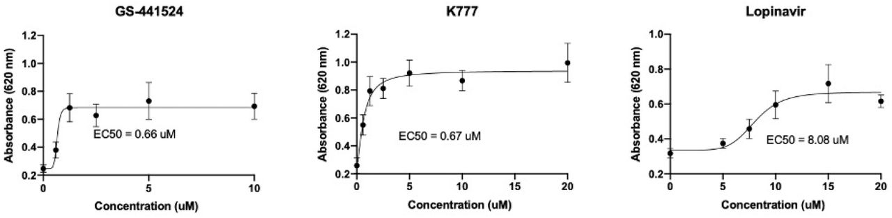
Figure 3
Representative examples of non-linear EC50 regression assays for compounds with anti-FIPV activity.

Serial dilutions of each compound with anti-FIPV activity were performed to identify half-maximal effective concentration (EC50). The GS-441524 results reported herein represent a compound derived from MedChemExpress.

Cytotoxicity safety profiles

Cytotoxicity safety profiles (CSPs) were determined for ten different antiviral compounds in CRFK cells. At 5 μM, the seven test compounds showed essentially no cytotoxicity, while two of the antivirals, amodiaquine and toremifene, had 11 and 12% cytotoxicity, respectively (Fig. 4; Table 2). The 50% cytotoxic concentration (CC50) for GC376 is reported as> 150μM [32]. Interestingly, based on the Promega CellTox-Green Cytotoxicity assay, the cytotoxicity of both EIDD compounds was essentially undetectable up to 100μM. However, visual inspection of the EIDD wells just prior to fluorescent dye application and matrix reading revealed differences in cell morphology (cytopathic effect) between untreated CRFK cells and treated cells. Untreated CRFK cells showed adherent spindle morphology in a single monolayer, while EIDD wells showed a marked decrease in confluence compared to variable cell morphology, including cell rounding (cytopathic effect). The discrepancy between the subjective visual evaluation of the EIDD wells and the fluorescence assay is a mystery. It is possible that an overall reduction in the number of cells in the EIDD wells led to the loss and degradation of the nucleic acid necessary for fluorescent binding and detection in the CellTox assay.

CompoundDrug category5μM10μM25μM50μM100μM
ElbasvirNS5A Inhibitor0.670.461.424.9
K777 / K11777PI0.610.292.3916
Toremifene citrateOther *1222233539
AmodiaquineOther **1112192326
LopinavirPI0.6715131618
RitonavirPI0.490.8423326
GrazoprevirPI0.5500.813.919
EIDD1931NPI1.20.940.60.782.8
EIDD2801NPI00.950.631.70.8
GS-441524+NPI0.210000
Table 2
Percent cytotoxicity by compound and concentration.
PI = Protease inhibitor; NPI = Nucleoside polymerase inhibitor
* Selective estrogen receptor modulator
** 4-Aminoquinoline
+ NMPharmTech
Figure 4
Representative cytotoxicity profiles.
Bar graphs of percent cytotoxicity +/- standard deviation (SD) for four compounds with anti-FIPV activity. Percent cytotoxicity values were determined by normalizing cytotoxicity for control wells with positive toxicity (set to 100% cytotoxicity) and untreated CRFK cells (set to 0% baseline cytotoxicity).

Quantification of inhibition of viral RNA production in monotherapy

A real-time RT PCR assay was used to measure the ability of each antiviral agent to inhibit coronavirus replication in monotherapy (Viral RNA knock-down assay). The compounds demonstrating the greatest inhibition of FIPV RNA production were GC376, a 3C-like coronavirus protease inhibitor, GS-441524, EIDD-1931 and EIDD-2801, the last three being nucleoside analogs (Fig. 5, Table 3). Substances with the least inhibitory effect on viral RNA production include elbasvir, nelfinavir and ritonavir. Ritonavir, a protease inhibitor, is used in combination with lopinavir to treat HIV-1 infection (Kaletra, AbbVie). Lopinavir monotherapy has unsatisfactory oral bioavailability in humans, but when used in combination, ritonavir has been shown to significantly improve lopinavir plasma concentrations [33]. Therefore, despite the relatively minimal inhibition of FIPV identified with ritonavir as monotherapy, this compound has been further tested, including combined anti-cancer evaluation.

CompoundVirus titer reduction fold
GC376 (20μM)25000
GC3767300
GS-441524 (NMPharmTech)5280
EIDD-19313700
GS-441524 (MedChemExpress)3500
EIDD-28012110
Lopinavir309
Toremifene10
K7777
Grazoprevir5
Amodiaquine4
Elbasvir2
Nelfinavir mesylate1
Ritonavir1
Table 3
Multiple reduction in viral RNA copy number for anti-FIPV compounds on monotherapy
* Unless otherwise indicated, all compounds were used at 10 μM.
Figure 5
Multiple reduction in FIPV RNA copy number using antiviral compounds as monotherapy.

FIPV-infected CRFK cells were incubated for 24 hours with compounds with detected anti-FIPV activity. The viral copy number was then determined by RT-qPCR and normalized to feline GAPDH copy number to determine the fold reduction effect for each compound. All compounds were tested at 10μM unless otherwise noted. All experimental treatments were performed in triplicate wells, and the fold decrease was calculated by dividing the average experimental normalized FIPV copy number by the average normalized FIPV copy number determined for untreated FIPV-infected wells.
1GS-441524 - NMPharmTech (China).
2GS-441524 - MedChemExpress (China).

Quantification of inhibition of viral RNA production in cACT

To identify drug combinations with synergistic antiviral activity versus monotherapy, combinations of two or more compounds were selected based on (i) established combinations used in other viral infections such as HIV-1 and HCV, (ii) drugs with different mechanisms of action, (iii) potential changes in the systemic distribution of the compound (eg ability to cross the blood-brain or blood-eye barrier according to chemical classification) and (iv) minimal cytotoxicity (based on CSP). For each cACT, any resulting reduction in FIPV copy number in excess of the calculated additive effect for each drug used in the monotherapy regimen was considered synergistic (Table 4). The combination of GC376 and amodiaquin achieved the greatest synergistic effect with the highest overall fold reduction in viral RNA with a 76-fold reduction in viral RNA compared to the additive effect (Fig. 6). This particular synergistic combination was one of the surprising results, given that amodiaquin alone showed only limited inhibition of FIPV viral RNA copies as determined by qRT-PCR.

Compound 1Compound 2Compound 3Virus titer reductionAddcACT / add
GC376 (20 μM)Amodiaquine—18970002500476
GC376 (20 μM)AmodiaquineToremifene2560002501410
GC376 (20 μM)K777—2480002500710
GC376 (20 μM)Toremifene—128000250105.1
GC376 (20 μM)Nelfinavir mesylate—91100250013.6
Elbasvir (5 μM)Lopinavir—1400031145
Elbasvir (5 μM)GC376 (20 μM)—12600250020.50
GC376 (10 μM)Amodiaquine—1170073041.6
GC376 (10 μM)Grazoprevir—829073051.1
GC376 (20 μM)GS-441524 (NMPharmTech)8260302800.27
GC376 (10 μM)AmodiaquineElbasvir (5 μM)757073061.0
GC376 (10 μM)GS-441524 (MedChem)—6910108000.64
GC376 (20 μM)Ritonavir—6560250010.26
K777Lopinavir—553031618
GC376 (10 μM)GS-441524 (NMPharmTech)—4340125800.34
GC376 (20 μM)Lopinavir—3400253090.13
LopinavirRitonavir—313031010
LopinavirRitonavirToremifene17403205.4
GC376 (10 μM)EIDD-2801—25594100.03
K777Toremifene—25171.5
Table 4
Multiple reduction in FIPV viral RNA copy number in combination therapy (cACT).
The expected additive effect reflects the sum of the fold reductions in viral RNA of each compound used in monotherapy (Table 3).
* Unless otherwise indicated, all compounds were used at 10μM.
cACT / add - ratio of FIPV titer reduction in combination therapy versus the sum of fold reduction titers in monotherapy
Add - the sum of fold reduction titers in monotherapy
Figure 6
Selected examples of fold FIPV RNA copy reduction using combination therapy (cACT).
The bars represent the mean fold decrease in three wells of treated CRFK cells compared to the mean fold decrease in three untreated wells infected with FIPV. All compounds were tested at 10μM unless otherwise noted.

Given the strong anti-FIPV activity of GC-376, as well as its potential availability for advancement in in vivo pharmacokinetic studies, clinical trials, and promising use in cACT, this compound was selected for a series of “viral RNA knock-down” assays in monotherapy and combination therapy (Fig. 7). Overall, GC376 demonstrated excellent anti-FIPV activity at 20μM both in monotherapy and combination therapy in vitro. The most significant reduction in FIPV RNA occurred when GC376 was combined at 20μM with amodiaquine at 10μM. The experiment combining GC376 with amodiaquine was repeated, and both results are shown for comparison in Fig. 7C.

Figure 7
GC376 antiviral activity in monotherapy and in combination therapy at 10 and 20 μM

(A) FIPV RNA reduction quantified by RT-qPCR using GC376 as monotherapy at 10 and 20 μM. There is a significant difference between the two concentrations, with 20μM being better than 10μM. (unpaired t-test; p <0.0001).
(B) Combination in vitro therapy using GC376 at 10 μM.
(C) Combination in vitro anti-FIPV therapy using GC376 at 20 μM.

Discussion

Because there is currently no effective vaccine against FIP, there is a strong clinical and worldwide need for effective antiviral treatment options for FIPV-infected cats. We tested 89 compounds, which resulted in the identification of 25 antiviral agents with antiviral activity and strong safety profiles against feline coronavirus, FIPV. We also identified combinations of antiviral agents (cACT) that resulted in greater efficacy or synergism over monotherapy alone. Of particular interest was the finding regarding the use of elbasvirus, which repeatedly demonstrated excellent protection of CRFK against CPE-induced FIPV at concentrations below 1 μM based on multiple assays (EC50 0.16 μM). In principle, however, no difference in viral RNA copy number was found between infected cells treated with or without elbasvirus. Further visual analysis of FIPV-infected CRFK cells treated with elbasvirus revealed an atypical cell morphology relative to uninfected cells, which was characterized by variable enlargement, cell rounding, and partial cell detachment (cytopathic effect). These "atypical cells" were rarely detached from the culture plate, and as a result, absorbance values were comparable to uninfected control wells. This dichotomous result between platelet analysis and viral RNA knock-down assay suggests that the antiviral effect of elbasvir occurs after viral replication and, as a result, elbasvir may not protect cells from viral RNA accumulation. Elbasvir is used to treat patients infected with hepatitis C virus (HCV) and is thought to target the HCV NS5A protein, which prevents replication and also to complete virions [34]. Although no NS5A homolog has been identified for FIPV, it is possible that elbasvir exhibits a similar antiviral effect by preventing the assembly of FIPV virions without blocking viral RNA synthesis in CRFK cells. Additional evaluation of treated FIPV-infected CRFK cells by transmission electron microscopy may shed light on the effect of elbasvirus on protecting CRFK from FIPV-related damage and death.

Co-administration of ritonavir with lopinavir has been shown to significantly increase lopinavir plasma concentrations in rats, dogs and humans [33]. Ritonavir is a potent inhibitor of CYP3A, which is the primary enzyme responsible for protease inhibitor metabolism, and therefore its co-administration with other protease inhibitors results in increased systemic concentrations of co-administered protease inhibitors such as lopinavir [38, 39]. The increase in the antiviral effect of lopinavir associated with ritonavir was relatively minimal in the viral RNA elimination assays with only a 10-fold inhibition of FIPV over the additive effect. This may be the result of an in vitro testing artifact on a feline kidney cell line (ie CRFK cells) that lacks the enzyme CYP3, an enzyme that typically occurs at sites of high protease inhibitor metabolism at the first pass effect (ie enterocytes). and hepatocytes) [39]. These results suggest that in vitro tests alone may not fully predict the effect of antiviral agents in FIPV-infected cats in vivo.

Grazoprevir, a serine protease inhibitor NS3 / 4, has been used in combination with elbasvirus, an NS5A inhibitor, to treat HCV-infected patients (Zepatier, Merck) [40]. Here, we demonstrated that grazoprevir has anti-FIPV activity when used as monotherapy. The cysteine protease inhibitor K777 / K11777 has been investigated for its ability to block coronavirus (MERS-CoV and SARS-CoV-1) and ebolavirus entry and has been found to completely inhibit coronavirus infection, but only in target cell lines without virus-activating serine proteases. [41]. For other cell lines, K777 inhibited coronavirus cell entry in combination with a serine protease inhibitor [41]. It is possible that limited inhibition of FIPV RNA K777 production could be increased if combined with a serine protease inhibitor.

Amodiaquine is an antimalarial drug and belongs to the class of 4-aminoquinoline drugs. Amodiaquine, along with related 4-aminoquinolines such as chloroquine and hydroxychloroquine, was originally developed to treat malaria [42], and like chloroquine and hydroxychloroquine, it has a wide range of anatomical distributions, including the eyes and brain [43-49]. Penetration of antiviral agents into the CNS and / or ocular compartments is particularly important in FIPV-infected cats with neurological and / or ocular disorders. While several studies have defined the antiviral properties of chloroquine and hydroxychloroquine [28, 50, 51], the antiviral activity of amodiaquin has also been investigated with the identification of antiviral activity against dengue virus, Ebola virus and severe fever with viral thrombocytopenia syndrome (SFTS) [52–55]. The mechanism of action of amodiaquine may involve an increase in cytoplasmic lysosomal and / or endosomal pH, which prevents the release of viable virions into the cytoplasm [56]. Due to its unique drug class status and presumed ability to cross the blood-brain barrier [57], amodiaquine is a promising candidate for the combined treatment of neurological and / or ocular forms of FIP.

Toremifene citrate, a selective estrogen receptor modulator (SERM), is used to treat metastatic breast cancer in human patients. Recently, toremifene has been evaluated for its antiviral properties and has demonstrated anticorrosive activity against zoonotic coronaviruses, Middle East Respiratory Syndrome (MERS-CoV) and SARS-CoV-1 coronaviruses [58]. Toremifene has also been shown to be active against Ebola virus (EBOV) [59, 60]. Although the exact mechanism of antiviral action is not defined, the antiviral effect of toremifene against EBOV appears to be to destabilize the EBOV glycoprotein [59].

Interestingly, GC376 demonstrated confusing differences between 10 μM and 20 μM in combination therapy. When used at 10 μM with other compounds, synergism and in some cases a decrease in antiviral effect compared to additive values ranging from 0.03 to 1.6 were absent (Table 4). When used at 20 μM, there were still cases where combination with another compound resulted in a reduced antiviral effect compared to GC376 used as monotherapy at 20 μM. However, many more variations in the 20 μM combinations showed a fold effect compared to additive values ranging from 0.13 to 76 (Table 4). A specific example is the contrast between GC376 at 20 μM compared to GC376 at 10 μM combined with amodiaquine at 10 μM. The first caused the greatest inhibition of viral RNA as well as the greatest multiple of the additive (synergistic) effect, while the second caused almost a loss of synergism with a value of the additive multiple of 1.6.

The identification of effective antiviral strategies for the treatment of FIPV-infected cats has translational implications for the ongoing SARS-CoV-2 pandemic. FIPV infection in cats resembles coronavirus infection in ferrets [61, 62] and is compared with the pathogenesis of other chronic macrophage-dependent diseases such as tuberculosis [63]. Because the clinical and pathogenic details of SARS-CoV-2 infection in humans are still emerging, there appears to be some overlap with FIPV in anatomical distribution, clinical manifestations, and likely response to certain antiviral therapies. In cats, the feline enteric coronavirus biotype (FECV) is restricted to the gastrointestinal tract due to enterocyte tropism. Clinical symptoms in FECV-infected cats range from mild gastrointestinal disease (diarrhea) to the absence of symptoms. The mutated FIPV coronavirus feline biotype acquires macrophage tropism and preferentially targets serous abdominal and thoracic surfaces with a subset of cats that demonstrate CNS or eye involvement [7]. Similarly, in patients with COVID-19, there are reports of diarrhea and a subset of patients with CNS involvement [14]. Although the cellular receptor for SARS-CoV-2 has been identified as ACE2 [64], the cellular receptor for FIPV serotype I has yet to be determined. The cellular receptor for less clinically relevant FIPV serotype II has been identified as feline aminopeptidase peptidase (fAPN) [4]. A study using RNAseq to evaluate gene expression profiles of ascites cells obtained from cats with FIP did not identify ACE2 expression, suggesting that ACE2 is unlikely to be a FIPV serotype I receptor [63]. A more detailed examination of the identity of the FIPV serotype I receptor is needed.

Clinical successes with GS-441524 or GC-376 in cats with experimental and naturally occurring FIPs indicate that FIP can be effectively treated, but treatment of dry (granulomatous), neurological and ocular FIPs remains a challenge. The protease inhibitor 3C-like protease, GC-376, appears to be relatively effective in the treatment of FIPV effusion infection limited to body cavities, but may be less effective in the treatment of neurological or ocular forms of the disease [24]. These different results may be the result of ineffective penetration of the blood-brain and blood-eye barriers, making GC-376 a promising candidate for combination therapy with a CNS-penetrating antiviral drug.

Materials and methods

FIPV inoculation for in vitro experiments

Crandell-Reese cat kidney cells (CRFK, ATCC) were cultured in T150 flasks (Corning), seeded with FIPV serotype II (WSU-79-1146, GenBank DQ010921) and propagated in 50 ml of Dulbecco's modified Eagle's medium (DMEM) with 4 , 5 g / l glucose (Corning) and 10% fetal bovine serum (Gemini Biotec). After 72 hours of incubation at 37 ° C, extensive cytopathic effect (CPE) and large cell clearing / separation areas were noted. The flasks were then flash frozen at -70 ° C for 8 minutes, thawed briefly at room temperature, and the cells and supernatant were then centrifuged at 1500 g for 5 minutes, followed by a second centrifugation step at 4000 g for 5 minutes to isolate cell - free viral volumes. The supernatant containing the virus base was divided into 0.5 and 1.0 ml aliquots in 1.5 ml cryotubes (Nalgene) and archived at -70 ° C. After freezing, one tube was allowed to thaw and the virus titer was determined using biological assays (TCID50) and real-time RT PCR methods (below).

The tissue culture dose-50 infectious dose (TCID50) was determined using a viral plaque assay. CRFK cells were grown in a 96-well tissue culture plate (Genesee Scientific) until CRFK cells reached approximately 75-85% confluence. Serial 10-fold dilutions were prepared from FIPV stock solution and 200 μl samples from each dilution were added to 10-well replicates. 72 hours after infection, cells were fixed with methanol and stained with crystal violet (Sigma-Aldrich). Individual wells were visually evaluated for virus-induced CPE, evaluated as CPE positive or negative, and TCID50 was determined based on the log equation.10TCID50 = [total number of # wells CPE positive / # replicates] + 0.5 to reflect infectious virions per milliliter of supernatant [68].

Quantification of FIPV by qRT-PCR

Cell-free viral RNA was isolated from the starting virus using the QIAamp Viral RNA Mini Kit (Qiagen) according to the manufacturer's instructions. The isolated RNA was treated with DNase (Turbo DNase, Invitrogen) and then reverse transcribed using the High-Capacity RNA-to-CDNA Kit (Applied Biosystems) according to the manufacturers' protocols. Copy numbers of FIPV and feline GAPDH cDNA were determined using the Applied Biosystems' QuantStudio 3 Real-Time PCR System and PowerUp SYBR Green Master Mix according to the manufacturer's protocol for a 10 μL reaction. Each PCR reaction was performed in triplicate with aqueous template as a negative control and plasmid DNA as a positive control. A control reaction excluding reverse transcriptase was included in each set of real-time PCR assays. cDNA templates were amplified using the FIPV forward primer, 5'-GGAAGTTTAGATTTGATTTGGCAATGCTAG, and the FIP reverse primer, 5'-AACAATCACTAGATCCAGACGTTAGCT (terminal part of the FIPV 7b gene) [25]. Real-time PCR for the feline housekeeping gene GAPDH was performed simultaneously using primers, 5'-GAPDH, 5'-AAATTCCACGGCACAGTCAAG, and 3'-GAPDH, 5'-TGATGGGCTTTCCATTGATGA. Cycling conditions for both FIPV and GAPDH amplicons were as follows: 50°C for 2 min, 95°C for 2 min, followed by 40 cycles of 95°C for 15 s, 58°C for 30 s, 72°C for 1 min. The final step included a dissociation curve to evaluate the specificity of primer binding. FIPV and GAPDH copy numbers were calculated based on standard curves generated in our laboratory. FIPV cDNA copies determined by real-time RT PCR were normalized to 106 copies of feline GAPDH cDNA.

Development of anti-helicase chemical fragments

The drugs studied and described in this study were already known antiviral agents. In contrast, the helicase enzyme FIPV was cloned, expressed and used as a target for coronavirus and enzyme-specific viral discovery. The AviTag-FIP Helicase-HisTag target DNA sequence was optimized and synthesized. The synthesized sequence was cloned (Adeyemi Adedeji) into the Avi-His tagged pET30a vector to express the protein in E. coli. E. coli strain BL21 (DE3) was transformed with a recombinant plasmid. One colony was inoculated into 1 liter of auto-induced medium containing the antibiotic and the culture was incubated at 37 ° C at 200 rpm.

When the OD600 reached about 3, the cell culture temperature was changed to 15 ° C for 16 hours. Cells were harvested by centrifugation. The cell pellets were resuspended in lysis buffer followed by sonication. The centrifuge precipitate was dissolved with a denaturing agent. The target protein was obtained by one-step purification on a Ni column. The target protein was sterilized with a 0.22 μm filter. The yield was 7.2 mg at 0.90 mg / ml and was stored in PBS, 10% glycerol, 0.5 mM L-arginine, pH 7.4. The concentration was determined by the Bradford protein assay with BSA as a standard. Protein purity and molecular weight were determined by SDS-PAGE with Western blot confirmation.

Surface plasmon resonance (SPR) fragments were screened on a ForteBio Pioneer FE SPR platform. A HisCap sensor chip containing an NTA surface matrix was used. Channels 1 and 3 were filled with 100 μM NiCl 2, followed by injection of 50 μg / ml FIP protein. Channel 2 was left protein-free as well as NiCl 2 as a reference. Channel 1 was immobilized to a density of 88000 RU, while channel 3 contained approximately 12000 RU. Channel 1 was used. The buffer used for immobilization was 10 mM HEPES, pH 7.4, 150 mM NaCl and 0.1% Tween-20. DMSO was added to a final concentration of 4% for this assay. The proprietary compound library was diluted in the same DMSO-free buffer to a final DMSO concentration of 4% DMSO. Compounds from the library were tested at a concentration of 100 μM using the OneStep gradient injection method. The findings were selected based on RU and kinetics and used for cell screening.

Viral plaque test

To screen for antiviral activity of compounds, infected CRFK cells were treated with compounds in six-well replicates and compared to positive control wells (infected cells), negative controls (uninfected cells) and treatment controls (infected cells treated with a known active antiviral compound) simultaneously on each tissue plate. culture. CRFK cells were grown in 96-well tissue culture plates (Genesee Scientific) containing 200 μl of culture medium. At 7575-85% cell confluence, the medium in uninfected control wells was aspirated and replaced with 200 μl of fresh medium. The medium in the infected wells was aspirated and replaced with FIPV inoculated medium at a multiplicity of infection (MOI) of 0.004 infectious virion per cell. The tissue culture plate was incubated for 1 hour with periodic gentle agitation ("number eight" manipulations) every 15 minutes to facilitate virus-cell interaction. One hour after infection, each putative antiviral compound was added to six wells infected with FIPV (to determine the antiviral activity of the compound) and six uninfected control wells (to screen for cytotoxicity of the compound in CRFK cells). All compounds were initially screened at 10 μM, except for the "chemical fragment" compounds supplied by M. Olsen (Midwestern University), which were evaluated at 50 μM. Tissue culture plates were incubated at 37 ° C for 72 hours and then fixed with methanol and stained with crystal violet. Plates were scanned for absorbance at 620 nm using an ELISA plate reader (FilterMax F3, Molecular Devices; Softmax Pro, Molecular Devices). For each treatment condition, individual well absorbance values ​​were recorded along with the mean absorbance value and mean error of the mean for 6-well experimental replicates.

For substances that demonstrated antiviral efficacy at initial screening at 10 or 50μM (protected from CPE-associated virus), the EC50 was determined by performing a series of progressive 2-fold dilutions of the compounds in a viral plaque assay. To determine the EC50, CRFK cells were grown in 96-well tissue culture plates similar to the antiviral screening assay. Except for uninfected control wells, all remaining wells were infected with FIPV as described above. A two-fold dilution series ranged from 20μM to 0μM and each concentration was performed in six well replicates. The number of dilution steps ranged from 6 to 14 and was compound dependent. Six well replicates of uninfected CRFK cells served as a control for normal CRFK cells; six FIPV-infected CRFK cell replicates served as untreated FIPV-infected control wells; and six well replicates of FIPV-infected CRFK cells treated with GS-441524 served as control wells for protection against virus-induced cell death based on published data on the efficacy of using GS-441524 in vitro in CRFK cells [26].

Tissue culture plates were incubated for 72 hours and then fixed with methanol, stained with crystal violet, and the absorbance at 620 nm was scanned using an ELISA plate reader. Individual absorbance values along with the mean absorbance value and standard deviation for 6-well experimental replicates were recorded for each treatment condition. The EC50 was calculated by plotting a non-linear regression equation (dose-response curve) using Prism 8 software (GraphPad).

Viral RNA knock-down test

Real-time RT-PCR assays were used to quantify inhibition of viral RNA production by the compound. CRFK cells were cultured in a 6-well tissue culture plate (Genesee Biotek). At approximately 75-85% cell fusion, the culture medium was replaced with fresh medium and the cells were infected with FIPV serotype II at an MOI of 0.2 (MOI based on TCID50 bioassay / pfu). The plates were incubated for one hour with periodic gentle shaking every 15 minutes. Wells infected with FIPV were treated with one (monotherapy), two or three (combined anti-cancer therapy) antiviral compounds; each experimental treatment was performed three times. The compound dose was based on the EC50 of the compounds and ranged from 0.001 to 20 μM. For each experimental set, three culture wells with FIPV-infected and untreated CRFK cells served as virus-infected controls. Infected cell cultures were then incubated for 24 hours and total RNA associated with the cells was isolated using a PureLink-RNA mini kit (Invitrogen). RNA was treated with DNAse (TurboDNAse, Ambion), reverse transcribed into cDNA using the High-Capacity RNA-to-cDNA Kit (Applied Biosystems), and FIPV cIPNA and feline GAPDH cDNA were measured by real-time qRT-PCR as described above. . The fold reduction in viral titer was determined by dividing the normalized mean FIPV RNA copy number for untreated FIPV-infected CRFK cells into the normalized mean FIPV RNA copy number for CRFK treated cells with the desired compound (s). The expected additive effect was determined by adding a fold reduction for each monotherapy used in combination. The composite additive effect was determined by dividing the predicted additive effect by the combined multiple reduction value for a particular combination therapy.

Determination of cytotoxicity safety profiles (CSP).

The cytotoxicity of the compound in feline cells was assessed using a commercially available kit (CellTox Green Cytotoxicity Assay, Promega) according to the manufacturer's instructions. Untreated CRFK cells were used as negative controls and the cells were treated with a cytotoxic solution provided by the manufacturer as positive toxicity controls. Briefly, in addition to control wells, CRFK cells were plated in 96-well tissue culture plates (Genesee Scientific) in four well replicates with 5, 10, 25, 50 or 100 μM concentrations of the desired compound and incubated for 72 hours. After 72 hours, all wells were stained with the DNA kit, incubated at 37 ° C protected from light for 15 minutes, and the fluorescence intensity at 485-500 nm Ex / 520-530 nm EM was subsequently determined using a plate reader (FilterMax F3, Molecular Devices; Softmax Pro, Molecular Devices). The cytotoxicity of a compound at a particular concentration was thought to be proportional to the fluorescence intensity based on the selective penetration and binding of the dye to the DNA of degenerated, apoptotic or necrotic cells. The extent of cytotoxicity was determined by adjusting the fluorescence value for cells treated with the positive control reagent to 100% and untreated feline cells as 0% cytotoxicity. The average fluorescence value for the four wells containing each compound concentration was then interpolated as a percentage (percent cytotoxicity) ranging from 0 to 100%.

Conflict of interests

The authors declare that there has been no conflict of interest.

We appreciate funding provided by the Winn Feline Foundation (MTW 17-020; MTW 19-026) and the University of California, Davis, Center for Companion Animal Health (CCAH; 2018-92-F; 2018-94-FE) through multi-FIP research donations individual donors and organizations (SOCK FIP, Davis, CA) and foundations (Philip Raskin Fund, Kansas City, KS).

Additional information

Complete list of compounds tested in vitro for FIPV activity

Nucleoside polymerase inhibitors

12x GS Nuc AnalogsNucleoside analogNPI
GS-441524 (China-sourced)Adenosine analog nucleosideNPI
3-Deazaneplanocin A HydrochlorideAdenosine analog nucleosideNPI
AdefovirAdenosine analog nucleosideNPI
GalidesivirAdenosine analog nucleosideNPI
GS-441524 (Manufactured in China)Adenosine analog nucleosideNPI
MK-0608Adenosine analog nucleosideNPI
NITD008Adenosine analog nucleosideNPI
DidanosineAdenosine analog nucleosideNPI
Tenofovir alafenamideAdenosine analog nucleosideNPI
Tenofovir disoproxil fumarateAdenosine analog nucleosideNPI
EIDD 1931Nucleoside analog Cytidine 
EIDD 2801Nucleoside analog Cytidine 
2′-C-methylcytidineNucleoside analog CytidineNPI
Gemcitabine HydrochlorideNucleoside analog CytidineNPI
2-C-methylguanosineNucleoside analogue GuanosineNPI
7-methylguanosineNucleoside analogue GuanosineNPI
EntecavirNucleoside analogue GuanosineNPI
MizoribineNucleoside analogue GuanosineNPI
RibavirinNucleoside analogue GuanosineNPI
PSI-6206Nucleoside analog UridineNPI
6-AzauridineNucleoside analog UridineNPI
BalapiravirNucleoside analog CytidineNPI
SofosbuvirNucleoside analog UridineNPI
FavipiravirNucleoside analog PurineNPI
Total 36

Protease inhibitors

GrazoprevirNS3 / 4A protease inhibitorPI
Rupin trivirRhinoviral 3CP inhibPI
LopinavirAntiretroviral PIPI
RitonavirAntiretroviral PIPI
NelfinavirAntiretroviral PIPI
Disulfiram (tetraethyliuram disulfide)Papain-like protease inhibPI
K777 / K11777Cysteine protease inhibitorPI
TelaprevirNS3 / 4A protease inhibitorPI
Camostat mesylateSerine protease inhibitorPI
ParitaprevirSerine protease inhibitorPI
GC376Coronavirus protease inhibitorPI
Total 11

NS5A inhibitors

VelpatasvirNS5A InhibitorNS5A Inhibitor
Ravidasvir / PPI-668NS5A InhibitorNS5A Inhibitor
LedipasvirNS5A InhibitorNS5A Inhibitor
OmbitasvirNS5A InhibitorNS5A Inhibitor
PibrentasvirNS5A InhibitorNS5A Inhibitor
DaclatasvirNS5A InhibitorNS5A Inhibitor
ElbasvirNS5A InhibitorNS5A Inhibitor
Total 7

NNPI

DasabuvirNon-nucleoside polymerase inhibitorNNPI
Total 1

other

MonensinIonophoreother
Phenazopyridine hydrochlorideCrystalline solidother
Pyrvinium pamoate hydrateAndrogen receptor inhibitorother
Toremifene citrateSelective estrogen receptor modulator other
AM580Retinobenzoic derivativeother
HomoharringtonineTranslation elongation inhibother
Amodiaquine4-aminoquinoloneother
Total 7

Midwestern Chemical Fragments

F0472-0017Midwestern
F6190-0257Midwestern
F6279-0675Midwestern
F2167-1080Midwestern
F6190-0740Midwestern
F6438-2155Midwestern
F3411-5663Midwestern
F6233-0011Midwestern
F9995-2543Midwestern
F2124-0890Midwestern
F2711-2577Midwestern
F2130-0055Midwestern
F2493-3358Midwestern
F2459-0974Midwestern
F2124-0465Midwestern
F1899-2269Midwestern
F2185-1982Midwestern
F2189-0717Midwestern
F2147-0158Midwestern
F1371-0192Midwestern
F2156-0057Midwestern
F9995-2431Midwestern
F3349-0218Midwestern
F2156-0059Midwestern
F2156-0070Midwestern
F5856-0194Midwestern
F2147-0975Midwestern
Total 27

References

  1. Chang HW, de Groot RJ, Egberink HF, et al. Feline infectious peritonitis: Insights into feline coronavirus pathobiogenesis and epidemiology based on genetic analysis of the viral 3c gene. J Gen Virol 2010; 91: 415–420.CrossRefPubMedWeb of ScienceGoogle Scholar
  2. Pedersen NC, Liu H, Dodd KA, et al. Significance of coronavirus mutants in feces and diseased tissues of cats suffering from feline infectious peritonitis. Viruses 2009; 1: 166–184.CrossRefPubMedWeb of ScienceGoogle Scholar
  3. Vennema H, Poland A, Foley J, et al. Feline infectious peritonitis viruses arise by mutation from endemic feline enteric coronaviruses. Virology 1998; 243: 150–157.CrossRefPubMedWeb of ScienceGoogle Scholar
  4. Rottier PJM, Nakamura K, Schellen P, et al. Acquisition of Macrophage Tropism during the Pathogenesis of Feline Infectious Peritonitis Is Determined by Mutations in the Feline Coronavirus Spike Protein. J Virol 2005; 79: 14122–14130.Abstract / FREE Full TextGoogle Scholar
  5. Diaz JV., Poma R. Diagnosis and clinical signs of feline infectious peritonitis in the central nervous system. Can Vet J 2009; 50: 1091–1093.PubMedGoogle Scholar
  6. Foley JE, Lapointe JM, Koblik P, et al. Diagnostic features of clinical neurologic feline infectious peritonitis. J Vet Intern Med 1998; 12: 415–423.CrossRefPubMedGoogle Scholar
  7. Pedersen NC. An update on feline infectious peritonitis: Diagnostics and therapeutics. Vet J 2014; 201: 133–141.CrossRefPubMedGoogle Scholar
  8. Stiles J. Ocular manifestations of feline viral diseases. Vet J 2014; 201: 166–173.CrossRefPubMedGoogle Scholar
  9. Pedersen NC. A review of feline infectious peritonitis virus infection: 1963-2008. J Feline Med Surg 2009; 11: 225–258.CrossRefPubMedGoogle Scholar
  10. Hohdatsu T, Yamada M, Tominaga R, et al. Antibody-Dependent Enhancement of Feline Infectious Peritonitis Virus Infection in Feline Alveolar Macrophages and Human Monocyte Cell Line U937 by Serum of Cats Experimentally or Naturally Infected with Feline Coronavirus. J Vet Med Sci 1998; 60: 49–55.CrossRefPubMedGoogle Scholar
  11. Takano T, Yamada S, Doki T, et al. Pathogenesis of oral type I feline infectious peritonitis virus (FIPV) infection: Antibodydependent enhancement infection of cats with type I FIPV via the oral route. J Vet Med Sci 2019; 81: 911–915.CrossRefPubMedGoogle Scholar
  12. Vennema H, de Groot RJ, Harbor DA, et al. Early death after feline infectious peritonitis virus challenge due to recombinant vaccinia virus immunization. J Virol 1990; 64: 1407–1409.Abstract / FREE Full TextGoogle Scholar
  13. Fenner's Veterinary Virology. 2017. Epub ahead of print 2017. DOI: 10.1016 / c2013-0-06921-6.CrossRefGoogle Scholar
  14. Wei X, Wang X, Peng W, et al. The Lancet Respiratory Medicine Clinical characteristics of SARS-CoV-2 infected pneumonia with diarrhea. 4.Google Scholar
  15. Hosoda T, Sakamoto M, Shimizu H, et al. SARS-CoV-2 enterocolitis with persisting to excrete the virus for about two weeks after recovering from diarrhea: A case report. Infect Control Hosp Epidemiol 2020; 1-4.Google Scholar
  16. Hamming I, Timens W, Bulthuis MLC, et al. Tissue distribution of ACE2 protein, the functional receptor for SARS coronavirus. A first step in understanding SARS pathogenesis. 2004; 631–637.Google Scholar
  17. Zhang J, Wu Y, Wang R, et al. Bioinformatic analysis of reproductive system Bioinformatic analysis reveals that the reproductive system is potentially at risk from SARS-CoV-2. 2020; 1-15.Google Scholar
  18. Zheng YY, Ma YT, Zhang JY, et al. COVID-19 and the cardiovascular system. Nat Rev Cardiol. Epub ahead of print 2020. DOI: 10.1038 / s41569-020-0360-5.CrossRefGoogle Scholar
  19. Chen L, Deng C, Chen X, et al. Ocular manifestations and clinical characteristics of 534 cases of COVID-19 in China: A cross-sectional study. medRxiv 2020; 2020.03.12.20034678.Google Scholar
  20. Mao L, Wang M, Chen S, et al. Neurological Manifestations of Hospitalized Patients with COVID-19 in Wuhan, China: a retrospective case series study. medRxiv 2020; 2020.02.22.20026500.Google Scholar
  21. Chen C, Zhou Y, Wang DW. SARS-CoV-2: a potential novel etiology of fulminant myocarditis. Heart 2020; 10-12.Google Scholar
  22. Li YC, Bai WZ, Hashikawa T. The neuroinvasive potential of SARS-CoV2 may be at least partially responsible for the respiratory failure of COVID-19 patients. J Med Virol 2020; 24-27Google Scholar
  23. Shi J, Wen Z, Zhong G, et al. Susceptibility of ferrets, cats, dogs, and other domesticated animals to SARS-coronavirus 2. Science. Epub ahead of print 2020. DOI: 10.1126 / science.abb7015.Abstract / FREE Full TextGoogle Scholar
  24. Pedersen NC, Kim Y, Liu H, et al. Efficacy of a 3C-like protease inhibitor in treating various forms of acquired feline infectious peritonitis. J Feline Med Surg 2018; 20: 378–392.CrossRefGoogle Scholar
  25. Murphy BG, Perron M, Murakami E, et al. The nucleoside analog GS-441524 strongly inhibits feline infectious peritonitis (FIP) virus in tissue culture and experimental cat infection studies. Vet Microbiol 2018; 219: 226–233.Google Scholar
  26. Pedersen NC, Perron M, Bannasch M, et al. Efficacy and safety of the nucleoside analog GS-441524 for the treatment of cats with naturally occurring feline infectious peritonitis. J Feline Med Surg 2019; 21: 271–281.Google Scholar
  27. Holshue ML, DeBolt C, Lindquist S, et al. First case of 2019 novel coronavirus in the United States. N Engl J Med 2020; 382: 929–936.CrossRefPubMedGoogle Scholar
  28. Wang M, Cao R, Zhang L, et al. Remdesivir and chloroquine effectively inhibit the recently emerged novel coronavirus (2019-nCoV) in vitro. Cell Res 2020; 30: 269–271.CrossRefPubMedGoogle Scholar
  29. Nakagawa F, May M, Phillips A. Life expectancy living with HIV: Recent estimates and future implications. Curr Opin Infect Dis 2013; 26: 17–25.CrossRefPubMedGoogle Scholar
  30. Simonetti FR, Kearney MF. Review: Influence of ART on HIV genetics. Current Opinion in HIV and AIDS. Epub ahead of print 2015. DOI: 10.1097 / COH.0000000000000120.CrossRefGoogle Scholar
  31. Arts EJ, Hazuda DJ. HIV-1 antiretroviral drug therapy. Cold Spring Harb Perspect Med; 2. Epub ahead of print 2012. DOI: 10.1101 / cshperspect.a007161.Abstract / FREE Full TextGoogle Scholar
  32. Kim Y, Liu H, Kankanamalage ACG. Reversal of the Progression of Fatal Coronavirus Infection in Cats by a Broad-Spectrum Coronavirus Protease Inhibitor. 2016; 1-18.Google Scholar
  33. Kumar GN, Jayanti VK, Johnson MK, et al. Metabolism and disposition of the HIV-1 protease inhibitor lopinavir (ABT-378) given in combination with ritonavir in rats, dogs, and humans. Pharm Res 2004; 21: 1622–1630.CrossRefPubMedWeb of ScienceGoogle Scholar
  34. Sulejmani N, Jafri SM, Gordon SC. Pharmacodynamics and pharmacokinetics of elbasvir and grazoprevir in the treatment of hepatitis C. Expert Opin Drug Metab Toxicol 2016; 12: 353–361.Google Scholar
  35. Combination Antiretroviral Therapy for HIV Infection - American Family Physician, https://www.aafp.org/afp/1998/0601/p2789.html (accessed 6 April 2020).Google Scholar
  36. Julg B. Atripla ™ - HIV therapy in one pill. 2008; 4: 573–577.Google Scholar
  37. Clay PG, Taylor TAH, Glaros AG, et al. “One pill, once daily”: what clinicians need to know about Atripla ™. 2008; 4: 291–302.Google Scholar
  38. Hill A, Van Der Lugt J, Sawyer W, et al. How much ritonavir is needed to boost protease inhibitors? Systematic review of 17 dose-ranging pharmacokinetic trials. Aids 2009; 23: 2237–2245.CrossRefPubMedWeb of ScienceGoogle Scholar
  39. A. H, GR G, RJ B. Ritonavir: Clinical pharmacokinetics and interactions with other anti-HIV agents. Clin Pharmacokinet 1998; 35: 275–291.CrossRefPubMedWeb of ScienceGoogle Scholar
  40. Kwo P, Gane EJ, Peng C, et al. Effectiveness of Elbasvir and Grazoprevir Combination, With or Without Ribavirin, for Treatment-Experienced Patients With Chronic Hepatitis C Infection. Gastroenterology 2017; 152: 164–175.e4.Google Scholar
  41. Zhou Y, Vedantham P, Lu K, et al. Protease inhibitors targeting coronavirus and filovirus entry. Antiviral Res 2015; 116: 76–84.CrossRefPubMedGoogle Scholar
  42. Alessandro SD, Scaccabarozzi D, Signorini L, et al. The Use of Antimalarial Drugs against Viral Infection. 2020; 1-26.Google Scholar
  43. Rynes RI, Bernstein HN. Ophthalmologic safety profile of antimalarial drugs. In: Lupus. 1993. Epub ahead of print 1993. DOI: 10.1177 / 0961203393002001051.CrossRefGoogle Scholar
  44. Briceño E, Reyes S, Sotelo J. Therapy of glioblastoma multiforme improved by the antimutagenic chloroquine. Neurosurg Focus. Epub ahead of print 2003. DOI: 10.3171 / foc.2003.14.2.4.CrossRefPubMedGoogle Scholar
  45. Ekong M, Igiri A, Ekanem T, et al. The effect of amodiaquine on some brain macromolecules of Wistar rats. Int J Biol Chem Sci. Epub ahead of print 2009. DOI: 10.4314 / ijbcs.v2i4.39762.CrossRefGoogle Scholar
  46. Mackenzie AH. Pharmacologic actions of 4-aminoquinoline compounds. Am J Med. Epub ahead of print 1983. DOI: 10.1016 / 0002-9343 (83) 91264-0.CrossRefPubMedWeb of ScienceGoogle Scholar
  47. Kuroda K. Detection and distribution of chloroquine metabolites in human tissues. J Pharmacol Exp Ther.Google Scholar
  48. Maguire A, Kolb H. THE EFFECT OF A SYNTHETIC ANTIMALARIAL (AMODIAQUINE) ON THE RETINA. Br J Dermatol. Epub ahead of print 1964. DOI: 10.1111 / j.1365-2133.1964.tb15487.x.CrossRefPubMedWeb of ScienceGoogle Scholar
  49. McAnally D, Siddiquee K, Goma A, et al. Repurposing antimalarial aminoquinolines and related compounds for treatment of retinal neovascularization. PLoS One. Epub ahead of print 2018. DOI: 10.1371 / journal.pone.0202436.CrossRefGoogle Scholar
  50. Keyaerts E, Li S, Vijgen L, et al. Antiviral activity of chloroquine against human coronavirus OC43 infection in newborn mice. Antimicrob Agents Chemother 2009; 53: 3416–3421.Abstract / FREE Full TextGoogle Scholar
  51. Savarino A, Boelaert JR, Cassone A, et al. Effects of chloroquine on viral infections: An old drug against today's diseases? Lancet Infect Dis 2003; 3: 722–727.CrossRefPubMedWeb of ScienceGoogle Scholar
  52. Azuma P, Massaquoi M, Job D, et al. Effect of Artesunate – Amodiaquine on Mortality Related to Ebola Virus Disease. 2016; 23–32.Google Scholar
  53. Boonyasuppayakorn S, Reichert ED, Manzano M, et al. HHS Public Access. 2015; 125–134.Google Scholar
  54. Baba M, Toyama M, Sakakibara N. Establishment of an antiviral assay system and identification of severe fever with thrombocytopenia syndrome virus inhibitors. 2017; 25: 83–89.Google Scholar
  55. Savarino A, Lucia MB, Rastrelli E, et al. Anti-HIV Effects of Chloroquine. JAIDS J Acquir Immune Defic Syndr. Epub ahead of print 2004. DOI: 10.1097 / 00126334-200403010-00002.CrossRefPubMedWeb of ScienceGoogle Scholar
  56. Al-bari AA. Targeting endosomal acidification by chloroquine analogs as a promising strategy for the treatment of emerging viral diseases. 2017; 5: 1–13.Google Scholar
  57. Terauchi M. Pharmacokinetics of selective estrogen receptor modulators (SERMs). Clin Calcium 2016; 26: 1571–1581.Google Scholar
  58. Agents A, Dyall J, Diseases I, et al. Repurposing of Clinically Developed Drugs for Treatment of Middle. Epub ahead of print 2014. DOI: 10.1128 / AAC.03036-14.Abstract / FREE Full TextGoogle Scholar
  59. Zhao Y, Ren J, Harlos K, et al. glycoprotein virus. Nature 2016; 535: 169–172.CrossRefPubMedGoogle Scholar
  60. Dyall J, Nelson EA, Dewald LE, et al. Identification of Combinations of Approved Drugs With Synergistic Activity Against Ebola Virus in Cell Cultures. 2018; 22908: 672–678.Google Scholar
  61. Michimae Y, Mikami S, Okimoto K, et al. The First Case of Feline Infectious Peritonitis-like Pyogranuloma in a Ferret Infected by Coronavirus in Japan. 2010; 99–101.Google Scholar
  62. Ramis A, Amarilla SP. Coronavirus Infection in Ferrets: Antigen Distribution and Inflammatory Response. 2016; 53: 1180–1186.Google Scholar
  63. Watanabe R, Eckstrand C, Liu H, et al. Characterization of peritoneal cells from cats with experimentally-induced feline infectious peritonitis (FIP) using RNA-seq. Vet Res 2018; 1-15.Google Scholar
  64. Inhibitor P, Hoffmann M, Kleine-weber H, et al. SARS-CoV-2 Cell Entry Depends on ACE2 and TMPRSS2 and Is Blocked by a Clinically Proven Article SARS-CoV-2 Cell Entry Depends on ACE2 and TMPRSS2 and Is Blocked by a Clinically Proven Protease Inhibitor. 2020; 1-10.Google Scholar
  65. Zhang B, Zhou X, Zhu C, et al. Immune phenotyping based on neutrophil-to-lymphocyte ratio and IgG predicts severity disease and outcome for patients with COVID-19. medRxiv. Epub ahead of print 2020. DOI: 10.1101 / 2020.03.12.20035048.Abstract / FREE Full TextGoogle Scholar
  66. Zhao J, Yuan Q, Wang H, et al. Antibody Responses to SARS-CoV-2 in Patients of Novel Coronavirus Disease 2019. SSRN Electron J. Epub ahead of print 2020. DOI: 10.2139 / ssrn.3546052.CrossRefGoogle Scholar
  67. Cao X. COVID-19: immunopathology and its implications for therapy. Nat Rev Immunol; 2019. Epub ahead of print 2020. DOI: 10.1038 / s41577-020-0308-3.CrossRefPubMedGoogle Scholar
  68. Ramakrishnan MA. Determination of 50% endpoint titer using a simple formula. World J Virol 2016; 5:85.CrossRefPubMedGoogle Scholar

The Neurological Form of Feline Infectious Peritonitis and GS-441524 treatment

1.6.2021
Niels C. Pedersen, DVM PhD
Original article: The Neurological Form of Feline Infectious Peritonitis and GS-441524 treatment

Introduction

Neurological impairment occurs in about 5–10% cases of FIP. This may vary from region to region, as the author's experience suggests that Turkish street cats are more prevalent. The age of onset is similar to other forms of FIP, with most cases occurring within 3 years.

Neurological FIP is considered a form of dry FIP, and typical dry FIP lesions in the abdomen, chest, or eyes occur in about half of the neurological cases of FIP. Neurological symptoms are only visible in about 5% cats with manifestations of wet FIP.1 However, in cats, there was a significant increase in the incidence of neurological FIP either during treatment with GS-441524 or in the form of post-treatment relapse periods in cats that were originally treated for non-neurological FIP.

Clinical signs

Neurological FIP occurs in two forms, primary and secondary. Abnormal neurological symptoms are present in cats with primary disease. However, general symptoms of ill health are also common, such as failure to thrive, weight loss, lethargy and anorexia. The fever may be overt or covert. About half of cats with primary neurological FIP will also have identifiable lesions outside the CNS and typical blood test results. However, cats with no obvious signs of CNS damage will often have normal or near-normal levels on the CBC and in their serum.

Early neurological symptoms, recognizable prospectively or retrospectively, include licking the floor or walls, sporadic muscle twitching, and indeterminate behavior and cognitive abnormalities. Anisocoria is another early sign. Suspicion of neurological FIP increases as clinical symptoms become more functional. The earliest sign is usually a gradual loss of coordination and balance (ataxia). The reluctance to jump up or down from high places is one of the first signs of incoordination. Incoordination is initially most noticeable on the hind legs, but quickly becomes general. In some cases, seizures of the grand mal type or psychomotor type may also occur. Grand mal seizures are manifested by a brief loss of consciousness, strong rhythmic muscle cramps affecting the whole body. Psychomotor epilepsy is associated with varying degrees of consciousness and uncontrolled or partially controlled body movements.

Cats with secondary neurological FIP show signs of extra-intestinal disease and CNS involvement occurs at a later stage of the disease. It often occurs during antiviral treatment of other forms of FIP and is a common cause of relapse in cats treated with other forms of FIP. These relapses usually occur within the first 1-4 weeks after successful treatment.

Spinal cord involvement is often overlooked in neurological FIP, although more than 50% cats with inflammatory spinal cord disease have FIP.2 Spinal cord involvement leads to fecal and / or urinary incontinence of varying severity. Paralysis of the tail or hind limbs are also symptoms of spinal cord disease. Spinal cord involvement is likely to lead to permanent neurological deficits and then to brain disease.

Diagnosis

The sudden onset of neurological abnormalities in cats less than 5-7 years of age is strong evidence of FIP on the basis of probability alone, as few other diseases will have similar symptoms in this age group. However, there is a tendency among veterinarians to include systemic toxoplasmosis on their diagnostic list above, especially when ocular or CNS symptoms are observed. Systemic toxoplasmosis in cats is a rare disease compared to FIP and often occurs in immunocompromised hosts, including hosts with FIP. 15-17 This is understandable because cats are the definitive host of Toxoplasma gondii in nature and have developed a state of facultative symbiosis. In addition, the main clinical manifestation of systemic toxoplasmosis is characteristic pneumonia, sometimes associated with hepatitis, pancreatic necrosis, myositis, myocarditis, and dermatitis.3-8 FIP-like uvitis occurs in approximately 10% cats with systemic toxoplasmosis, 4 and encephalitis is even less common.7,17 The diagnostic test for systemic toxoplasmosis is based on a comparison of IgG and IgM antibody titers using the indirect fluorescent antibody (IFA) procedure. 3 High IgG titers in the absence of IgM antibodies indicate previous toxoplasma exposure, which can reach up to 50% in feral cat populations.4 The presence of high titers of IgM antibodies is an indication of the systemic spread of the organism from the intestine to other tissues and is one of the requirements for the diagnosis of systemic disease. However, many cats with ocular and neurological signs are inappropriately treated for systemic toxoplasmosis only on the basis of high IgG titers.

The diagnosis of typical forms of FIP is usually made by combining information on the age and origin of the cat, historical and physical signs (eg ill health, fever, abdominal or thoracic effusions, palpable abdominal mass) with certain laboratory abnormalities in the complete blood count (anemia; high white blood cell count, low lymphocyte count and high neutrophil count), serum biochemical panel (high total protein, high globulin, low albumin and low A: G ratio), effusion tests, if present (exudate or modified exudate, yellow tint) and determining the likelihood that these findings can best be explained by the FIP. A definitive diagnosis can be obtained by identifying coronavirus proteins or RNA in effusions or tissue samples by PCR or immunohistochemistry. However, it is possible that cats that develop neurological FIP during or after treatment with a non-neurological form will lack many or all of these clinical signs.

Diagnosis of neurological FIP, especially in the primary form, is usually made in three ways: 1) consider all historical, clinical, and laboratory findings and estimate the likelihood of FIP, 2) examine the brain for FIP by magnetic resonance imaging (MRI), and / or cerebrospinal fluid (CSF) analysis, 8,9 and 3) treat on the assumption that it is a neurological FIP, and hope for a positive response to antiviral therapy.

Contrast-enhanced magnetic resonance imaging is increasingly being used in the diagnosis of neurological FIP. Dilation (hydrocephalus) of one or more ventricles is a common lesion in the brain.8,9 Similar dilatations in the form of syringomyelia can be observed in the spinal cord. Dilatations are secondary to inflammation of the surrounding ependyma. The ependyma ensures the excretion, circulation and maintenance of CSF homeostasis. Therefore, the severity of FIP secondary obstructive hydrocephalus is proportional to the degree of ependymal inflammation and the associated increase in contrast. Discrete lesions of the parenchyma are not identified. MRI significantly increases the cost of diagnosis, anesthesia increases the risk of death in seriously ill cats, and expertise and equipment are not always available. Therefore, the final diagnosis often falls in response to a specific antiviral treatment. The drug of choice for FIP neurological cases is GS-441524.9,12

CSF analysis is an alternative way to quantify the nature and severity of inflammation in the ependymus and meninges. CSF protein levels and cell numbers are elevated in cats with FIP, and it is often possible to obtain suitable samples for the detection of infected macrophages by IHC or PCR.10,11 CSF analysis is associated with a low risk of anesthesia and needle puncture into the magna tank.

Treatment

Neurological FIP can be cured if a sufficient amount of antiviral drug crosses the blood-brain barrier and the virus does not acquire drug resistance.9,12 Field tests with the GS376 viral protease inhibitor were the first to show that neurological symptoms could be significantly suppressed, but the infection could not be cured. The reason was considered the inability to reach sufficiently high levels of GC376 in the CNS. Greater success in treating cats with neurological FIP has been achieved with the nucleoside analog and viral RNA transcription inhibitor GS-441524.9,12 GS-441524 was shown to enter cerebrospinal fluid (CSF) at concentrations from 7-21% blood, depending on the cat tested. 13 These differences in the blood-brain barrier between cats are likely to explain the variable doses of GS-441524 from 4 to 10 mg / kg per day required for the treatment of naturally occurring cases of neurological FIP.9,12

The current starting dose for GS-441524 was based on recent findings7 set at 10 mg / kg daily by the subcutaneous route. Although it is possible to treat some cats at lower doses, 9,12 There is no easy way to measure the strength of the blood-brain barrier, so use the lowest dosage that will have a healing effect for most cats. Treatment success is measured by both improvement in clinical symptoms and improvement in critical blood test abnormalities. Weight gain and coat quality are also important quality traits that need to be observed. Sequence analyzes of MRI and CSF will provide more direct evidence of response to treatment,9 but in most cases they are impractical.

Improvement in general health and neurological symptoms usually appear within 24-48 hours, and most cats destined for complete recovery will return to normal within 4-6 weeks. However, a significant proportion of cats will respond more slowly and require a reassessment of their clinical condition and blood test status every 4 weeks. Slowing down the course of treatment, either clinically or in the form of a reversal in the initial abnormalities of the blood test, will require an increase in the dose from +2 to +5 mg / kg per day.9,12

Discontinuation of treatment, which is usually after 84 days, is not always easy to confirm. Typical blood test abnormalities used in most other forms of FIP either do not occur at the time of diagnosis or return to normal before treatment is stopped. Persistent neurological abnormalities may persist after the infection has healed, making clinical evaluation difficult. Without magnetic resonance and / or cerebrospinal fluid analysis to confirm that the disease has passed, the only option left is to stop treatment and hope that there will be no relapse.

Complications of neurological FIP

Relapses in cats treated for neurological FIP usually occur within a few days of stopping treatment and are caused by either inappropriate dosing and / or the acquisition of drug resistance. The incidence of relapses appears to be slightly higher than after treatment of forms of FIP without CNS involvement. Underdosing may be the result of a stronger blood-brain barrier in some cats compared to others, a poor quality antiviral drug, or incorrect dose calculation. However, it is common for cats to recover from re-treatment until drug resistance has occurred.

The acquisition of drug resistance is well known in antiviral drugs used in humans for diseases such as HIV / AIDS. It has also been reported with GC37611,14 also GS-441524 in cats.12 Drug resistance can occur by mutations in either the native FECV or its wild-type FIP biotype14, and manifested by an insufficient initial response to treatment, but this is not a common phenomenon.12 Resistance is more likely to occur during treatment and is due to both chronic drug exposure and lower sub-inhibitory drug levels. Drug resistance is usually partial and can often be overcome by increasing the dose. Drug resistance may worsen over time, and further dose increases will have no effect.

Cats with neurological FIP may show residual brain and / or spinal cord damage and permanent consequences after cessation of treatment. Disabilities include varying degrees of incoordination, behavioral changes, and dementia. The most problematic consequences are associated with spinal cord injury. The spinal cord is enclosed in a bone tube that does not allow for large expansion in the event of inflammation or some form of syringomyelia. Spinal cord involvement in FIP is often manifested by varying degrees of fecal and / or urinary incontinence. Paralysis of the hind limbs and tail is also observed, but is less common. Unfortunately, these clinical abnormalities are often permanent, especially if the neurological disease is not treated for a long time.

One of the most common negative antiviral treatment outcomes in cats with neurological FIP is failure to cure, although continuing high-dose treatment still allows for a sustainable quality of life (ie, management of disease symptoms without cure). This situation suggests that inhibition of virus replication by antiviral drugs may not be sufficient to cure the infection, and that an effective immune response is also required. This phenomenon of "treatment without cure" has in many cases led many owners to continue treatment at all costs for more than a year. It has also led to many experiments with ultra-high doses of GS441524 (> 15 mg / kg daily), divided doses, switching from injections to oral therapy, concomitant oral and injectable therapy, combination antiviral therapy (eg GS-441424 plus GC376) and antiviral support. treatment with high doses of corticosteroids and other immunosuppressants. Treatment with such treatments is occasionally required, but the result has been unfavorable for most of these cats.

There is circumstantial evidence that the host's immunity to FIP is shared between the CNS and other parts of the body. The incidence of CNS involvement appears to be increased when GS-441524 inhibits infection outside the CNS. Therefore, active disease outside the CNS appears to have an inhibitory effect on CNS disease. Cats with pure neurological disease often do not show abnormal blood test values on the CBC panel or in the serum, even with significant inflammatory changes in the cerebrospinal fluid.8 Compared to other forms of FIP, cats with neurological FIP often have the highest serum, ie the highest CSF antibody titers.8 These are all evidence of "compartmentalization" of the infection on either side of the blood-brain barrier.

References

  1. Pedersen NC. 2009. A review of feline infectious peritonitis virus infection: 1963-2008. J Feline Med Surg 11: 225–58.
  2. Marioni-Henry K. Feline spinal cord diseases. Vet Clin No Am Small Anim Pract. 2010; 40: 1011–1028.
  3. Pedersen NC. Toxoplasmosis. In: Feline Infectious Diseases. American Veterinary Publications, Inc., Goleta, CA, USA, 1988, pp 372-380.
  4. Petrak M, Carpenter J. Feline toxoplasmosis. J Am Vet Assoc. 1965; 146:728734. 
  5. Jokelainen P, Simola O, Rantanen E, Nareaho A, Lohi H, Sukura A. Feline toxoplasmosis in Finland: Cross-sectional epidemiological study and case series study. J Vet Diagn Invest. 2012; 24: 1115–1124.
  6. Henriksen P, Dietz HH, Henriksen SA. Fatal toxoplasmosis in five cats. Vet Parasitol. 1994;55:15-20.
  7. Holzworth J. Encephalitic toxoplasmosis in a cat. J Am Vet Assoc. 1954; 124:313-316.
  8. Foley JE, Lapointe JM, Koblik P, Poland A, Pedersen NC Diagnostic features of clinical neurologic feline infectious peritonitis. J Vet Intern Med. 1998;12:415-423.
  9. Dickinson PJ, Bannasch M, Thomasy SM, Murthy VD, Vernau KM, Liepnieks M, Montgomery E, Knickelbein KE, Murphy B, Pedersen NC. Antiviral treatment using the adenosine nucleoside analogue GS ‐ 441524 in cats with clinically diagnosed neurological feline infectious peritonitis. J Vet Intern Med. 2020; 34: 1587–1593.
  10. Ives EJ, Vanhaesebrouck AE, Cian F. 2013. Immunocytochemical demonstration of feline infectious peritonitis virus within cerebrospinal fluid macrophages. J Feline Med Surg. 2013; 15: 1149–1153. 
  11. Pedersen NC, Kim Y, Liu H, Galasiti Kankanamalage AC, Eckstrand C, Groutas WC, Bannasch M, Meadows JM, Chang KO. Efficacy of a 3C-like protease inhibitor in treating various forms of acquired feline infectious peritonitis. J Feline Med Surg. 2018;20:378-392.
  12. Pedersen NC, Perron M, Bannasch M, et al. Efficacy and safety of the nucleoside analog GS-441524 for the treatment of cats with naturally occurring feline infectious peritonitis. J Feline Med Surg. 2019;21:271-281. 
  13. Murphy BG, Perron M, Murakami E, et al. The nucleoside analog GS-441524 strongly inhibits feline infectious peritonitis (FIP) virus in tissue culture and experimental cat infection studies. Vet Microbiol. 2018;219:226-233. 
  14. Perera KD, Rathnayake AD, Liu H, Pedersen NC, Groutas WC, Chang KO, Kim Y. Characterization of amino acid substitutions in feline coronavirus 3C-like protease from a cat with feline infectious peritonitis treated with a protease inhibitor. Vet Microbiol. 2019;237:108398. 
  15. Ward, BC and Pedersen, NC: Infectious Peritonitis in Cats.  J Am Vet Med. 1969; 154: 26-35.  
  16. Toomey JM, Carlisle-Nowak MM, Barr SC, Lopez JW, French TW, Scott FW, Hoose W, Pizano S, Dubey JP. Concurrent toxoplasmosis and feline infectious peritonitis in a cat. J Am Anim Hosp Assoc. 1995;31:425-428.
  17. Zandonà L, Brunetta R, Zanardello C, Vascellari M, Persico L, Mazzolini E. Cerebral toxoplasmosis in a cat with feline leukemia and feline infectious peritonitis viral infections. Can Vet J. 2018;59:860-862

Rapid Resolution of severe feline infectious peritonitis using natural products

5.4.2021
Yue Zhang, Yuanfeng Zhang, Lili Zhu, Fang Lu, Xin Du, Xin Zhao, Tao Lu
Original article: Rapid Resolution of severe feline infectious peritonitis using natural products

Abstract

This is the first report of the successful treatment of a serious case of feline infectious peritonitis (FIP) with Lianhuaqingwen Capsule (LH), a Chinese natural products medicine that has also been used to treat Covid-19. The one-year-old cat was diagnosed with FIP - strong ascites, aphasia, inflammation, difficulty breathing and weight loss. After 22 days of treatment with LH capsules, all signs of the cat returned to normal and the ascites gradually disappeared. This case report is the first description of the clinical recovery of a cat with systemic FCoV infection using LH capsules.

KEY WORDS: coronavirus, inflammation, feline infectious peritonitis, FIP, natural products, TCM

Basic facts

Feline coronavirus (FCoV), RNA alpha coronavirus, is a highly infectious enteric virus that is epidemiologically a member of the Coronaviridae family of the Nidovirales family. [1,2], and can infect cats of all ages. Most cases occur in cats aged 1-2 years and older than 11 years. May lurk in the intestines for a long time and cause feline infectious peritonitis (FIP) in a weakened immune or stress state [3], which is known to cats as an "incurable disease" with almost 100% mortality.

Although treatment of FIP with new therapies has been published [4], further investigation is needed into the efficacy and safety of FCoV-targeted broad-spectrum antiviral drugs. GS441524 is a great promise in the treatment of cats with FIP [5], but is not currently FDA approved. What's more, the cost of FIP treatment costs $ 5,000 to $ 9,000, which is a big financial burden for most ordinary families. The LH capsule, one of the natural products of TCM (Traditional Chinese Medicine), was made to inhibit SARS-CoV-2 replication. [6]. The LH capsule exerts its anti-influenza activity by regulating the immune response in order to influence both viral and host responses. [7]. The LH capsule was approved by the National Board of Health for Covid-19, a prospective randomized study involving 284 patients conducted in a multicenter environment suggests that treatment with LH capsules for 14 days significantly improved the rate of symptom relief. [8].

Case Study

"Big Tangerine" is a one-year-old neutered Chinese domestic cat (Felis catus) from a household of four cats (2 Felis catus, 2 British Shorthair). "Big Tangerine" developed an appetite loss and weight loss lasting several weeks. The other cats showed no signs of disease. On November 15, 2020, the cat was transferred to Guaiguai Pet Hospital (Tianjin, China). Abdominocentesis showed a large amount of ascites. FCoV was detected by RT-PCR in ascites and was thus diagnosed with FIP. Symptoms included aphasia (inability to swallow), diarrhea, ascites, lethargy, weight loss and difficulty breathing (Figure 1)

Figure 1:
Cat body weight before, during and after recovery from FIP and timeline. Graphically represented cat weight: Day 0 represents the FIP diagnosis at the clinic. LH treatment was performed between 10 and 31 days. The cat lost 1.5 kg of weight during LH treatment (days 1 to 15). After the body returned to normal, the cat's weight increased rapidly and increased steadily even after the end of treatment, until it stabilized at about 7.2 kg.

RT-PCR of ascites showed a high FCoV load (Ct value 21.73). Laboratory test results showed elevated levels of white blood cells (WBC) and neutrophilic granulocytes (Neu), indicating severe inflammation (Table 1). In addition, the immune response was altered: lymphocyte levels (Lym%) were significantly reduced, indicating virus-induced lymphocyte depletion. SAA (marker of inflammation and tissue damage) was tested for cat diarrhea and respiratory distress (Fig. 2A). The patient received saline and protein as supportive care. Due to the clinical condition, the veterinarian recommended euthanasia or the experimental drug GS-441524 for the cat. Due to the high cost of GS-441524 treatment, cat owners decided to look for other therapy options.

TitleDay 0Day 30Reference rangeUnits
Leukocytes (WBC)44.813.855.5-19.510 ^ 9 / L
Neutrophilic granulocytes (Neu%)94.176.238-80%
Lymphocytes (Lym%)2.217.212-45%
Monocytes (Mon%)2.32.61.00-8.00%
Eosinophils (Eos%)1.43.91.00-11.00%
Basophils (Bas%)00.10.00-1.20%
Neutrophilic granulocytes (Neu#)42.1610.563.12-12.5810 ^ 9 / L
Lymphocytes (LYM#)0.982.380.73-7.8610 ^ 9 / L
Monocytes (Mon#)1.040.360.07-1.3610 ^ 9 / L
Eosinophils (Eos#)0.620.540.06-1.9310 ^ 9 / L
Basophils (Bas#)00.010.00-0.1210 ^ 9 / L
Erythrocytes (RBC)5.837.234.62-10.2010 ^ 12 / L
Hemoglobin (HGB)8511785.0-153.0g / L
Hematocrit24.834.626.00-47.00%
Mean erythrocyte volume (MCV)42.547.938.00-54.00fL
Mean erythrocyte hemoglobin (MCH)14.616.111.89-18.00pg
Mean erythrocyte hemoglobin (MCHC) concentration342337290.0-360.0g / L
Erythrocyte distribution width - coefficient of variation (RDW-CV)20.6166.50-23.00%
Erythrocyte distribution width - standard deviation (RDW-SD)40.535.519.30-43.10fL
Platelets (PLT)187333100.0-518.010 ^ 9 / L
Mean platelet volume (MPV)1111.79.90-16.30fL
Platelet distribution width (PDW)10.214.712.00-17.50 
Platelet hematocrit (PTC)0.210.390.09-0.70%
Table 1. Laboratory data related to FIP treatment monitoring

The cat's caregiver sought help in traditional Chinese medicine. Due to feline coronavirus infection and respiratory distress, he was recommended an LH capsule, which was prescribed for the treatment of respiratory diseases and approved by the National Animal Health Commission for the treatment of covid-19. FCoV and SARS-CoV-2 share partially identical RNA sequences, so antivirals targeting SARS-CoV-2 are likely to be effective against FCoV as well.

Figure 2. A, B, C. The SAA value on day 0 is 21.93 mg / l <0.5 mg / l on day 30. D, Abdominal swelling and ascites on day 0. E, ascites disappeared on day 21.

On 17 November 2020, 'Big Tangerine' began receiving 4 LH capsules (Table 2) three capsules a day (0.35 g per capsule) in combination with antipyretics; no side effects were observed. Shortly afterwards (November 18, day 1), shortness of breath improved. At that time, he received a liquid diet for his ongoing anorexia and diarrhea. 48 hours after taking the drug (November 19, day 2), the patient's condition was largely stabilized and the first soft stool appeared; his mental condition also improved. He was able to sit up, lick his fur while he was still lying on the ground a few days ago, gasping for breath. 3 days after starting treatment (November 20), he started eating on his own (albeit in small amounts). During the next 7 days of treatment, his appetite improved and the effusion began to decrease. During this time he had soft stools. In early December, the cat underwent a palpation examination, which showed that the effusion had almost completely disappeared. As shown in Figure 1, with ascites disappearing, it was estimated to be about 1.5 kg lighter, which was the lowest weight throughout the treatment. On December 2, all symptoms subsided. The cat started running and playing with the other cats.

IngredientComponent%
Forsythia suspensa (Zlatovka overhang)Dried fruit12.7
Lonicera japonica (Japanese earthworm)Dried flower bud or flower12.7
Ephedra sinica (Chinese wormwood)Dried herb stem4.2
Isatis indigotica (Boryt dyeing)Dried root12.7
Pogostemon cablin (Patchouli)Dried above ground part4.2
Rheum palmatum (RhubarbDried root and rhizome2.5
Glycyrrhiza uralensis (Ural Malt)Dried root and rhizome4.2
Dryopteris crassirhizomaDried rhizomes and leaves12.7
Rhodiola crenulata (Rozchodnica)Dried root and rhizome4.2
Houttuynia cordata (Hutinia)Dried above ground part12.7
Prunus sibirica (Siberian apricot)Dried ripe seed4.2
gypsumCaSO4 · 2H2O12.7
1-mentholC10H20O0.4
Table 2. Capsule composition (granules) Lianhuaqingwen
* LH capsules have been manufactured according to the Pharmacopoeia of the People's Republic of China.

Result and next steps

The patient continued to take LH capsules every day until December 9. The treatment period lasted 3 weeks and the symptoms did not return after discontinuation of the drug. On January 3, he was examined at a pet clinic in Guaiguai and his blood count and SAA levels were already in the normal range, except for total protein (8.2 g / dl, normally 5.4 to 8.2) and urea (42 mg / dl, normally 10-30). (Table 3 and Figure 2). The samples were subsequently tested by FCoV RT-PCR with a negative result, which means that the FCoV viral load was below the limit of detection. The symptoms of the FIP did not recur until the report was submitted (March 17).

TitleDay 30Reference rangeUnits
ALB albumin42.2-4.0g / dL
Alkaline phosphatase ALP3810-90U / L
Alanine aminotransferase ALT6020-100U / L
AMY amylase1082300-1100U / L
Total bilirubin TBIL0.50.1-0.6mg / dL
Urea BUN4210.0-30.0mg / dL
Calcium CA11.78.0-11.8mg / dL
Phosphorus PHOS4.53.4-8.5mg / dL
Creatinine CRE1.50.3-2.1mg / dL
Glucose GLU10170-150mg / dL
NA +146142-164mmol / L
K +43.7-5.8mmol / L
Total TP protein8.25.4-8.2g / dL
GLOB globulin4.21.5-5.7g / dL
Table 3: Laboratory values after FIP treatment

Discussion

FIP is a fatal disease in cats caused by FCoV, which lurks in the intestinal system. FIP is still a terminal disease with a variable death time, with more than 95% feline cats dying several days to months after diagnosis [9]. The symptoms of FIP in cats take two forms: dry and moist[10]. The most prominent symptom of wet FIP is effusion in the abdomen and thoracic cavity [11]. Glucocorticoids such as prednisolone are used to relieve symptoms. After diagnosing FIP ascites, a veterinarian may recommend euthanasia because there are few cases of successful treatment.

There are currently several effective therapies. For example, the successful treatment of FIP with GS441524 has recently been published [12], GC376 [13] or an oral adenosine nucleoside analog [14]. GS441524 in combination with antibiotic treatment, recommended by many veterinarians, has shown a good therapeutic effect. GS441524 is a precursor of remdesivir used in the treatment of COVID-19. GS441524 treatment lasts for 1-3 months, which can mean approximately $ 5,000 to $ 9,000 in China. Unfortunately, due to price and availability (GS441524 not yet officially approved), many cat owners are giving up this proven and effective treatment option.

In this case, the veterinarian feared that without the use of GS441524, the cat would not survive even a week. Cat owners hoped to find another therapy. LH Capsule, a Chinese patent drug composed of 13 herbs, is a commonly used broad-spectrum antiviral drug developed on the example of traditional Chinese medicine [15]. It is used to treat a series of influenza viruses, such as viral rhinitis, pneumonia and the like. [16, 17] Pharmacological and clinical studies have shown that it can significantly alleviate the inflammatory response by inhibiting viral replication and reducing the release of cytokines from host cells. [17]. It is also recommended as one of the supportive drugs in the treatment of COVID-19 [6, 18, 19]. Therefore, we tried to use this drug to alleviate the inflammatory response and symptoms caused by FIP. Surprisingly, there was a significant daily improvement during the first week of treatment. Therefore, we have recorded this case in detail for scientific purposes.

Several strengths of this case study need to be mentioned. First, it is a cheap and readily available drug for cat owners, which can benefit more cats. Especially in China, where the pet population has risen sharply in the last decade, while current pet clinics do not have sufficient capacity to meet the medical care needs of pets. Many pets have to face euthanasia because their owners cannot afford expensive drugs. Second, LH will take effect within a week and the FIP will heal in about a month, while the recommended duration of administration of GS-441524 is 84 days.

Although the decision to administer LH was based on the worsening of the cat's clinical condition and the optimistic results achieved with LH use, we should handle it with caution. Further studies are needed to determine the safety and efficacy of LH in the treatment of feline feline infections. Notably, although the samples were negative for FCoV, the TP and BUN values were slightly higher than normal. We cannot yet be sure whether the cause was LH capsules or a virus. Previous clinical studies have not shown whether the LH capsule has side effects causing liver damage in humans. Clearly, further research and data on BUN levels are needed in future cases. In order to find out whether this was not just an isolated case, 3 other cats also underwent experimental treatment and all of them recovered after about a month. Due to incomplete test data for some cats, we were only able to list them in supplementary materials.

Conclusions

To our knowledge, this is the first use of natural products to reverse (or cure) a cat with FIP. A study with multiple cats undergoing this treatment protocol is required.

Abbreviations

FCoV: feline coronavirus;
FIP: Infectious feline peritonitis
LH capsule: Lianhuaqingwen capsule
TCM: Traditional Chinese Medicine

References

  1. Pedersen NC, Sato R, Foley JE, Poland AM. Common virus infections in cats, before and after being placed in shelters, with emphasis on feline enteric coronavirus. Journal of feline medicine and surgery. 2004;6(2):83-88.
  2. Felten S, Hartmann K. Diagnosis of Feline Infectious Peritonitis: A Review of the Current Literature. Viruses. 2019;11(11).
  3. Rissi DR. A retrospective study of the neuropathology and diagnosis of naturally occurring feline infectious peritonitis. Journal of veterinary diagnostic investigation: official publication of the American Association of Veterinary Laboratory Diagnosticians, Inc. 2018;30(3):392-399.
  4. Kim Y, Shivanna V, Narayanan S, et al. Broad-spectrum inhibitors against 3C-like proteases of feline coronaviruses and feline caliciviruses. Journal of virology. 2015;89(9):4942-4950.
  5. Ko WC, Rolain JM, Lee NY, et al. Arguments in favor of remdesivir for treating SARS-CoV-2 infections. International journal of antimicrobial agents. 2020;55(4):105933.
  6. Runfeng L, Yunlong H, Jicheng H, et al. Lianhuaqingwen exerts anti-viral and anti-inflammatory activity against novel coronavirus (SARS-CoV-2). Pharmacological research. 2020;156:104761.
  7. Zhang ZJ, Morris-Natschke SL, Cheng YY, Lee KH, Li RT. Development of anti-influenza agents from natural products. Medicinal research reviews. 2020;40(6):2290-2338.
  8. Hu K, Guan WJ, Bi Y, et al. Efficacy and safety of Lianhuaqingwen capsules, a repurposed Chinese herb, in patients with coronavirus disease 2019: A multicenter, prospective, randomized controlled trial. Phytomedicine: international journal of phytotherapy and phytopharmacology. 2020:153242.
  9. Dickinson PJ, Bannasch M, Thomasy SM, et al. Antiviral treatment using the adenosine nucleoside analogue GS-441524 in cats with clinically diagnosed neurological feline infectious peritonitis. Journal of veterinary internal medicine. 2020;34(4):1587-1593.
  10. Hartmann K. Feline infectious peritonitis. The Veterinary clinics of North America Small animal practice. 2005; 35 (1): 39-79, vi.
  11. Giori L, Giordano A, Giudice C, Grieco V, Paltrinieri S. Performances of different diagnostic tests for feline infectious peritonitis in challenging clinical cases. The Journal of small animal practice. 2011;52(3):152-157.
  12. Pedersen NC, Perron M, Bannasch M, et al. Efficacy and safety of the nucleoside analog GS-441524 for the treatment of cats with naturally occurring feline infectious peritonitis. Journal of feline medicine and surgery. 2019;21(4):271-281.
  13. Pedersen NC, Kim Y, Liu H, et al. Efficacy of a 3C-like protease inhibitor in treating various forms of acquired feline infectious peritonitis. Journal of feline medicine and surgery. 2018;20(4):378-392.
  14. Sharun K, Tiwari R, Dhama K. Protease inhibitor GC376 for COVID-19: Lessons learned from feline infectious peritonitis. Annals of medicine and surgery (2012). 2021;61:122-125.
  15. Addie DD, Curran S, Bellini F, et al. Oral Mutian®X stopped faecal feline coronavirus shedding by naturally infected cats. Research in veterinary science. 2020;130:222-229.
  16. Ding Y, Zeng L, Li R, et al. The Chinese prescription lianhuaqingwen capsule exerts anti-influenza activity through the inhibition of viral propagation and impacts immune function. BMC complementary and alternative medicine. 2017;17(1):130.
  17. Liu L, Shi F, Tu P, et al. Arbidol combined with the Chinese medicine Lianhuaqingwen capsule versus arbidol alone in the treatment of COVID-19. Medicine. 2021; 100 (4): e24475.
  18. Duan ZP, Jia ZH, Zhang J, et al. Natural herbal medicine Lianhuaqingwen capsule anti-influenza A (H1N1) trial: a randomized, double blind, positive controlled clinical trial. Chinese medical journal. 2011;124(18):2925-2933.
  19. Ren JL, Zhang AH, Wang XJ. Traditional Chinese medicine for COVID-19 treatment. Pharmacological research. 2020;155:104743.

Feline Infectious Peritonitis in Pregnant Cats

24.5.2021
Niels C. Pedersen, DVM. PhD
Original article: Feline Infectious Peritonitis in Pregnant Cats

Introduction - Pregnancy is an interesting immunological paradox. Although organs and tissues from individuals of the same (allograft) or other species (xenograft) are considered foreign and destroyed, this is not the case for a fetus that is an allograft. The prevention of fetal rejection is accompanied by events related to the forthcoming complex hormonal changes. 1-4 The effects of pregnancy affect many functions of the immune system. 1-4 However, pregnancy has the main effect on T cell immunity. T-cell immunity is mediated by a class of thymus-affected lymphocytes that are active in identifying infected host cells evaluated as foreign due to their interaction with pathogens. T-cell immunity also plays an important role in autoimmune diseases.

The modulation of immunity that occurs during pregnancy can variously affect maternal immunocompetence. Pregnancy is known to ameliorate or exacerbate autoimmune diseases, increase the postpartum incidence of autoimmune diseases, and increase susceptibility to many common bacterial, fungal, and viral diseases. 4-7 The incidence of these diseases is greatest in the third trimester, when estradiol and progesterone levels are highest in the blood.4

Although mothers may be more susceptible to certain infections during pregnancy, the fetus remains protected from maternal infections through a placental barrier that separates the mother's and fetus's bloodstream. Although this barrier is highly effective in retaining infectious agents, it is permeable to most drugs. Therefore, the role of the placental barrier in resisting mainly pathogens differs from the blood-brain barrier, which is a barrier for both drugs and pathogens.

FIP and pregnancy - Only limited information has been published on FIP in pregnant females. 8-10 Based on the author's further experience with the small number of cases observed during FIP treatment with GS-441524, the affected females appear to be in the subclinical or preclinical stage of FIP at the time of pregnancy. The immunosuppressive effect of pregnancy then allows the infection to progress to a clinical stage, usually in the last trimester. The most common clinical form of FIP in pregnant cats is abdominal and moist, and clinical signs usually appear in the late stages of pregnancy, at or shortly thereafter.

FIP in females also affects kittens, depending on the severity of the infection and its timing. Kittens either die at the beginning of pregnancy when they are resorbed, aborted at a later stage of pregnancy, or they may be born sick and die soon after birth. 8-10 Some may even survive for weeks before succumbing to FIP. However, some litters are born alive and remain healthy during foster care or bottle feeding. It is not clear what role infection plays in this mortality. Are early fetal resorptions and abortions due to fetal infection from maternal monocytes / macrophages? Are fetal deaths caused by the non-specific effect of the disease and the associated cytokine storm? It is likely to be a combination of both. However, there is no doubt that some kittens may be infected in late pregnancy, be relatively healthy and may develop confirmed FIP in the first weeks of life.

The advent of FIP treatment with GS-441524 significantly affected FIP during pregnancy, the fate of fetuses and newborn kittens. Females treated in the early stages of the disease often give birth to healthy kittens, and if they respond quickly to treatment, they can take care of them in the usual way. No studies have been performed to determine how much GS-441524 reaches the fetus from the mother's bloodstream, but it is a small molecule and should easily cross the placental barrier. GS-441524 has been shown to have no adverse effects on fetal or kitten development when administered to a female during the second or third trimester and / or during the neonatal period. GS-441524 is thought to pass easily from females to kittens in colostrum and milk.

The current recommendation is to treat pregnant females as if they were not pregnant and not to interfere with pregnancy or neonatal care unless necessary. Successive ultrasound examinations will provide an accurate picture of what is happening to the fetus. Some will be dead and absorbed in utero, others are weak and miscarried, others have a normal appearance and are healthy after birth. Females usually respond quickly to GS-441524 and most of them are healthy enough to care for and care for their kittens. In addition to the treatment itself, females should be allowed to interact normally with their kittens. Kittens do not need individual treatment, as a sufficient amount of GS-441524 is probably provided by breast milk. Kittens that are born healthy and not breastfed can be artificially fed and their weight monitored daily. GS-441524 treatment should only be used if a lack of weight gain and activity indicate that it may be necessary.

References

  1. Robinson DP, Klein SL. Pregnancy and pregnancy-associated hormones alter immune responses and disease pathogenesis. Horm Behav. 2012;62:263-271.
  2. Aghaeepour N, Ganio EA, Mcilwain D, et al. An immune clock of human pregnancy. Science Immunology 2017; 2 (15): eaan2946
  3. Pazos M, Sperling RS, Moran TM, Kraus TA. The influence of pregnancy on systemic immunity. Immunol Res. 2012;54:254-261.
  4. Kourtis AP, Read JS, Jamieson DJ. Pregnancy and Infection. N Engl J Med. 2014;370(23):2211-2218.
  5. Ledan S Infectious Diseases in Pregnancy. US Pharm. 2020;45(9):22-26.
  6. Arora N, Sadovsky Y, Dermody TS, Coyne CB. Microbial Vertical Transmission during Human Pregnancy. Cell Host Microbe. 2017;21(5):561-567. 
  7. Racicot K, Mor G. Risks associated with viral infections during pregnancy. J Clin Invest. 2017;127(15):1591-1599.
  8. Pastoret PP, Henroteaux M. Epigenetic transmission of feline infectious peritonitis. Comp Immunol Microbiol Infect Dis. 1978;1:67-70.
  9. Addie DD, Jarrett O. A study of naturally occurring feline coronavirus infection in kittens. Vet Rec, 1992; 130: 133–137.
  10. Addie DD, Jarrett O. Feline coronavirus infections. In: Greene CE, editor. Infectious diseases of the dog and cat. WB Saunders; Philadelphia: 1990. pp. 300– 312. 

FIP - Differential diagnoses 2

Read about diseases that are often confused with FIP. Free continuation of the article on differential diagnoses of FIP.

Toxoplasmosis Transmission / epidemiology: Obtained by vertical transfer (in young cats) or by hunting or eating raw meat
Clinical symptoms: Cats may have involvement of the liver, lungs, nervous system, muscles and / or pancreas. Symptoms include lethargy, anorexia, dyspnoea (pneumonia and / or pleural effusion), jaundice, abdominal discharge, uveitis (especially posterior) and / or neurological symptoms
Diagnostic examination: Clinical toxoplasmosis is less common than FIP and is usually not associated with severe hyperglobulinemia or a decreased albumin / globulin ratio, which often occurs in FIP. Hyperbilirubinaemia may occur. Serology (high IgM titer or rising IgG titer) may be useful in diagnosis. The organisms can be found by sampling and microscopic examination of, for example, the lung or lymph nodes. PCR can also be performed on such samples to detect the presence of Toxoplasma gondii DNA. In cases with neurological symptoms, cerebrospinal fluid (CSF) PCR may be performed.
Treatment: If toxoplasmosis is suspected, trial treatment with clindamycin may be initiated to determine if a positive reaction occurs.
Lymphocytic cholangitis (LC) Epidemiology: It has been thought that Persian cats may be predisposed to the disease, but recent studies have not confirmed the predisposition to this breed.
Clinical signs: Often associated with jaundice. Some cats with LC have abdominal discharge
Diagnostic examination: The nature of the effusion is similar in terms of protein concentration to FIP (i.e. high), although the number of cells in LC is usually higher than the number observed in FIP. Hyperglobulinemia can also be observed in LC. Both LC and FIP cases can be hyperbilirubinemic. In contrast to FIP, LC is also usually associated with a marked increase in liver enzymes, especially cholestatic markers (i.e., alkaline phosphatase and gamma-glutamyltransferase), compared to the milder or moderate increase that occurs in FIP cats. Furthermore, cats with LC are usually not as ill as those with FIP; for example, they may be polyphagic rather than anorexic
Neoplasia (eg lymphoma, abdominal cancer) Epidemiology: Lymphoma can affect young cats, but occurs in cats of all ages. Older cats usually have additional neoplasms. Focal lesions of FIP in the gut or (especially mesenteric) lymph nodes can manifest themselves very similarly to cases with apparently solitary neoplasms of these organs.
Clinical symptoms: Lymphoma can affect multiple body organs and, like FIP, can lead to lymphadenopathy and / or biquavian effusions. Cats are often systemically ill
Diagnostic examination: Sampling of affected tissues or effusions followed by cytology may confirm the diagnosis of lymphoma rather than mixed inflammatory cells, which usually occur in cytological sampling of FlP-affected tissues. Other neoplastic lesions (eg carcinomas) can be diagnosed by effusion cytology
Pancreatitis Clinical symptoms: Cats may experience anorexia, jaundice and weight loss. Severe fever is not a common symptom, although it can occur in cases of acute pancreatitis that are associated with severe pain and / or sepsis.
Diagnostic examination: Hyperbilirubinaemia may occur. In acute cases, a small amount of abdominal fluid is sometimes present (usually with high protein and high cell counts [non-degenerate neutrophils], as opposed to high protein and low cell effusion in FIP). Pancreatitis can be diagnosed by ultrasonography and pancreatic measurements. feline pancreatic lipase.
Treatment: Trial treatment with antiemetics and analgesics may be used
Retroviral infection Epidemiology: Feline leukemia virus (FeLV) and feline immunodeficiency virus (FIV) infections are more common in adults than in young cats, but age-related immunity plays a role in FeLV infection, with younger cats being more susceptible to infection. Both cats are more likely to have external viruses. FeLV affects both males and females, while males have an increased risk of FIV infection.
Clinical signs: Both FeLV and FIV infection may be associated with pyrexia, lethargy, lymphadenopathy and / or uveitis.
Diagnostic examination: FIV infection can be associated with severe hyperglobulinemia. Please note that retroviral positivity may be a risk factor for the development of FIP
Mycobacterial infection including tuberculosis (TB) Epidemiology: There are geographical differences in prevalence and infection is usually associated with outdoor access and hunting
Clinical symptoms: Lymphadenopathy, respiratory symptoms and / or uveitis, as well as wet non-healing wounds, may occur. Affected cats may be in relatively good condition despite the disease, and pyrexia and anorexia are not common symptoms, although acute manifestations such as shortness of breath may occur. Mycobacterial infections that affect the lungs usually affect the lung parenchyma compared to pleural effusion as in FIP.
Diagnostic examination: Mycobacterial infection is usually not associated with the severe hyperglobulinemia or decreased albumin / globulin ratio observed in FIP. Hypercalcaemia may occur. The cytology of the affected lymph nodes or organs shows inflammatory changes (marked macrophages, but inflammation may be similar to FIP with pyogranulomatous changes). Ziehl-Neelsen staining of cytology or biopsy specimens may be positive and specimens may be sent for culture (although positive culture results may take up to weeks in slow-growing organisms, and cultivation may be impossible with some mycobacterial species). An interferon gamma test is available, which is performed on blood samples and helps diagnose suspected cases of feline TB.
Pyothorax Clinical symptoms: It may be associated with pyrexia. Pleural effusion is seen
Diagnostic examination: Chest effusion analysis reveals a very high number of cells due to marked neutrophilic inflammation with degenerative changes and the possible presence of intracellular bacteria, although any previous antibiotic treatment may cause the presence of bacteria not to be confirmed. Unique effusion.
Sepsis It can be associated with many conditions; for example with septic peritonitis, pyothorax, pneumonia, pyelonephritis
Clinical symptoms: Pyrexia (although low temperatures may occur in cats), tachycardia or bradycardia, tachypnoea, and other symptoms associated with the source of sepsis may occur in cats.
Diagnostic examination: Leukocytosis, neutrophilia and hyperbilirubinaemia (in the absence of increased liver enzymes) may be present.
Septic peritonitis Clinical symptoms: They may be associated with pyrexia. Presence of abdominal effusion
Diagnostic examination: Abdominal effusion analysis reveals a very high number of cells due to marked neutrophilic inflammation with degenerative changes and possibly intracellular bacteria, although any previous antibiotic treatment may cause bacteria not to be detected. Unique effusion. Glucose concentration in abdominal effusion is lower than blood concentration (> 1.1 mmol / l)
Congestive heart failure (CHF) Clinical symptoms: Bicavitary effusions are possible in the pleural and peritoneal spaces, although pleural effusions are much more common in abdominal CHFs than abdominal effusions, and separate abdominal effusions are very rare in feline CHFs. The presence of gallop, arrhythmia and possibly heart murmur increases the suspected CHF index. Jugular vein distension may be present in right-sided CHF.
Diagnostic examination: The fluid is a modified low protein transudate, unlike the fluid observed in FIP. Echocardiography confirms heart disease and CHF.
Rabies In countries where rabies is endemic, it must be considered a differential diagnosis in unvaccinated cats with neurological symptoms, especially acute behavioral changes and progressive paralysis.
Edited by Tasker S and Dowgray with permission from BSAVA Publications, Gloucester

Synopsis of Feline leukemia virus infection and its relationship to feline infectious peritonitis

21.5.2021
Niels C. Pedersen, DVM, PhD
Original article: Synopsis of Feline leukemia virus infection and its relationship to feline infectious peritonitis

Basic facts - Feline leukemia virus (FeLV) is a retrovirus related to the murine leukemia virus that existed among feral cats for tens of thousands of years before its discovery in 1964 (Jarrett et al., 1964). FeLV infection occurs mainly in cats less than 3-8 years of age (Pedersen, 1998, 1991). Cats in the asymptomatic stage of the infection are the main source of infection. The virus is secreted in all secretions and bodily secretions and spreads by close contact (Pedersen et al., 1977). FeLV infection usually occurs in nature after cats are old enough to socialize, and the primary phase of the infection is either asymptomatic or transient and ends with a long-lasting immune response in 95% or more cats. Only a small proportion of infections in nature lead to chronic viremia. FeLV-associated diseases occur predominantly in a small group of cats with persistent infection.
FeLV disease in feral cats did not arouse increased interest prior to its discovery, and any associated mortality remained unnoticed among the spectrum of diseases that affect feral cats. What we know about the pathogenesis of FeLV infection in feral cats therefore originally came from studies conducted in the 1970s and 1980s on groups of domestic cats and laboratory infections (Review Pedersen, 1998, 1991).

Epizootiology - FeLV appears to have inadvertently migrated from the wild to the domestic cat population sometime before the 1960s, and the incidence has increased rapidly since then. The first indications that the virus may be behind the disease came in 1964 with the identification of intracellular particles resembling the murine leukemia virus in the cat's home with multiple cases of lymphosarcoma (Jarrett et al., 1964). Our understanding of the severity of FeLV infection and its relationship to diseases other than lymphosarcoma began in 1969 in research laboratories (Hardy et al., 1969). The main spectrum of FeLV-related diseases arose from the commercial application of the Indirect Fluorescence Antibody Test (IFA) for the detection of viremic cats, beginning in 1972 (Hardy, 1973; Hardy and Zuckerman, 1991). Rapid domestic ELISA-based FeLV assays followed (Lutz et al., 1979).

FeLV was retrospectively identified as the leading cause of domestic cat disease in the 1960s and prospectively in the 1970s and 1980s. What caused the panzootic of FeLV infection and diseases in domestic cats? It was later found that a human-controlled environment with a large population of cats, especially where young kittens were in contact with older infected kittens and cats, proved to be ideal for cat-to-cat transmission. The severity of these exposures, along with young age (Hoover et al., 1976) and other environmental stressors, has significantly increased the incidence of persistent infections compared to transient infections (Pedersen et al., 1977). While only a small percentage of cats in the wild become permanently viremic, one-third or more cats exposed in a controlled laboratory environment have developed persistent viremia.

The end of the FeLV panzootic came with extensive "testing and segregation" of viremic cats, as documented by Weijer et al. (1986). Testing and segregation were later supplemented by effective vaccines. Remarkably, FeLV infection is no longer the leading cause of disease in households with multiple cats. Again, it exists as a natural infection, with positivity only in 1-5% wild cats. However, several viremic cats, especially younger ones, continue to appear among cats moved from the wild to shelters and temporary homes.

Pathogenesis - primary FeLV infection is largely insignificant in nature and terminated by a strong immune response and lifelong immunity (Review Pedersen, 1991). However, if the extent of exposure is large enough and / or the feline's immunity is reduced in some way, primary and transient disease may occur. This stage can be manifested by fever, generalized lymphadenopathy, low platelet and WBC counts, and mild anemia. This stage is often followed by long-lasting and largely asymptomatic viremia lasting months and years. Cats with persistent FeLV infection eventually develop several primary and secondary diseases, which are usually fatal. The usual mortality estimate for viremic cats is around 50% per year (Pedersen, 1988), which means that only 12.5% of them will survive after three years. Because infection in nature occurs mainly in younger cats and most of them die within 3 years, few cases of FeLV infection are observed in nature in cats older than 5-8 years.

Primary FeLV diseases are associated with various mutants of the infecting strain and include aplastic anemia, various myeloproliferative disorders, and lymphoma, which is usually generalized, ocular, or neurological (Summary Pedersen 1988, 1991). FeLV-associated secondary disease is caused by several common feline infectious agents, which are usually not very pathogenic, but which are exacerbated by suppression of FeLV-associated T cell immunity.

Relationship between FeLV and FIP infection – It was found that one-third to one-half of cats with FIP during the 1970s and 1980s were infected with FeLV (Cotter et al., 1973; Pedersen et al., 1977). The association between the two infections was demonstrated when young laboratory cats infected with enzootic feline enteric coronavirus (FECV) were housed with FeLV carriers (Pedersen et al., 1977). When these young cats became persistently infected with FeLV, their coronavirus antibody titers began to rise and within weeks or months clinical signs of FIP appeared. FIP occurred in only one-third of cats with FeLV viremia, and two-thirds of cats that became immune to FeLV did not develop FIP. In a later study, cats chronically infected with a laboratory strain of feline immunodeficiency virus (FIV) were experimentally infected with FECV (Poland et al., 1996). Two of 19 cats in the chronic immunosuppressive stage of FIV infection developed FIP, whereas none of 20 FIV-uninfected “housemates” became ill. Studies similar to this one with immunosuppression of FeLV and FIV were important in concluding that FIP virus (FIPV) was a commonly occurring mutant of FECV and a minimal pathogen in healthy immunocompetent cats.

What does FeLV infection mean for the treatment of FIP? I admit that I am not in favor of FIP treatment in FeLV-positive cats. We know that FeLV infection induces the type of immunosuppression that leads to the development of FIP. I also suspect that successful treatment with FIP antivirals such as GS-441524 is based on restoring the protective immune response to FIPV. If true, FeLV-induced immunosuppression may interfere with FIPV immunity and reduce the cure rate in GS treatment, or interfere with any long-term protective immunity elicited by successful antiviral therapy. Two other problems with the treatment of such cats need to be considered. Breeding FeLV-infected cats causes financial and physical difficulties in terms of routine veterinary care and quarantine of susceptible cats. It is also known that only 10% cats infected with FeLV survive for more than three years. These facts raise questions about the best use of the resources of temporary / rescue groups and individuals - for cats with only FIP, cats with other treatable diseases or for healthy cats waiting for home?

Conclusion - Foster parents / rescuers are unlikely to choose not to treat FIP in FeLV positive cats. However, the decision to treat FIP in such cats must be based primarily on the accuracy of the initial FeLV test. About 5 of the 100 positive in-house FeLV ELISAs in a healthy cat population will be false positive. If FeLV is present in the 1% street cat population, false positive results will be five times more common than true positives. Therefore, it is important that positive ELISAs are confirmed by another test, such as PCR. Owners should, whenever possible, ensure that these cats do not have other FeLV-related diseases, such as aplastic anemia, lymphoma or myeloproliferative disorders. Cat owners who opt for GS treatment should also share the short-term and long-term results of their efforts in order to obtain a better prognosis in this FIP group.

References

  • Cotter SM, Gillmore CE, Rollins C. 1973. Multiple cases of feline leukemia and feline infectious peritonitis in a household. J Am Vet Med Assoc 162:1054-1058.
  • Hardy WD Jr., et al. Feline leukemia virus: occurrences of viral antigen in the tissues of cats with lymphosarcoma and other diseases. Science 1969, 166:1019-1021. 
  • Hardy WD Jr. Horizontal transmission of feline leukemia virus in cats. Nature 1973, 244:266269. 
  • Hardy WD Jr., Zuckerman EE. Ten-year study comparing enzyme-linked immunosorbent assay with the immunofluorescent antibody test for detection of feline leukemia virus infection in cats. J Am Vet Med Assoc. 1991, 199:1365-73.
  • Hoover EA, et al. Feline leukemia virus infection: Age-related variation in response of cats to experimental infection. J Natl Cancer Inst. 1976, 57:365-369. 
  • Jarrett WFH, et al., Leukemia in the cat. A virus-like particle associated with leukemia (lymphosarcoma). Nature 1964, 202:567-56.
  • Lutz H, et al. The Demonstration of Antibody Specificity by a New Technique. The Gel
  • Electrophoresis-Derived Enzyme-Linked Immunosorbent Assay (GEDELISA) and its Application to Antibodies Specific for Feline Leukemia Virus. J Histochem Cytochem. 1979, 27: 1216-1218.
  • Pedersen NC. Feline Infectious Diseases. American Veterinary Publications, Inc., Goleta, CA, USA, 1988.  
  • Pedersen NC. Feline leukemia virus infection. In: Feline Husbandry. Disease and management in the multiple-cat environments. American Veterinary Publications, Inc., Goleta, CA, USA, 1991, pp210-228. 
  • Pedersen NC, Theilen G, Keane MA, Fairbanks L, Mason T, Orser B, Che CH, Allison C. 1977. Studies of naturally transmitted feline leukemia virus infection. Am J Vet Res 38: 1523–31.
  • Poland AM, Vennema H, Foley JE, Pedersen NC. 1996. Two related strains of feline infectious peritonitis virus isolated from immunocompromised cats infected with feline enteric coronavirus. J Clin Micro 34: 3180–3844. Weijer K, de Haag U, Osterhaus A. 1986. Control of feline leukemia virus infection by a removal program. Vet Rec, 119, 555–556.

The Story About Supplements for Pets like Cats

6. April 2021
Niels C. Pedersen, DVM PhD
Original article: The Story About Supplements for Pets like Cats

The use of supplements for dogs and cats is becoming more common, which reflects the trend of using supplements for humans. I have also noticed that many cat owners use numerous supplements to support the treatment of FIP with GS-441524. I strongly feel that these supplements have no effect and cost the owners a huge amount of money. Some owners decide to take the supplements themselves, but in some cases they use them on the advice of their veterinarians. People also rely on supplements in those parts of the world where there is a lack of veterinary care. In many cases, they are "prescribed" to prevent, slow down or reverse specific medical conditions. In fact, they are often used just to do something, and even if they don't work, at least they don't hurt. In some cases, it is sufficient to indicate that a particular authority "needs some form of assistance". A significant proportion of test panels performed even in healthy animals will show one or more suspicious values, especially in the blood, liver or kidneys. Such values should not be used as a reason for prescribing or selling supplements. As a person who believes in scientific methods and clinical trials aimed at ensuring safety and efficacy, I cannot in good conscience recommend owners to use untested over-the-counter supplements that they claim to prevent, alleviate or treat diseases.

I am aware of the many testimonies that exist on the web that prove the effectiveness of a wide range of products. However, there are also many articles from reputable sources that support my beliefs. I quote excerpts from such articles below.


McKenzie B. Top Ten Pet Supplements: Do They Work? Science Based Medicine, May 19, 2011. https://sciencebasedmedicine.org/the-top-ten-pet-supplements-do-they-work/.

The torment of the rich

"Much has been written about the nutritional supplements business, the billions of dollars-long industry with strong political ties, and the deplorable inadequacy of regulation, which allows for extensive marketing of supplements without a solid scientific basis or scientific evidence. …………. . The marketing used to promote these supplements, of course, goes beyond anything justified by real scientific evidence and is almost generally unreliable. Likewise, testimonies and anecdotes about their effects, whether from patients, pet owners, veterinarians or Nobel laureates, are just stories that have almost no probative value. And since most good medical ideas will not eventually become a real and effective clinical therapy, it is likely that many of these even more attractive products will prove ineffective or cover unknown risks. Without adequate supporting evidence and effective quality control, regulation and post-market surveillance, we can never be sure that by using them, we are helping and not harming our patients. "


Finno CJ. Veterinary supplements for animals and nutraceuticals. Nutr. today. 2020; 55 (2): 97-101. doi: 10.1097 / nt, 0000000000000399. https://www.ncbi.nlm.nih.gov/pmc/articles/PMC7802882/.

"Claims about the effectiveness of many pet supplements and nutraceuticals are often based on subjective evaluation methods, including owner reviews, when they have not been rigorously tested in well-designed clinical trials and published in professional journals and should therefore be viewed with skepticism. Although the results extrapolated from studies performed in humans or rodent models are valuable for interspecies comparisons, they do not take into account the different pharmacokinetics and pharmacodynamics of different species. Furthermore, many pet supplements and nutraceuticals are consumed orally, and the bioavailability of orally administered drugs varies greatly between species. Consumers should also be advised to be skeptical of in vitro test-based marketing claims. For example, many of the declared benefits of joint supplements for pets are based on in vitro tests, which often rely on very high doses applied directly to cartilage explants or cultured chondrocytes. "


Pet Nutrition Alliance. How are animal nutritional supplements regulated? https://petnutritionalliance.org/site/pnatool/how-are-dietary-supplements-for-animals-regulated/.

Answers:

  • As far as animals are concerned, the Food and Drug Administration (FDA), the Center for Veterinary Medicine (CVM) in the United States, regulates two classes of products: food and medicine. Depending on the intended use, the animal nutritional supplement is considered to be a food or medicine. There is no separate category for "supplements" for animals.
  • The Health Supplements Act (DSHEA) of 1994 defined the term 'food supplement', but did not specify whether the definition applied to humans, animals or both. The main benefit of DSHEA has been the reclassification of certain regulated food ingredients into food additives that require approval before being placed on the market.
  • In 1996, CVM published a notice in the Federal Register explaining that DSHEA did not apply to animal products.
  • Federal laws and regulations simply do not know the category of animal products called "nutritional supplements." Depending on the intended use, the product is either a food or a drug regulated by the FDA.
  • Many owners buy products for their pets for human use. It is important to know that manufacturers of human nutritional supplements may not provide the FDA with evidence that their supplements are effective or safe. However, they are not allowed (knowingly) to place dangerous or ineffective products on the market.
  • As soon as a nutritional supplement is placed on the market, the FDA must prove that the product is not safe in order to limit or withdraw it from the market.
  • On the contrary, before manufacturers can get a drug on the market, they must obtain FDA approval by providing convincing evidence that it is safe and effective.
  • Some supplements that are safe for humans can be toxic to dogs or cats. Therefore, it is essential that pet owners consult their veterinarians before administering the supplement to their pet.

FIP - Differential diagnoses 1

Original article: Feline Infectious Peritonitis Differential Diagnoses

“My cat was just diagnosed with FIP. My vet says the disease is fatal and there is no cure. How is this possible? What can I do? I feel helpless. I love my cat, I can’t bear the thought of losing her… Please help.”

There are no words to describe the feeling of utter despair and helplessness. We know, we experienced it too.

You are not alone and you are not completely helpless. There are important steps you need to take now to make sure everything has been done to keep your cat healthy.

The very first step is to determine the correct diagnosis. Why? Because mistakes are common. Making a misdiagnosis can have more devastating consequences than no diagnosis. It is estimated that in 80% cats diagnosed with non-fusion (dry) FIP, the condition is caused by another, usually treatable disease. The probability of error in the case of effusion (wet) FIP is lower, but it is still very variable depending on the age and breed of the cat.

Making sure you have the right diagnosis means talking to a veterinarian, even if it should call into question his or her findings. It can save your cat's life, so just do it. Don't be stopped by the fear that you will not offend your veterinarian or that you have no medical or veterinary education, especially if your veterinarian recommends euthanasia so that your cat does not suffer unnecessarily. If it was your child, brother or yourself, would you accept the diagnosis without reservation?

The most common cause of misdiagnosis is a positive FCoV titer. Feline 95% is a carrier of FCoV (feline coronavirus). However, this does not mean that they will develop FIP. Only 5% eventually develop feline infectious peritonitis.

We often hear "My cat tested positive for FIP".

Let's be clear: there is no FIP test. It just doesn't exist. This is not how you are used to FIV or FeLV. Many people over and over again - including some veterinarians - will tell you that a cat has FIP only based on a positive FCoV titer. Even worse, some recommend euthanasia on the spot. Don't let this be the case for your cat. Let's clarify this mistake forever: A positive FCoV titer DOES NOT mean that your cat has a FIP.

FIP is a difficult disease to diagnose and the diagnosis is based more on an excretory process, especially in the dry (non-fusion) form.

Below is a table of differential diagnoses for both dry and wet forms of FIP. This list is not exhaustive. Talk to your veterinarian about these options.

Wet FIP

EffusionDifferential diagnosis
Peritoneal (abdominal) dischargeBacterial peritonitis
Congestive heart failure
Cystic kidney
Glomerulonephritis
Liver disease
Lymphocytic cholangitis
Malabsorption
Neoplasia
Pancreatitis
Pansteatitis
Parasitism
Pregnancy
Pyometra
Toxoplasmosis
Trauma
Tuberculosis
Pleural effusionHeart failure
Chylothorax
Cryptococcosis
Diaphragmatic hernia
Torsion of the pulmonary lobe
Neoplasia (lymphoma)
Pyothorax

Dry FIP

Clinical signsDifferential diagnosis
loss of appetite
ataxia
matte coat
enlarged mesenteric lymph nodes
jaundice
lethargy
eye lesions
pyogranulomas
pyrexia
weight loss
anemia
neurological symptoms
…
Not all symptoms are listed
CNS disease
Cholangitis
Chronic upper respiratory tract infection
Deep fungal infections:
- cryptococcosis
- coccidioidomycosis
- histoplasmosis
- blastomycosis
FeLV
FIV
IBD
Liver disease
Mycoplasma (infectious anemia of cats)
Neoplasia (lymphoma)
Pancreatitis
Kidney failure
Systemic infectious diseases:
- toxoplasmosis
- nocardiosis
- actinomycosis

I recommend free continued of this article with a detailed description of some differential diagnoses.

Russia unveils world’s first coronavirus vaccine for dogs, cats and other animals

31.3.2021, Robyn Dixon and Miriam Berger
Original article: Russia unveils world's first coronavirus vaccine for dogs, cats and other animals

Russia has registered the world's first coronavirus vaccine for dogs, cats, minks, foxes and other animals, the local agricultural supervisory authority said on Wednesday.

The vaccine, called Carnivak-Cov, was developed by scientists from the Federal Service for Veterinary and Phytosanitary Surveillance, also known as Rosselkhoznadzor, the Russian agency Tass News reported.

Since many scientists claim that the virus that caused Covid-19 originally jumped from the bat to humans, or even through another intermediary, infections in animals, from zoos to mink farms, have been reported around the world.

It remains unclear how easily the virus can be transmitted between animals and humans. However, after repeated infections among minks on farms in Denmark and elsewhere in Europe, millions of fur animals were killed as a precaution to prevent possible transmission. The researchers were particularly concerned about possible mutated variants of the virus developed in minks and other animals that could infect humans.

Russia has already conditionally approved three coronavirus vaccines for human use. Rosselkhoznadzor's deputy head, Konstantin Savenkov, said on Wednesday that it would be the first approved vaccine in the world for a wide range of animal vaccinations.

The vaccine could be mass-produced in April, although the agency did not say when it would be available on the market. It will be produced by the Russian Federal Center for Animal Health, which is the largest producer of veterinary products in Russia.

"Carnivak-Cov, a sorbate-inactivated coronavirus infection vaccine," is the first and only product in the world to prevent Covid-19 in animals, "Savenkov told Tass News.

Two U.S. companies, a New Jersey-based veterinary pharmaceutical company, Zoetis and Medgene Labs of South Dakota, are also developing coronavirus vaccines for use in minks and other animals.

Russian researchers launched clinical trials in October 2020, and tested the vaccine on dogs, cats, minks and foxes, as well as polar foxes, among other animals. According to Savenko, mass production of the vaccine could begin in April 2021.

"The research results give us reason to assume that the vaccine is safe and has a strong immunogenic effect," Savenkov said.

The vaccine is expected to develop antibody resistance that will last for at least six months.

Savenkov told Tass News that "domestic breeders and trading companies from Greece, Poland and Austria" are planning to buy the vaccine, while companies from the United States, Canada and Singapore have also shown interest in it.

The Russian coronavirus vaccine for human use has not yet been approved in either the United States or Europe.

Earlier this year, Zoetis was given a trial trial of nine infected apes at the San Diego Zoo, who have recovered ever since.

FELINE CORONAVIRUS AND FELINE INFECTIOUS PERITONITIS IN NONDOMESTIC FELID SPECIES

1.4.2021, Translation 8.5.2021
Stout, Alison E., André, Nicole M., and Whittaker, Gary R.
Original article: FELINE CORONAVIRUS AND FELINE INFECTIOUS PERITONITIS IN NONDOMESTIC FELID SPECIES

Abstract

Feline coronavirus (FCoV) is reported worldwide and is known to cause disease in both domestic and non-domesticated feline species. Although FCoV often leads to mild or subclinical disease, a small subset of cats succumb to the deadly systemic disease, feline infectious peritonitis (FIP). The outbreak of FIP in cheetahs (Acinonyx jubatus) in the zoo's collection highlighted the devastating effect of introducing FCoV into a group of animals. In addition to cheetahs, FIP has also been recorded in European wild cats (Felis silvestris), tigers (Panthera tigris), mountain lions (Puma concolor) and lions (Panthera leo). This article provides an overview of reported cases of FIP in non-domesticated cat species and highlights FCoV in non-domesticated cat populations.

Introduction

Infectious diseases pose a significant threat to wildlife conservation.74 Soil fragmentation has the potential to allow higher populations to grow in natural habitats than usual, and urban sprawl may increase the interaction of wild cats with domestic cats.15 Recently, the COVID-19 pandemic also highlighted undelivered felines when the coronavirus SARS-CoV-2 was first detected in a tiger (Panthera tigris) at the Bronx Zoo in New York, NY, with subsequent cases diagnosed in other cats kept in captivity in the same zoo; 122 although the condition of these animals has improved since diagnosis, coronaviruses remain a concern for the health of cats in both domestic and non-domestic environments.

Feline coronavirus infections are currently widespread in the domestic cat population. 17 In most domestic cats, the disease is either asymptomatic or manifests as self-limiting mild diarrhea. 83 However, a small subset of cats experience an "internal mutation" that leads to macrophage tropism46, wherein these cats succumb to a systemic disease known as FIP. 9,60,99 FCoV classified as alpha-coronavirus (species alpha-coronavirus-1) can be divided into two biotypes: benign feline enteric coronavirus or feline infectious peritonitis virus (FIPV).82 Furthermore, the virus can be classified according to serotype as feline coronavirus type I or type II.52 These viral types originally differed based on a different antibody response, but it has recently been proposed that they be considered different genetic strains based on their spike glycoproteins.68 Type II viruses are much less common in natural infections, yet they are more easily isolated and propagated in cell culture systems, and have been characterized in more detail. The type II virus is considered to be derived from double homologous recombination between the native FCoV type and canine coronavirus.46

The viral spike protein is the driving mechanism of cellular tropism and pathogenesis. The FCoV type II receptor is known to be feline aminopeptidase (fAPN), 114 while type I viruses have a different and as yet unknown receptor. 21 Another feature distinguishing between the two virus types is the fusion activation process, the type I virus contains two proteolytic activation points (at sites S1 / S2 and S29), while the type II virus has a single cleavage point S29.52 Although both serotypes are known to cause FIP, many questions remain as to the natural behavior of each and the host's response to infection.

LocationYearSeroprevalenceReferences
South Africa1977–19781.1251
19823.528,45
19890/40and105
1999–2001105/10757
Serengeti / Tanzania19858.258,45
-24/3926
Kenya19854.128,25,45
Namibia1999–200119/3957
1992–199821/7272
2002-20043/88b113
Botswana2008–20112.611
Winston, Oregon (epidemic)Before June 19820/258,45
198335/358,45
198516/168,45
North America198631/908,45
198749/918,45
198942/10226
1990106/16426
199124/3926
England19851.128,25,45
Table 1. FCoV seroprevalence in cheetahs that move freely and in captivity.
a) Paired samples of a total of 40 animals (80 samples).
b) Repeated sampling has taken place

Antibody responses to both type I and type II FCoV have been reported in captive cats in the United States, with reports specifically reporting a response to FCoV type I in cheetahs, African lions (Panthera leo) and Bengal tigers (Panthera tigris tigris). ), and the response against FCoV type II in cheetahs, jaguars (Panthera onca), African lions, Bengal tigers, lynxes (Lynx canadensis), snow leopards (Panthera uncia) and African leopards (Panthera pardus pardus).56 In some cases, type II FCoV is considered to be more transmissible compared to type I virus in domestic cats, 123 although due to the presence of both types of viruses in the study facility, this could not be confirmed. The situation in feral cats is unknown.

Clinically, there are two classic forms of the disease in cats, namely effusion or non-fusion ("wet" or "dry"). The wet form of FIP is characterized by effusion in the abdominal or thoracic cavity, or both, or in the pericardial cavities, and the dry form by the presence of pyogranulomatous lesions. However, the clinical symptoms associated with the FIPV biotype can be quite variable and non-specific. FCoV and FIP remained a major global challenge. Vaccination is not recommended, in part due to concerns about negative outcomes due to exacerbation of antibody-dependent infection in the presence of non-neutralizing antibodies.125 Despite promising initial studies 73,84 and other treatment options still do not have approved antiviral therapies. Prevention is difficult and pre-mortem diagnosis is challenging. Current PCR and serological testing methodologies do not distinguish between pathological forms of the virus and antibody titers are unpredictable. In feline populations, FCoV secretion is often observed in non-FIP positive animals. Likewise, antibody titers only provide evidence of previous exposure and a negative antibody test in domestic cats does not rule out the possibility of FCoV secretion.29 Depending on the antibody assay, sensitivity and specificity may provide variable assistance in the diagnosis of FIP, as found by Felten and Hartmann.28 Immunohistochemistry (IHC) currently remains the gold standard for the diagnosis of FIP.28 Managing the virus can be problematic in the management of captive species, and limited reference ranges for blood analysis, high stress scenarios, and possible new presentations are uncertain.

FIP and FCoV in non-domesticated species

Cheetah (Acinonyx jubatus): In 1979 Horzinek and Osterhaus51 demonstrated a serological response to FCoV in cheetahs for the first time (Table 1). In 1982, it became clear that cheetahs could suffer from the same serious illness as FCoV-infected domestic cats when a FIP outbreak in captive-bred cheetahs at Winston Zoo, Oregon. 27,88 The first individual (8.5 year old female) was recently introduced into the herd and had non-specific symptoms of the disease including lethargy, anorexia, dehydration, fever and jaundice with hematocrit 30%, hyperproteinemia (9.1 g / dl), leukocytosis and azotemia.88 Ascites was present at post-mortem examination, along with fibrinous lesions adjacent to the abdominal organs and fluid in the thoracic cavity.88 Petechization has been reported in several organs.88 In the months that followed between this initial case and other cases of FIP, occasional diarrhea was observed in other cheetahs.27 Pathology of tissue samples from other animals that died of FIP revealed multifocal necrotic lesions affecting the abdominal organs (liver, kidney, pancreas, spleen), lymph nodes, and thyroid gland. Facial erosions and ulcerative glossitis have also been observed.27 Although it is not possible to rule out calicivirus, which also causes these ulcers, there are a number of recent cases with human patients with COVID-19 who have experienced tongue ulcers due to infection.94 Longitudinal serological monitoring following the first case of cheetah FIP showed seroconversion, and by 1983 each cheetah in the park was serologically positive.6,27,45 During this epidemic, almost 60% cheetahs succumbed to FIP.25,45 Early characterization of the cheetah isolate involved inoculation of several cell lines, including feline Crandall-Rees kidney cells (CRFK), Felis catus-4 fetal cells (FCWF-4), and two kidney-derived cheetah cell lines (AjuKid234) or fibroblasts (AjuFib238).24 CRFK cells showed similar properties after inoculation with cheetah isolates compared to the two FCoV type I strains, while FCWF-4 cells were less permissive to the isolate. Across cheetah cell lines, AjuKid234 cell transmissibility was low.24

Other cases of FIP have also been reported in a litter of three caught, approximately 6-month-old wild cheetahs, co-infected with Toxoplasma gondii.119 Clinical pathology data revealed hypochromic anemia, neutrophilia, lymphopenia and eosinopenia with normal total protein but elevated alpha-2 globulin and gamma globulins.119 In addition to fibrinous peritonitis and lesions affecting the abdominal organs, pulmonary edema and gastric blood have also been reported.119 In this report, the authors point to a lack of diet; however, other predisposing factors may include relocation stress and genetic predisposition.119 In a pathological study of 31 dead cheetahs kept in captivity in North America, the FIP was considered the cause of death in two animals.71 Although FIP was undeniably considered a fatal disease in domestic cats, many hypotheses have been considered that have made cheetahs particularly susceptible to FIP, including genetic control of the immune response, especially the homogeneity of the majority histocompatibility complex (MHC). 25,79 Analysis of the cheetah-infected virus showed deletion mutations in gene 7a, although interestingly, genes 7a and 7b were intact in cheetahs with FIP.58 Since the first recorded case of cheetah FIP, the disease remains sporadic despite evidence of exposure. No further devastating losses have been observed since the initial outbreaks of the FIP epidemic in cheetahs, although research continues.

Following the outbreak of FIP in Oregon, FCoV monitoring revealed a common antibody response to the virus (Table 1). Initial tests have shown that faecal excretion and antibody titers are often inconsistent.45 In a study of farmed and wild South African cheetahs, an overall seroprevalence of 57% (169/298) was observed compared to 36% (66/182) in those who secreted the viral antigen, as assessed by nested reverse transcription polymerase chain reaction (RT / nCR). .57 In 49 animals that shed viral antigen, 46 showed an antibody response against FCoV type I, 45 showed an antibody response against FCoV type II, and three showed no antibody response.57 Whether this high seroprevalence against both type I and II FCoV was caused by a number of circulating viruses, cross-reactivity, or other factors remains unknown, but this finding is unique. In South African cheetahs that were observed longitudinally (n¼48), almost a fifth (n¼10) excreted antigen at more than one time point, even after a negative test after a positive test.57 In serological research, Munson et al. in Namibia72 were more seropositive males, with 5/14 (36%) juvenile males showing seroprevalence compared to 0/8 juvenile females and 13/37 (35%) adult males compared to 3/13 (23%) adult females.

Lifestyle and risk factors for wild and captive species may vary depending on the disease. In a study among captive cheetahs in the United States, three of the 22 healthy cheetahs shed viral antigen (RT / nPCR) (Table 2).55 Of the three viral antigen shedding animals, one animal was positive for anti-FCoV type I antibodies and the other two were seronegative.55 In another 19 animals, the antibody response was determined in 15 animals, of which 10 showed no antibodies, four animals were seropositive against type I virus and one animal was seropositive against both type I and type II viruses.55 However, in another study on 10 cheetahs that did not shed the virus, the antibodies were common against type II virus (5/10) compared to type I (4/10) or both (1/10). Discrepancies in the response of specific antibodies may be due to changes in management strategies, seasonal fluctuations, or differences in sample handling. In addition, the differences between the antibody response and faecal excretion pose a challenge to the actual disease state of the animal, both in captive cheetahs and in wild cheetahs. To provide guidance on the testing strategy, faecal samples were collected from 25 cheetahs in captivity in the United States for 30 days.38 Of these faecal samples, viral antigen was found in 13% (4/31 samples) fecal samples from one animal to 93% (26/28 samples) samples from one animal.38

Common nameGenus speciesAntigen frequencyReferences
European wild catFelis silvestris5/1544
African wild catFelis lybica0/257
Black-footed catFelis nigripes0/157
American cougarPuma concolor0/2037
Jaguarundi catPuma yagouaroundi0/1637
  0/143
CheetahAcinonyx jubatus22/9045
  66/18257
  3/2255
Bengal catPrionailurus bengalensis3/3412
Spanish lynxLynx pardinus0/2567
  0/6865
LynxLynx lynx0/2102
OcelotLeopardus pardalis0/843
  0/2137
  0/1and62
MargayLeopardus wiedii0/1443
  0/737
Pampas catLeopardus colocolo0/2237
Small-spotted catLeopardus [Oncifelis] geoffroyi0/143
  0/637
Ocelot treeLeopardus tigrinus0/2437
  0/2043
Rys caracalCaracal caracal0/257
African lionPanthera leo0/157
American JaguarPanthera onca0/6137
Spotted leopardPanthera pardus0/257
Table 2. Detection of FCoV antigen in non-domesticated cats.
(a) part of the diagnostic test

The role of persistent cheetah infections in the development of ulcerative colitis and other long-term consequences remains relatively unclear and represents a potential area for study.96,112 In 1993 Munson71 described in two dead cheetahs with lesions characteristic of FIP also pathological changes in the villi of the small intestine in one animal (FIP did not consider the cause of death in even one cheetah). In a sample of nine cheetahs with necrotizing colitis and diarrhea that ruled out FCoV, six animals showed antibodies only to FCoV type I, two animals showed antibodies to both type I and type II, and one animal was not positive for antibodies of any serotype.56 Furthermore, FCoV was diagnosed in cheetah pups showing acute hind limb paresis, but no apparent relationship to serology was demonstrated, and antibody titers varied from animal to animal during testing.120,121 Feline leukoencephalomyelopathy (LFL) in large cats has been identified in many cheetahs and two other feline species in the United States. The disease is characterized by progressive vision loss, disorientation and difficulty eating.7 In the process of understanding the etiology of LFL, 14 animals were tested immunohistochemically for FCoV, but no animals in this small subgroup showed positive.7

European wild cat (Felis silvestris): In 1993, Watt and colleagues124 reported an outbreak of FIP in European feral cats in a closed colony that had existed for almost 14 years and affected only males. Numerous clinical signs have been reported, along with multisystem organ involvement, including liver, kidney, intestine, brain, lung, heart, and lymphatic tissue.124 Respiratory symptoms were noted in two cats.124 It has not been established how cats became infected and it is considered unlikely that a stray cat could introduce the virus. Whether the source of the contamination could have been the nurse or the nurses at the zoo was not further investigated. It was also not determined whether there was long-term shedding of the virus. Interestingly, a previous study by McOrist et al.64 was unable to identify European feral kittens as serologically positive for FCoV exposure by indirect immunofluorescence assay (IFA) performed on plasma and directed against the Wellcome virus strain (Table 3). Most recently, however, Heddergott et al.44 reported 47.1% (16/34) seroprevalence in a sample of 34 European wild cats in Luxembourg and found that increasing age was a predictor of antibody status. Of the 16 seropositive cats, 75% were seropositive for at least one other common feline viral infection (feline leukemia virus, feline parvovirus, feline calicivirus, or feline herpesvirus).44 Paradoxically, another group of European feral cats was seronegative in Portugal (0/26), although a third (5/15) of faecal samples were positive for viral antigen (Tables 2, 3).19 Differences in sampling and processing may explain some inconsistencies; Possible additional differences include interactions with domestic and other felines and the nature of FCoV infections.

LocationYearSeroprevalenceReferences
European wild cat (Felis silvestris), Scotland1987–19890/2364
1992–19973/4914
France1996–19972/5161
Luxembourg2001–201616/3444
Portugal1992–20070/2619
American Puma (Puma concolor), USA1978–19914/21 (IFA) vs 0/32 (KELA)97a
1987–199016/5881
1990–200425 / 180b4
2005-20080/977
1990–200874/48033
BrazilUnknown3/5103
1998–20040/1830
Canada1996–20010/1589
African lion (Panthera leo), Botswana2003-20050/2191
1992–200016 / 146b1
2008–20110/1411
2012–20140/1363
Namibia1991–20015/2231
1999–200116/9857
South Africa1987–19900/32106
1992–200043/3121
Tanzania1984–1991177 / 311c47
Uganda1997–19990/1418
Zimbabwe1990–19980/411
Germany19890 / 1d116
Table 3. Seroprevalence in non - domesticated cat species by country and year.
a) Felis concolor coryi.
b) Estimate from the bar graph presented in the study.
c) Repeated sampling.
d) Part of the diagnostic examination.
IFA uno immunofluorescence test; KELA ¼ Kinetic enzyme-linked immunosorbent assay.

American Puma (Puma concolor): A case of FIP has been reported in a young male American cougar, moving freely in California, who, in addition to enlarged lymph nodes, also had lesions in the small and large intestine, heart, lungs and brain. Interestingly, the kidneys appeared histologically normal, but viral RNA was evident.108 This case report is unique in demonstrating FIP in a free-moving animal, especially in a species that often lives alone. The risk of further cases of FIP in free-ranging bombs remains unknown and other pathogens, such as the zoonotic disease Yersinia pseudotuberculosis, have been reported in bombs with pyogranulomatous liver lesions and peritonitis.3

According to serological research in California bombs, seroprevalence appears to fluctuate during the 18 years during which samples were taken.33 In 1992, only three samples were taken, but one animal was positive, while in the next 3 years no animal was positive (n ¼46). By 1996, seroprevalence began to rise and peaked in 2000–2001, and after another period of decline, the highest seroprevalence reached almost 50% (n ¼ 29).33 Longitudinal data provided by Foley et al.33 are important because previous years do not predict future seroprevalence. An additional study in the Rocky Mountains demonstrates a high degree of correlation between antibody levels in females and their pups, even when the pups were 4 months of age or older at the time of sampling.4 In addition, statistical modeling suggests that increasing age and population location are risk factors for FCoV exposure.4 Other seroprevalences have been reported in the United States (Table 3).

Tiger (Panthera tigris): FIP was reported in an 8-year-old male tiger in captivity in Romania, which initially showed non-specific clinical signs.49 The postmortem examination corresponded to FIP and revealed uveitis, abdominal discharge and, in addition to pericarditis, serous fluid in the pericardial space.49 Macrophage staining for FIP was consistent with the diagnosis, with infected macrophages being identified in the lung and other tissues.49 Amyloid deposits have also been reported.49 There was an increase in serum amyloid A in cats with FIP, potentially similar to human patients with SARS-CoV-2,39,69 Interestingly, amyloidosis is often observed in black-legged cats (Felis nigripes); thus, there may be other host factors that lead to the presence of amyloid plaques in the tiger.111 Finally, in a tiger with FIP, liquefied necrosis was detected in the spleen, although this may not be specific for FIP infection.49

In addition to the described case of FIP in tigers, one case of peritonitis in tigers was recorded in a survey of Italian zoos between 2004 and 2015; however, whether it was related to FCoV infection has not been established and is unlikely.101 A compilation of mortality data on captive-bred tigers in India identified peritonitis in addition to enteritis, gastritis, gastroenteritis, hepatitis and gastric ulcers.107 Although no viral etiology has been reported, gastrointestinal diseases have been one of the most common causes of mortality, even during the monsoon season.107

In Siberian or Amur tigers (Panthera tigris altaica), 43% animals (n-44) were positive for the presence of FCoV antibodies (Table 4).42 Neither sex nor age was associated with seroprevalence, although longitudinal monitoring led to three other tigers becoming seropositive.42 In one case report of a Siberian tiger that succumbed to a morbillivirus infection, IFA demonstrated the presence of anti-FCoV antibodies, although this was unlikely to have contributed to the animal's death.90 In the case report of an captured Amur tiger, which was originally presented with anorexia and lethargy, the IFA was negative for FCoV; further diagnosis revealed bacterial pyothorax.104 Virus shedding was observed in another Siberian tiger considered clinically healthy, but no antibody response.56

Of the three Bengal tigers housed in the same institution, two showed antibody responses to both type I and type II FCoV, and the third showed anti-type II antibodies, although none of these animals secreted viral antigen.56

Lion (Panthera leo): In 1970, Colby reported two cases of FIP in lions.13 Although this initial report did not provide any clinical details, the ocular form of FIP in a 15-year-old captive lion was recently described and confirmed to be blind.75 The patient's clinical pathology, including anisocytosis and hypochromasia, was non-specific for FIP, although PCV (HCT) was lower than normal (31%) and total plasma protein was higher than normal (7.8 g / dl), values commonly found in domestic Histopathology revealed bilateral panuveitis and retinal detachment along with an infiltrate of lymphocytes, plasma cells and several macrophages.75 In the anterior chamber, the exudate was characterized as eosinophilic and proteinaceous, and lesions were found in the brain.75

In free-ranging lions in East Africa, 57% serum samples were evaluated as evidence of an antibody response by IFA (Table 3). 47 FCoV secretion was previously observed in the captive African lion, but without an antibody response.56 Similarly, two other lions in different institutions did not actively secrete the virus, but showed antibody responses.56,57 Interestingly, one of these lions showed a low-level antibody response to both type I and type II FCoV.56. In the case of a series of lions with encephalitis, one lion was tested for antibodies to FCoV, but appeared negative.116 In a study of inbred lions compared to an outbred population, the relationship between inbreed status and FCoV antibodies was not statistically significant.115 A longitudinal study of lions in the Serengeti showed a high degree of seroprevalence variability each year, especially in younger animals.80 In addition, these data revealed possible effects on fertility and a possible link between the spread of the virus and higher population densities.80 The second longitudinal study showed a high degree of seroprevalence; in most age groups more than 50% animals demonstrated seroprevalence and a trend of increasing seroprevalence with increasing age, with a few exceptions34 (additional data from the study). In addition, FCoV seroprevalence may predict the occurrence of calicivirus or feline immunodeficiency virus (FIV).34

Common nameGenus speciesSeroprevalenceReferences
Desert catFelis margarita0/151
ManulOtocolobus manul0/376
  0/6and59
Amur catPrionailurus bengalensis euptilura0/151
  0/241
Iriomote catPrionailurus bengalensis iriomotensis14/1770
Flat-faced catPrionailurus planiceps0/151
JaguarundiPuma yagouaroundi0/131
  0/1103
OcelotLeopardus pardalis1/1103
  1/130
  0/1031
  0/1235
MargayLeopardus wiedii0/151
  4/4103
Pampas catLeopardus colocolo1/1103
Pampas catLeopardus geoffroyi0/931
  1/40118
Ocelot treeLeopardus tigrinus0/230
Rys caracalCaracal2/257
  3/3113
ServalLeptailurus serval0/251
JaguarPanthera onca3/4103
Spotted leopardPanthera pardus0/151
  1/257
  0/7113
  0/411
Sumatrian tigerPanthera tigris sondaica0/151
Ussuri TigerPanthera tigris altaica19/4442
Snow leopardPanthera uncia0/2b32
Table 4. Seroprevalence in other non-domesticated cat species.
(a) Immunodeficient animals from the Zoo. b) Part of a diagnostic run that involved two paired samples.

Serval (Leptailurus serval): Juan-Salle´sa et al.54 report two FIP cases in a group of four coexisting servers. Case 1, a two-year-old male, stopped entering the indoor area where the animals were routinely fed. It was not determined whether the cause was neurological behavioral changes or anorexia. Case 2 was an 8-year-old female with a melengesterol acetate implant, with non-specific clinical signs and mild abdominal distension.54 Exploratory laparotomy revealed abdominal effusion and FIP-like lesions, and clinical pathology demonstrated leukocytosis and anemia. Although no extensive lesions were reported in Case 1, histology revealed fibrinopurulent ventriculitis; accumulation of neutrophils, fibrin and macrophages in the brain; and severe leptomeningitis. Focal pyogranulomatous lesions were present in the kidneys.54 In case 2, in addition to pyogranulomatous necrotizing splenitis, pyogranulomatous lesions were also present in the mesentery, liver and gallbladder.54

Feature: VanRensburg and Silkstone119 mention personal communication about FIP that has affected at least one red lynx. Of the 25 red lynx in the Golden Gate National Recreation Area near San Francisco, California, the only urban red lynx was serologically positive for FCoV (Table 5).95 FCoV secretion was reported in two captive red lynx in the US Zoo, despite the absence of an antibody response.56 Similarly, in the second US zoological collection, one healthy feature in captivity secreted FCoV in the absence of an antibody response, while the other trait did not secrete virus but showed a low-level antibody response against FCoV type II.56

The Spanish lynx (Lynx pardinus) was once considered one of the most endangered wild carnivores, and as such, the risks of FCoV infection could be devastating. In one study of 22 Spanish traits evaluated by competitive enzyme-linked immunosorbent assay (ELISA) and 25 animals evaluated by IFA or rapid immunochromography, no animals showed signs of exposure or active FCoV infection.67. However, further studies have shown a seroprevalence of approximately 26%, with no apparent relationship between FCoV and FeLV.65 In a single sample of the Spanish lynx, 19/74 were positive for FCoV IFA exposure, although 0/68 animals shed virus in the faeces, as assessed by RT-PCR.65 In two other lynx features for which the cause of death could not be determined, neither animal was found to be IHC positive for FCoV, despite pancreatitis and fibrinous serositis in one of these animals.102 The effect of FCoV on the development of other disease pathologies, including membrane glomerulonephritis and lymphoid cell depletion, has also been investigated, but no study has shown an apparent link to disease development.53,86

Kind ofLocationYearSeroprevalenceReferences
Spanish Lynx (Lynx pardinus)Spain1989–20000/3798
  2003-200719/7465
  2004-20060/2267
Lynx lynxNetherlands1977–19780/151
 Sweden1993–19990/102100
Canadian Lynx (Lynx canadensis)North America1993–200112/2155
 Canada20000/589and
Red Lynx (Lynx rufus)USA1992–19951/2595
  Unknown0/1b87
Table 5. FCoV seroprevalence by lynx species species.
(a) Listed as a feature. b) Part of diagnostic processing.

Leopard (Panthera pardus): FIP has also been reported in leopards in Germany.117 No exclusion of FCoV was observed in three African leopards at two US zoos, but all three were serologically positive for type II FCoV.56 Of the other two African leopards in South Africa, only one animal was seropositive and none shed the virus (Tables 2, 4).57

Bengal wild cat (Prionailurus bengalensis): Between 2005 and 2006, 35 of a total of 1,453 rectal swab specimens obtained from Asian leopard cats in wildlife markets were positive for the new coronavirus, which found that the spike protein was phylogenetically similar to the spike protein of other alpha-coraviruses.16 However, this virus showed a phylogenetic relationship outside the group, suggesting that it separated at the beginning of the coronavirus evolutionary history, or probably represented an unidentified group of coronaviruses. Recent analysis suggests that the spike protein is related to a newly identified genus of deltacoronavirus.110 According to previously published results of an RT seminested PCR assay targeting viral polymerase, 8.8% (3/34) wild Bengal cats were positive for pancoronavirus, with a close phylogenetic relationship to FCoV.12 In Iriomote cats, subspecies (Prionailurus bengalensis iriomotensis), most animals (14/17) tested by ELISA against canine coronaviruses showed the presence of antibody (Tables 2, 4).70

Clouded Leopard (Neofelis nebulosa): In a survey of the causes of mortality of non-domesticated carnivores at Nandankanan Biological Park in Bhubaneswar, Orissa, India, peritonitis was thought to be the cause of death in one clouded leopard; 92 however, FCoV has not been studied.

Snow Leopard (Panthera uncia): FCoV secretion was observed in a healthy captive snow leopard, although no antibody response was evident.56 In contrast, virus excretion was not observed in two snow leopards at different institutions, but were positive for antibodies to FCoV type II. 56 In the case report of a 6-week-old captured bred male snow leopard with neurological disease, including cerebellar degeneration and encephalomyelopathy, FCoV was investigated as a potential cause, but was not identified.109

Ocelot (Leopardus pardalis): Philoni et al.30 reported one seropositive, caught, apparently healthy Amazonian ocelot, which was also serologically positive for feline herpesvirus1, feline calicivirus, feline parvovirus and Bartonella henselae. This single observation may have been an anomaly and the result of other underlying medical conditions. Interestingly, however, the anti-B. henselae antibody response was associated with the anti-FCoV antibody in domestic cats.40 It is not known whether a similar association exists for ocelots or other cats. Natural FCoV circulation in isolated ocelots nevertheless appears to be rare (Tables 2, 4).31,35

Swamp cat (Felis chaus): In a survey of non-domesticated feline carnivores in Nandankanan Biological Park, peritonitis was thought to be the cause of death in one jungle cat; 92 however, FCoV has not been studied.

American Jaguar (Panthera onca): FIP was considered a differential diagnosis in a case report of a newborn jaguar with respiratory difficulties that progressed to ascites.36 However, the confirmatory diagnosis did not take place and the pup recovered after extensive treatment.36 In two apparently healthy jaguars in captivity, FCoV secretion was observed; in addition, one of these animals showed an antibody response to FCoV type II. (Tables 2, 4)56

Caracal caracal: Four caracals showed an antibody response, although none animal excreted FCoV (Tables 2, 4).56,57

Discussion

In addition to other infectious diseases, feline coronavirus challenges the effort to preserve non-domesticated felines.74 Credible FIP records exist for seven non-domesticated species of felines and 15 species have shown seroprevalence. However, antigen release is observed relatively rarely. Screening and diagnostic testing are challenging and it is still difficult to determine the source of FCoV infection. In the cases reported by Watt et al.124 and Juan-Salle et al., 54 it was considered unlikely that the stray cat would be the source of the infection. Whether the virus was constantly circulating in these animals, or whether the virus was introduced in any other way, remains unknown. This leads to the need to always be vigilant with regard to biosecurity, especially in the case of endangered animals. The use of faeces to identify FCoV creates a strategy for easy sampling of both wild and captive animals; however, PCR from fecal contents may be inhibited.20 In addition, treacherous onset in some cases of FIP often requires an investigation of the potential or exposure to FCoV infection. The spectrum of clinical manifestations, in addition to the "standard clinical picture", also includes dermatological lesions, 10,93 myocarditis 22 and upper respiratory tract diseases.2

Reports of FIP outbreaks in non-domestic cats have shown that males are likely to be at higher risk than females, similar to those observed in domestic cats. FIP has only been found in European wild male cats, and through individual reports we know of FIP in mountain lion, lion and tiger, which only affected males. 49,75,108, 124 In domestic cats, males are also over-represented in FIP cases.78 In addition, in humans, SARSCoV-2 infection led to more serious consequences in men compared to women.66

While shelters and kennels are the optimal place for FCoV to spread, FCoV circulation in free-moving species can be improved by the solitary lifestyle of some of these species. The virus survives in the environment for a relatively short time, so that the transmission takes place mainly through direct or indirect interactions within the species or with other felines. In some cases, domestic cats can be a source of prey for larger non-domesticated felines. It is not known whether an infection could occur in this way, but it could be the cause of certain antibody responses or antigen secretion in the feces. Habitat changes, including fragmentation and habitat expansion, may, in addition to exposing domestic cats, lead to increased interactions within the same species in the wild.

Although antibody testing is useful for detecting previous exposure, even in non-domesticated species, a seronegative assay does not always indicate the absence of virus shedding.38,56 This discrepancy poses a challenge to understanding the true levels of FCoV circulating in feral cat populations. Several testing strategies have demonstrated the ability to detect virus shedding with a high degree of reliability, including the feasible possibility of obtaining five samples in a row.38 Taking into account critically endangered species, individuals must be adequately tested before being introduced to individuals of the same species, together with stress minimization, unnecessary anesthesia, etc. Sequential testing of faecal samples has been performed in captive animals, and similar strategies may be useful for understanding the presence or absence of FCoV in wild animal populations.

Acknowledgments: The authors greatly appreciate the members of the Whittaker Lab and the Cheetah Conservation Fund for their helpful discussion in preparing this manuscript. Jobs Dr. Stouta is supported by the National Institutes of Health comparative medicine training program (T32OD011000). The work in the author's laboratory (GRW) is partly funded by the Cornell Feline Health Center and the Winn Feline Foundation.

References

  1. Alexander KA, McNutt JW, Briggs MB, StandersPE, Funston P, Hemson G, Keet D, van Vuuren M. Multi-host pathogens and carnivore management in southern Africa. Comp Immunol Microbiol Infect Dis. 2010; 33 (3): 249–265.
  2. Andre´ NM, Miller AD, Whittaker GR. Feline infectious peritonitis virus-associated rhinitis in a cat. J Feline Med Surg Open Rep. 2020; 6 (1): 1–6.
  3. Bernard JM, Newkirk KM, McRee AE, Whittemore JC, Ramsay EC. Hepatic lesions in 90 captive nondomestic felids presented for autopsy. Vet Pathol. 2015; 52 (2): 369–376.
  4. Biek R, Ruth TK, Murphy KM, Anderson CR, Johnson M, DeSimone R, Gray R, Hornocker MG, Gillin CM, Poss M. Factors associated with pathogen seroprevalence and infection in rocky mountain cougars. J Wildl Dis. 2006; 42 (3): 606–615.
  5. Biek R, Zarnke RL, Gillin C, Wild M, Squires JR, Poss M. Serological survey for viral and bacterial infections in western populations of Canada lynx (Lynx canadensis). J Wildl Dis. 2002; 38 (4): 840–845.
  6. Briggs MB, Evermann JF, McKeirnan AJ. Felineinfectious peritonitis: an update of a captive cheetah population. Feline Pract. 1986; 16 (2): 13–16.
  7. Brower AI, Munson L, Radcliffe RW, Citino SB, Lackey LB, Van Winkle TJ, Stalis I, Terio KA, Summers BA, de Lahunta A. Leukoencephalomyelopathyof mature captive cheetahs and other large felids: a novel neurodegenerative disease that came and went? Vet Pathol. 2014; 51 (5): 1013–1021.
  8. Brown EW, Olmsted RA, Martenson JS, O'BrienSJ. Exposure to FIV and FIPV in wild and captive cheetahs. Zoo Biol. 1993; 12 (1): 135-142.
  9. Brown MA, Troyer JL, Pecon-Slattery J, RoelkeME, O'Brien SJ. Genetics and pathogenesis of feline infectious peritonitis virus. Emerg Infect Dis. 2009; 15 (9): 1445–1452.
  10. Cannon MJ, Silkstone MA, Kipar AM. Cutaneous lesions associated with coronavirus-induced vasculitis in a cat with feline infectious peritonitis and concurrent feline immunodeficiency virus infection. J Feline Med Surg. 2005; 7 (4): 233–236.
  11. Chaber AL, Cozzi G, Broekhuis F, Hartley R, JW McNutt. Serosurvey for selected viral pathogens among sympatric species of the African large predator guild in northern Botswana. J Wildl Dis. 2017; 53 (1): 170–175.
  12. Chen CC, Chang AM, Chen WJ, Chang PJ, Lai YC, Lee HH. Molecular survey for selected viral pathogens in wild leopard cats (Prionailurus bengalensis) in Taiwan with an emphasis on the spatial and temporal dynamics of carnivore protoparvovirus 1. BioRxiv [Internet]. 2020.02.21.960492; doi: https: // doi. org / 10.1101 / 2020.02.21.960492
  13. Colby ED. Feline infectious peritonitis. Vet MedSmall Anim Clin. 1970; 65 (8): 783–786.
  14. Daniels MJ, Golder MC, Jarrett O, MacDonaldDW. Feline viruses in wildcats from Scotland. J Wildl Dis. 1999; 35 (1): 121–124.
  15. Daszak P, Cunningham A, Hyatt A. Emerginginfectious diseases of wildlife — threats to biodiversity and human health. Science. 2000; 287 (5452): 443-449.
  16. Dong BQ, Liu W, Fan XH, Vijaykrishna D, TangXC, Gao F, Li LF, Li GJ, Zhang JX, Yang LQ, Poon LLM, Zhang SY, Peiris JSM, Smith GJD, Chen H, Guan Y. Detection of a novel and highly divergent coronavirus from Asian leopard Cats and Chinese ferret badgers in southern China. J Virol. 2007; 81 (13): 6920–6926.
  17. Drechsler Y, Alcaraz A, Bossong FJ, CollissonEW, Diniz PPVP. Feline coronavirus in multicat environments. Vet Clin North Am Small Anim Pract. 2011; 41 (6): 1133–1169.
  18. Driciru M, Siefert L, Prager KC, Dubovi E, Sande R, Princee F, Friday T, Munson L. A serosurvey of viral infections in lions (Panthera leo), from Queen Elizabeth National Park, Uganda. J Wildl Dis. 2006; 42 (3): 667–671.
  19. Duarte A, Fernandes M, Santos N, Tavares L.
    Virological survey in free-ranging wildcats (Felis silvestris) and feral domestic cats in Portugal. Vet Microbiol. 2012; 158 (3-4): 400-404.
  20. Dye C, Helps CR, Siddell SG. Evaluation of realtime RT-PCR for the quantification of FCoV shedding in the faeces of domestic cats. J Feline Med Surg. 2008; 10 (2-3): 167-174.
  21. Dye C, Temperton N, Siddell SG. Type I felinecoronavirus spike glycoprotein fails to recognize aminopeptidase N as a functional receptor on feline cell lines. J Gen Virol. 2007; 88 (Pt 6): 1753–1760.
  22. Ernandes MA, Cantoni AM, Armando F, Corradi A, Ressel L, Tamborini A. Feline coronavirusassociated myocarditis in a domestic longhair cat. JFMS Open Rep. 2019; 5 (2): 1–5.
  23. Evermann JF, Burns G, Roelke ME, McKeirnanAJ, Greenlee A, Ward AC, Pfeifer ML. Diagnostic features of an epizootic of feline infectious peritonitis in captive cheetahs. In: Proc Am Assoc Vet Lab Diagn; 1983. 26: 365–382.
  24. Evermann JF, Heeney JL, McKeirnan AJ, O'Brien SJ. Comparative features of a coronavirus isolated from a cheetah with feline infectious peritonitis. Virus Res. 1989; 13 (1): 15-27.
  25. Evermann JF, Heeney JL, Roelke ME,
    McKeirnan AJ, O'Brien SJ. Biological and pathological consequences of feline infectious peritonitis virus infection in the cheetah. Arch Virol. 1988; 102 (3-4): 155-171.
  26. Evermann JF, Laurenson MK, McKeirnan AJ, Caro TM. Infectious disease surveillance in captive and free-living cheetahs: an integral part of the species survival plan. Zoo Biol. 1993; 12 (1): 125-133.
  27. Evermann JF, Roelke ME, Briggs MB. Felinecoronavirus infections of cheetahs: clinical and diagnostic features. Feline Pract. 1986; 16 (3): 21-28.
  28. Felten S, Hartmann K. Diagnosis of felineinfectious peritonitis: a review of the current literature. Viruses. 2019; 11 (11): 1068.
  29. Felten S, Klein-Richers U, Hofmann-LehmannR, Bergmann M, Unterer S, Leutenegger CM, Hartmann K. Correlation of feline coronavirus shedding in feces with coronavirus antibody titer. Pathogens. 2020; 9 (8): 598.
  30. Filoni C, Cata˜o-Dias JL, Bay G, Durigon EL, Jorge RSP, Lutz H, Hofmann-Lehmann R. First evidence of feline herpesvirus, calicivirus, parvovirus, and Ehrlichia exposure in Brazilian Free-ranging felids. J Wildl Dis. 2006; 42 (2): 470–477.
  31. Fiorello CV, Noss AJ, Deem SL, Maffei L, Dubovi EJ. Serosurvey of small carnivores in the Bolivian Chaco. J Wildl Dis. 2007; 43 (3): 551–557.
  32. Fix AS, Riordan DP, Hill HT, Gill MA, EvansMB. Feline panleukopenia virus and subsequent canine distemper virus infection in two snow leopards (Panthera uncia). J Zoo Wildl Med. 1989; 20 (3): 273–281.
  33. Foley JE, Swift P, Fleer KA, Torres S, GirardYA, Johnson CK. Risk factors for exposure to feline pathogens in California mountain lions (Puma concolor). J Wildl Dis. 2013; 49 (2): 279–293.
  34. Fountain-Jones NM, Packer C, Jacquot M, Blanchet FG, Terio K, Craft ME. Endemic infection can shape exposure to novel pathogens: pathogen cooccurrence networks in the Serengeti lions. Ecol Lett. 2019; 22 (6): 904–913.
  35. Franklin SP, Kays RW, Moreno R, TerWee JA, Troyer JL, VandeWoude S. Ocelots on Barro Colorado Island are infected with feline immunodeficiency virus but not other common feline and canine viruses. J Wildl Dis. 2008; 44 (3): 760–765.
  36. Fransen DR. Feline infectious peritonitis in an infantile jaguar. In: Proc Am Assoc Zoo Vet; 1974. 261– 264.
  37. Furtado MM, Taniwaki SA, de Barros IN, Branda˜o PE, Cata˜o-Dias JL, Cavalcanti S, Cullen L, Filoni C, Almeida Ja´como AT de, Jorge RSP, Silva NDS, Silveira L, Ferreira Neto JS . Molecular detection of viral agents in free-ranging and captive neotropical felids in Brazil. J Vet Diagn Invest. 2017; 29 (5): 660– 668.
  38. Gaffney PM, Kennedy M, Terio K, Gardner I, Lothamer C, Coleman K, Munson L. Detection of feline coronavirus in cheetah (Acinonyx jubatus) feces by reverse transcription-nested polymerase chain reaction in cheetahs with variable frequency of viral shedding. J Zoo Wildl Med. 2012; 43 (4): 776–786.
  39. Giordano A, Spagnolo V, Colombo A, PaltrinieriS. Changes in some acute phase protein and immunoglobulin concentrations in cats affected by feline infectious peritonitis or exposed to feline coronavirus infection. Vet J. 2004; 167 (1): 38–44.
  40. Glaus T, Hofmann-Lehmann R, Greene C, Glaus B, Wolfensberger C, Lutz H. Seroprevalence of Bartonella henselae infection and correlation with disease status in cats in Switzerland. J Clin Microbiol. 1997; 35 (11): 2883–2885.
  41. Goncharuk MS, Kerley LL, Naidenko SV, Rozhnov VV. Prevalence of seropositivity to pathogens in small carnivores in adjacent areas of Lazovskii Reserve. Biol Bull Russ Acad Sci. 2012; 39 (8): 708–713.
  42. Goodrich JM, Quigley KS, Lewis JCM, AstafievAA, Slabi EV, Miquelle DG, Smirnov EN, Kerley LL, Armstrong DL, Quigley HB, Hornocker MG. Serosurvey of free-ranging Amur tigers in the Russian Far East. J Wildl Dis. 2012; 48 (1): 186–189.
  43. Guimaraes AMS, Branda˜o PE, Moraes W de, Cubas ZS, Santos LC, Villarreal LYB, Robes RR, Coelho FM, Resende M, Santos RCF, Oliveira RC, Yamaguti M, Marques LM, Neto RL, Buzinhani M, Marques R , Messick JB, Biondo AW, Timenetsky J. Survey of feline leukemia virus and feline coronaviruses in captive neotropical wild felids from southern Brazil. J Zoo Wildl Med. 2009; 40 (2): 360–364.
  44. Heddergott M, Steeb S, Osten-Sacken N, Steinbach P, Schneider S, Pir JP, Mu¨ller F, Pigneur LM, Frantz AC. Serological survey of feline viral pathogens in free-living European wildcats (Felis s. Silvestris) from Luxembourg. Arch Virol. 2018; 163 (11): 3131–3134.
  45. Heeney JL, Evermann JF, McKeirnan AJ, Marker-Kraus L, Roelke ME, Bush M, Wildt DE, Meltzer DG, Colly L, Lukas J. Prevalence and implications of feline coronavirus infections of captive and freeranging cheetahs (Acinonyx jubatus). J Virol. 1990; 64 (5): 1964–1972.
  46. Herrewegh AAPM, Smeenk I, Horzinek MC, Rottier PJM, Groot RJ de. Feline coronavirus type II strains 79-1683 and 79-1146 originate from a double recombination between feline coronavirus type I and canine coronavirus. J Virol. Ame Soc Microbiol J. 1998; 72 (5): 4508–4514.
  47. Hofmann-Lehmann R, Fehr D, Grob M, ElgizoliM, Packer C, Martenson JS, O'Brien SJ, Lutz H.
    Prevalence of antibodies to feline parvovirus, calicivirus, herpesvirus, coronavirus, and immunodeficiency virus and of feline leukemia virus antigen and the interrelationship of these viral infections in freeranging lions in east Africa. Clin Diagn Lab Immunol. 1996; 3 (5): 554–562.
  48. Holzworth J. Some important disorders of cats.Cornell Vet. 1963; 53: 157–160.
  49. Horhogea C, Lazar M. Immunohistochemicalmethods to diagnose atraumatic spleen rupture in feline infectious peritonitis of tiger (Panthera tigris). Rev Chim Buchar. 2017; 68 (5): 1055.
  50. Horzinek MC, Osterhaus AD. The virology andpathogenesis of feline infectious peritonitis. Brief review. Arch Virol. 1979; 59 (1-2): 1-15.
  51. Horzinek MC, Osterhaus ADME. Feline infectious peritonitis: a worldwide serosurvey. Am J Vet Res. 1979; 40 (10): 1487–1492.
  52. Jaimes JA, Millet JK, Stout AE, Andre´ NM, Whittaker GR. A tale of two viruses: the distinct spike glycoproteins of feline coronaviruses. Viruses. 2020; 12 (1): 83.
  53. Jime´nez A, Sa´nchez B, Pe´rez Alenza D, Garcı´a P, Lo´pez JV, Rodriguez A, Mun˜oz A, Martı´nez F, Vargas A, Pen˜a L. Membranous glomerulonephritis in the Iberian lynx (Lynx pardinus). Vet Immunol Immunopathol. 2008; 121 (1–2): 34–43.
  54. Juan-Salle´s C, Domingo M, Herra´ez P, Ferna´ndez A, Segale´s J, Ferna´ndez J. Feline infectious peritonitis in servals (Felis serval). Vet Rec. 1998; 143 (19): 535–536.
  55. Kennedy M, Citino S, Dolorico T, McNabb AH, Moffat AS, Kania S. Detection of feline coronavirus infection in captive cheetahs (Acinonyx jubatus) by polymerase chain reaction. J Zoo Wildl Med. 2001;
    32 (1): 25-30.
  56. Kennedy M, Citino S, McNabb AH, Moffatt AS, Gertz K, Kania S. Detection of feline coronavirus in captive Felidae in the USA. J Vet Diagn Invest. 2002; 14 (6): 520–522.
  57. Kennedy M, Kania S, Stylianides E, BertschingerH, Keet D, van Vuuren M. Detection of feline coronavirus infection in southern African nondomestic felids. J Wildl Dis. 2003; 39 (3): 529-535.
  58. Kennedy MA, Moore E, Wilkes RP, Citino SB, Kania SA. Analysis of genetic mutations in the 7a7b open reading frame of coronavirus of cheetahs (Acinonyx jubatus). Am J Vet Res. 2006; 67 (4): 627–632.
  59. Ketz-Riley CJ, Ritchey JW, Hoover JP, JohnsonCM, Barrie MT. Immunodeficiency associated with multiple concurrent infections in captive Pallas' cats (Otocolobus manul). J Zoo Wildl Med. 2003; 34 (3): 239-245.
  60. Kipar A, Meli ML. Feline infectious peritonitis:
    still an enigma? Vet Pathol. 2014; 51 (2): 505–526.
  61. Leutenegger CM, Hofmann-Lehmann R, RiolsC, Liberek M, Worel G, Lups P, Fehr D, Hartmann M, Weilenmann P, Lutz H. Viral infections in free-living populations of the European wildcat. J Wildl Dis. 1999; 35 (4): 678–686.
  62. Masot AJ, Gil M, Risco D, Jime´nez OM, Nu´nez˜ JI, Redondo E. Pseudorabies virus infection (Aujeszky's disease) in an Iberian lynx (Lynx pardinus) in Spain: a case report. BMC Vet Res. 2016; 13 (1): 6.
  63. McDermid KR, Snyman A, Verreynne FJ, Carroll JP, Penzhorn BL, Yabsley MJ. Surveillance for viral and parasitic pathogens in a vulnerable African lion (Panthera leo) population in the Northern Tuli Game Reserve, Botswana. J Wildl Dis. 2017; 53 (1): 54–61.
  64. McOrist S, Boid R, Jones TW, Easterbee N, Hubbard AL, Jarrett O. Some viral and protozool diseases in the European wildcat (Felis silvestris). J Wildl Dis. 1991; 27 (4): 693-696.
  65. Meli ML, Cattori V, Martínez F, Lo´pez G, Vargas A, Simo´n MA, Zorrilla I, Mun˜oz A, Palomares F, Lo´pez-Bao JV, Pastor J, Tandon R, Willi B , Hofmann-Lehmann R, Lutz H. Feline leukemia virus and other pathogens as important threats to the survival of the critically endangered Iberian lynx (Lynx pardinus). PLOS ONE. 2009; 4 (3): e4744.
  66. Meng Y, Wu P, Lu W, Liu K, Ma K, Huang L, Cai J, Zhang H, Qin Y, Sun H, Ding W, Gui L, Wu P.
    Sex-specific clinical characteristics and prognosis of coronavirus disease-19 infection in Wuhan, China: a retrospective study of 168 severe patients. PLOS Pathog. 2020; 16 (4): e1008520.
  67. Milla´n J, Candela MG, Palomares F, Cubero MJ, Rodrı´guez A, Barral M, de la Fuente J, Almerı´a S, Leo´n-Vizcaı´no L. Disease threats to the endangered Iberian lynx (Lynx pardinus). Vet J. 2009; 182 (1): 114– 124.
  68. Millet JK, Whittaker GR. Host cell proteases: critical determinants of coronavirus tropism and pathogenesis. Virus Res. 2015; 202 (2015): 120-134.
  69. Mo XN, Su ZQ, Lei CL, Chen DF, Peng H, Chen RC, Sang L, Wu HK, Li SY. Serum amyloid A is a predictor for prognosis of COVID-19. Respirology. 2020; 25 (7): 764–765.
  70. Mochizuki M, Akuzawa M, Nagatomo H. Serological survey of the Iriomote cat (Felis iriomotensis) in Japan. J Wildl Dis. 1990; 26 (2): 236-245.
  71. Munson L. Diseases of captive cheetahs (Acinonyx jubatus): results of the cheetah research council pathology survey, 1989–1992. Zoo Biol. 1993; 12 (1): 105–124.
  72. Munson L, Marker L, Dubovi E, Spencer JA,
    Evermann JF, O'Brien SJ. Serosurveyof viral infections in free-ranging Namibian cheetahs (Acinonyx jubatus). J Wildl Dis. 2004; 40 (1): 23–31.
  73. Murphy BG, Perron M, Bauer K, Park Y, Eckstrand C, Liepnieks M, Pedersen NC. The nucleoside analog GS-441524 strongly inhibits feline infectious peritonitis (FIP) virus in tissue culture and experimental cat infection studies. Vet Microbiol. 2018; 219 (2018): 226–233.
  74. Murray DL, Kapke CA, Evermann JF, FullerTK. Infectious disease and the conservation of freeranging large carnivores. Anim Conserv. 1999; 2 (4): 241–254.
  75. Mwase M, Shimada K, Mumba C, Yabe J,
    Squarre D, Madarame H. Positive immunolabelling for feline infectious peritonitis in an African lion (Panthera leo) with bilateral panuveitis. J Comp Pathol. 2015; 152 (2-3): 265-268.
  76. Naidenko SV, Pavlova EV, Kirilyuk VE. Detection of seasonal weight loss and a serologic survey of potential pathogens in wild Pallas' cats (Felis [Otocolobus] manul) of the Daurian steppe, Russia. J Wildl Dis. 2014; 50 (2): 188–194.
  77. Nicholson KL, Noon TH, Krausman PR. Serosurvey of mountain lions in southern Arizona. Wildl Soc Bull. 2012; 36 (3): 615–620.
  78. Norris JM, Bosward KL, White JD, Baral RM, Catt MJ, Malik R. Clinicopathological findings associated with feline infectious peritonitis in Sydney, Australia: 42 cases (1990–2002). Aust Vet J. 2005; 83 (11): 666–673.
  79. O'Brien SJ, Roelke ME, Marker L, Newman A, Winkler CA, Meltzer D, Colly L, Evermann JF, Bush M, Wildt DE. Genetic basis for species vulnerability in the cheetah. Science. 1985; 227 (4693): 1428–1434.
  80. Packer C, Altizer S, Appel M, Brown E, Martenson J, O'Brien SJ, Roelke-Parker M, HofmannLehmann R, Lutz H. Viruses of the Serengeti: patterns of infection and mortality in African lions. J Anim Ecol. 1999; 68 (6): 1161–1178.
  81. Paul-Murphy J, Work T, Hunter D, McFie E, Fjelline D. Serologic survey and serum biochemical reference ranges of the free-ranging mountain lion (Felis concolor) in California. J Wildl Dis. 1994; 30 (2): 205-215.
  82. Pedersen NC. A review of feline infectiousperitonitis virus infection: 1963–2008. J Feline Med Surg. 2009; 11 (4): 225–258.
  83. Pedersen NC, Allen CE, Lyons LE. Pathogenesisof feline enteric coronavirus infection. J Feline Med Surg. 2008; 10 (6): 529–541.
  84. Pedersen NC, Perron M, Bannasch M, Montgomery E, Murakami E, Liepnieks M, Liu H. Efficacy and safety of the nucleoside analog GS-441524 for the treatment of cats with naturally occurring feline infectious peritonitis. J Feline Med Surg. 2019; 21 (4): 271–281.
  85. Pedersen NC, Sato R, Foley JE, Poland AM.Common virus infection in cats, before and after being placed in shelters, with emphasis on feline enteric coronavirus. J Feline Med Surg. 2004; 6 (2): 83–88.
  86. Pen˜a L, Garcia P, Jime´nez MA´, Benito A, Alenza MDP, Sa´nchez B. Histopathological and immunohistochemical findings in lymphoid tissues of the endangered Iberian lynx (Lynx pardinus). Comp Immunol Microbiol Infect Dis. 2006; 29 (2): 114-126.
  87. Persky ME, Jafarey YS, Christoff SE, MaddoxDD, Stowell SA, NortonTM. Tick paralysis in a freeranging bobcat (Lynx rufus). J Am Vet Med Assoc.
    2020; 256 (3): 362-364.
  88. Pfeifer ML, Evermann JF, Roelke ME, GallinaAM, Ott RL, McKeirnan AJ. Feline infectious peritonitis in a captive cheetah. J Am Vet Med Assoc. 1983; 183 (11): 1317–1319.
  89. Philippa JDW, Leighton FA, Daoust PY, NielsenO, Pagliarulo M, Schwantje H, Shury T, Van Herwijnen R, Martina BEE, Kuiken T, Van de Bildt MWG, Osterhaus ADME. Antibodies to selected pathogens in free-ranging terrestrial carnivores and marine mammals in Canada. Vet Rec. 2004; 155 (5): 135–140.
  90. Quigley KS, Evermann JF, Leathers CW, Armstrong DL, Goodrich J, Duncan NM, Miquelle DG. Morbillivirus infection in a wild Siberian tiger in the Russian Far East. J Wildl Dis. 2010; 46 (4): 1252–1256.
  91. Ramsauer S, Bay G, Meli M, Hofmann-Lehmann R, Lutz H. Seroprevalence of selected infectious agents in a free-ranging, low-density lion population in the Central Kalahari Game Reserves in Botswana. Clin Vaccine Immunol. 2007; 14 (6): 808–810.
  92. Rao AT, Acharjyo LN. Etiopathology of mortality in Indian lesser cats at Nandankanan Biological Park. Indian Vet J. 1994; 71 (6): 550–553.
  93. Redford T, Al-Dissi AN. Feline infectious peritonitis in a cat presented because of papular skin lesions. Can Vet J. 2019; 60 (2): 183–185.
  94. Riad A, Kassem I, Hockova B, Badrah M, Klugar M. Tongue ulcers associated with SARS-CoV2 infection: a case series. Oral Dis. 2020. doi: 10.1111 / odi.13635
  95. Riley SPD, Foley J, Chomel B. Exposure tofeline and canine pathogens in bobcats and gray foxes in urban and rural zones of a national park in
    California. J Wildl Dis. 2004; 40 (1): 11-22.
  96. Robert N, Walzer C. Pathological disorders incaptive cheetahs. In: Vargas A, Breitenmoser-Wu¨rsten C, Breitenmoser U (eds.). Iberian Lynx ex situ conservation: an interdisciplinary approach]. Madrid (Spain): Biodiversity Fundation / IUCN Cat Specialist Group; 2009. p. 265–272.
  97. Roelke M, Forrester D, Jacobson E, Kollias G, Scott F, Barr M, Evermann J, Pirtle E. Seroprevalence of infectious disease agents in free-ranging Florida panthers (Felis concolor coryi). J Wildl Dis. 1993; 29 (1): 36-49.
  98. Roelke ME, Johnson WE, Milla´n J, Palomares F, Revilla E, Rodrí´guez A, Calzada J, Ferreras P, Leo´nVizcaı´no L, Delibes M, O'Brien SJ. Exposure to disease agents in the endangered Iberian lynx (Lynx pardinus). Eur J Wildl Res. 2008; 54 (2): 171–178.
  99. Rottier PJM, Nakamura K, Schellen P, VoldersH, Haijema BJ. Acquisition of macrophage tropism during the pathogenesis of feline infectious peritonitis is determined by mutations in the feline coronavirus spike protein. J Virol. 2005; 79 (22): 14122–14130.
  100. Ryser-Degiorgis M, Hofmann-Lehmann R, Leutenegger CM, Segerstad CH, Morner T, Mattsson R, Lutz H. Epizootiologic investigations of selected infectious disease agents in free-ranging Eurasian lynx from Sweden. J Wildl Dis. 2005; 41 (1): 58–66.
  101. Scaglione FE, Biolatti C, Pregel P, Berio E, Cannizzo FT, Biolatti B, Bollo E. A survey on zoo mortality over a 12-year period in Italy. PeerJ. 2019; 7: e6198.
  102. Schmidt-Posthaus H, Breitenmoser-Wu¨rsten C, Posthaus H, Bacciarini L, Breitenmoser U. Causes of mortality in reintroduced Eurasian lynx in Switzerland. J Wildl Dis. 2002; 38 (1): 84–92.
  103. Schmitt AC, Reischak D, Clavac CL, MonforteCHL, Couto FT, Almeida ABPF, Santos DGG, Souza L, Alves C, Vecchi K. life and knowledge of the Pantanal matogrossense region. [Feline leukemia and feline peritonitis virus infection in captive and wildcats from Mato Grosso swamp region]. Acta Sci Vet. 2003; 31 (3): 185-188.
  104. Schrader GM, Whiteside DP, Slater OM, BlackSR. Conservative management of pyothorax in an Amur tiger (Panthera tigris altaica). J Zoo Wildl Med. 2012; 43 (2): 425–429.
  105. Spencer JA. Lack of antibodies to coronaviruses in a captive cheetah (Acinonyx jubatus) population. JS Afr Vet Assoc. 1991; 62 (2): 124–125.
  106. Spencer JA. Survey of antibodies to felineviruses in free-ranging lions. South Afr J Wildl Res, 1991; 21 (2): 59–61.
  107. Srivastav A, Chakrabarty B. Seasonal distribution of deaths of tigers (Panthera tigris) in Indian zoos. Zoos Print J. 2002; 17 (3): 741–743.
  108. Stephenson N, Swift P, Moeller RB, Worth SJ, Foley J. Feline infectious peritonitis in a mountain lion (Puma concolor), California, USA. J Wildl Dis. 2013; 49 (2): 408–412.
  109. Stidworthy MF, Lewis JCM, Penderis J, PalmerAC. Progressive encephalomyelopathy and cerebellar degeneration in a captive-bred snow leopard (Uncia uncia). Vet Rec. 2008; 162 (16): 522–524.
  110. Stout AE, Andre´ NM, Jaimes JA, Millet JK, Whittaker GR. Coronaviruses in cats and other companion animals: where does SARS-CoV-2 / COVID-19 fit? Vet Microbiol. 2020; 247: 108777. doi: 10.1016 / j. vetmic.2020.108777
  111. Terio KA, O'Brien T, Lamberski N, FamulaTR, Munson L. Amyloidosis in black-footed cats (Felis nigripes). Vet Pathol. 2008; 45 (3): 393–400.
  112. Terio KA, Walzer ME, Schmidt-Kuntzel A, Marker L, Citino S. Diseases impacting captive and free-ranging cheetahs. In: Marker L, Boast, LK, Schmidt-Kuntzel A. Cheetahs: biology and conservation. Biodiversity of the world: conservation from genes to landscapes. Cambridge (MA): Academic Press; 2018. p. 349–364.
  113. Thalwitzer S, Wachter B, Robert N, Wibbelt G, Mu¨ller T, Lonzer J, Meli ML, Bay G, Hofer H, Lutz H. Seroprevalences to viral pathogens in free-ranging and captive cheetahs (Acinonyx jubatus) on Namibian farmland . Clin Vaccine Immunol. 2010; 17 (2): 232–238.
  114. 114. Tresnan DB, Levis R, Holmes KV. Feline aminopeptidase N serves as a receptor for feline, canine, porcine, and human coronaviruses in serogroup I. J Virol. 1996; 70 (12): 8669-8674.
  115. Trinkel M, Cooper D, Packer C, Slotow R.
    Inbreeding depression increases susceptibility to bovine tuberculosis in lions: an experimental test using an inbred-outbred contrast through translocation. J Wildl Dis. 2011; 47 (3): 494–500.
  116. Truyen U, Stockhofe-Zurwieden N, KaadenOR, Pohlenz J. A case report: encephalitis in lions. Pathological and virological findings. Dtsch Tierarztl Wochenschr. 1990; 97 (2): 89-91.
  117. Tuch K, Witte KH, Wu¨ller H. Feststellung der Felinen Infektio¨sen Peritonitis (FIP) bei Hauskatzen und Leoparden v Deutschland [Determination of feline infectious peritonitis (FIP) in domestic cats and leopards in Germany]. J Vet Med B Infect Dis Vet
    Public Health. 1974; 21 (6): 426–441.
  118. Uhart MM, Rago MV, Marull CA, Ferreyra Hdel V, Pereira JA. Exposure to selected pathogens in Geoffroy's cats and domestic carnivores from central Argentina. J Wildl Dis. 2012; 48 (4): 899–909.
  119. Van Rensburg IB, Silkstone MA. Concomitantfeline infectious peritonitis and toxoplasmosis in a cheetah (Acinonyx jubatus). JS Afr Vet Assoc. 1984; 55 (4): 205–207.
  120. Walzer C, Ku¨bber-Heiss A, Gelbmann W, Suchy A, Weissenblo¨ck H. Acute hind limb paresis in Cheetah (Acinonyx jubatus) cubs. In: Proc Eur Assoc Zoo Wildl Vet; 1998. p. 267–274.
  121. Walzer C, Url A, Robert N, Ku¨bber-Heiss A, Nowotny N, Schmidt P. Idiopathic acute onset myelopathy in cheetah (Acinonyx jubatus) cubs. J Zoo Wildl Med. 2003; 34 (1): 36–46.
  122. Wang L, Mitchell PK, Calle PP, Bartlett SL, McAloose D, Killian ML, Yuan F, Fang Y, Goodman LB, Fredrickson R, Elvinger F, Terio K, Franzen K, Stuber T, Diel DG, Torchetti MK. Complete genome sequence of SARS-CoV-2 in a tiger from a US zoological collection. Microbiol Resour Announc. 2020; 9 (22): e00468-20.
  123. Wang Y, Su B, Hsieh L, Chueh L. An outbreacof feline infectious peritonitis in a Taiwanese shelter: molecular and epidemiological evidence for horizontal transmission of a novel type II feline coronavirus. Vet Res. 2013; 44 (1): 57.
  124. Watt NJ, MacIntyre NJ, McOrist S. An extended outbreak of infectious peritonitis in a closed colony of European wildcats (Felis silvestris). J Comp Pathol. 1993; 108 (1): 73–79.
  125. Weiss RC, Scott FW. Antibody-mediated enhancement of disease in feline infectious peritonitis: comparisons with dengue hemorrhagic fever. Comp Immunol Microbiol Infect Dis. 1981; 4 (2): 175-189.
  126. Zook BC, King NW, Robison RL, McCombs
    HL. Ultrastructural evidence for the viral etiology of feline infectious peritonitis. Pathol Vet. 1968; 5 (1): 91-95.
en_GBEN