2024 has been a remarkable year for FIP treatment and research. Although many thousands of cats have been cured of FIP in the past 5 years, antiviral drugs such as GS-441524 and molnupiravir have until now only been available on the unapproved market. Fortunately, both drugs are now available legally in many countries at a price comparable to unapproved sources. The full approval of Remdesivir, Molnupirvir, and Paxlovid for human use against COVID-19 has allowed veterinarians to prescribe them to cats with FIP, but at the price of a human prescription. The price of the unapproved and approved drug GS-441524 has also dropped significantly over the past few years, making it much more affordable for cat owners, cat rescue groups, catteries, and shelters.
SOCK FIP Friends continued their long-term support of FIP research at the University of California, Davis (UCD) School of Veterinary Medicine (SVM), and the ongoing studies were quite diverse. Clinical trials of the treatment, led by Dr. Krystle Reagan, included GS-441524 by oral and subcutaneous routes (equally effective); GS-441524 vs. Molnupiravir (equally effective); GS-441524 vs. Remdesivir (also equally effective); 1, 2 and Paxlovid (very promising preliminary results). Ongoing studies in owned cats have also served as a crucial source for other types of studies. Approximately 20 % cats with FIP die within the first few days of treatment, and the causes of these deaths have been investigated by Dr. Brian Murphy and his team.3 In addition to the typical lesions of severe FIP, cats that died early often had signs of secondary bacterial sepsis (supporting pneumonia, hepatitis) and severe heart disease (myodegeneration, myocarditis/pericarditis). Immune cell phenotypes from blood and lymph nodes and levels of various cytokines in body fluids were analyzed by Dr. Amir Kol and colleagues to determine how the immune system responds to infection. Preliminary results show that immunity is much more complex than previously thought, and that lymph node enlargement and cellular changes persist for a very long time after recovery. A third research team, led by Drs. Patty Pesavento and Teresa Brostoff, set out to investigate how vaccines could help prevent FIP, which, if effective, would be a desirable adjunct to treatment. A messenger RNA vaccine against the core protein of feline coronavirus was developed and has been shown to be highly immunogenic in mice. 4 and these studies will now be extended to laboratory and field cats.
Discovery of an effective treatment for FIP, as first announced in 2018 (GC376)5 and 2019 (GS-441524)6 , has led to a renaissance of clinical knowledge and interest in FIP research. FIP research has also intensified significantly in many countries outside the United States, such as China, Japan, and countries in Southeast Asia and Europe. Recently, the severity of FIP in Mediterranean countries, particularly in feral and rescue cat populations, has been documented, leading to a renewed focus on cats in this region of the world. The ability to effectively treat FIP with antiviral drugs has also stimulated not only knowledge of FIP but also interest in feline medicine among veterinarians worldwide. SVM UC Davis is proud of our contribution to this renewed interest in FIP, and we hope that SOCK FIP contributors are equally proud of the support they have provided to this effort.
On behalf of the entire SOCK FIP board, I would like to wish our supporters happy holidays and a happy and productive year 2025. We look forward to an even more productive year 2025.
–Niels C. Pedersen
References
Cosaro, E.; Pires, J.; Castillo, D.; Murphy, BG; Reagan, KL Efficacy of Oral Remdesivir Compared to GS-441524 for Treatment of Cats with Naturally Occurring Effusive Feline Infectious Peritonitis: A Blinded, Non-Inferiority Study. Viruses2023, 15, 1680. https://doi.org/10.3390/v15081680
Reagan KL, Brostoff T, Pires J, Rose A, Castillo D, Murphy BG. Open label clinical trial of orally administered Molnupiravir as a first-line treatment for naturally occurring effusive feline infectious peritonitis. J Vet Intern Med. 2024; 38(6), 3087. https://doi.org/10.1111/jvim.17187
Murphy, BG; Castillo, D.; Neely, NE; Kol, A.; Brostoff, T.; Grant, CK; Reagan, KL Serologic, Virologic and Pathologic Features of Cats with Naturally Occurring Feline Infectious Peritonitis Enrolled in Antiviral Clinical Trials. Viruses2024, 16, 462. https://doi.org/10.3390/v16030462
Brostoff, T.; Savage, HP; Jackson, KA; Dutra, JC; Fontaine, JH; Hartigan-O'Connor, DJ; Carney, RP; Pesavento, PA Feline Infectious Peritonitis mRNA Vaccine Elicits Both Humoral and Cellular Immune Responses in Mice. Vaccines2024, 12, 705. https://doi.org/10.3390/vaccines12070705
Pedersen NC, Kim Y, Liu H, Galasiti Kankanamalage AC, Eckstrand C, Groutas WC, Bannasch M, Meadows JM, Chang KO. Efficacy of a 3C-like protease inhibitor in treating various forms of acquired feline infectious peritonitis. J Feline Med Surg. 2018 20, 378. https://doi.org/10.1177/1098612X17729626.
Pedersen NC, Perron M, Bannasch M, Montgomery E, Murakami E, Liepnieks M, Liu H. Efficacy and safety of the nucleoside analog GS-441524 for treatment of cats with naturally occurring feline infectious peritonitis. J Feline Med Surg. 2019 Apr;21(4):271-281. https://doi.org/10.1177/1098612X19825701.
When discussing feline coronavirus (FCoV) infection in a multi-cat environment, it is important to understand the correct nomenclature. The term FCoV is a collective term for two historically named viruses. The coronavirus was eventually identified as the causative agent of feline infectious peritonitis (FIP) in cats and named FIP virus or FIPV (Ward, 1970; Zook et al., 1968). FIPV was subsequently found to be a mutant form of FCoV that was present in cats infected with the widespread and minimally pathogenic enteric coronavirus and was named feline enteric coronavirus (FECV) (Pedersen et al., 1981). To avoid misunderstanding, this author prefers to refer to the form of FCoV that is relevant to the discussion. Therefore, it is appropriate to use the term FIPV when discussing the form of FCoV that is found in a specific type of white blood cell (monocyte/macrophage) in affected tissues and body fluids of cats with FIP. The term FECV is used when referring to the form of FCoV that causes chronic and intermittent infections of the epithelium in the lower intestine of healthy cats and is shed in large quantities in the feces. Enzootic is the correct term for infections that are maintained at a low and variable level in an animal population, while endemic is the corresponding term used for humans. Epizootic refers to a sudden and significant outbreak of a new infection, usually with rapid direct spread to animals of all ages. The human equivalent of epizootic is epidemic. Clinical “signs” are what veterinarians and doctors observe during a physical examination or what owners/parents convey to them, while symptoms are what people recognize in themselves and tell their doctors about.
FECV, like other feline mucosal pathogens, is maintained in the population as a persistent or recurrent latent infection (ie, enzootic). FECV is first shed in faeces from around 9–10 weeks of life, coinciding with the loss of maternal immunity (Pedersen et al., 2008). Infection occurs via the faecal-oral route and targets the intestinal epithelium, and the primary symptoms of enteritis are mild or mild, transient and rarely chronic or severe (Pedersen et al., 2008; Vogel et al., 2010). Subsequent excretion of feces is from the large intestine and usually stops after several weeks or months (Herrewegh et al., 1997; Pedersen et al., 2008; Vogel et al., 2010) with the development of immunity. The resulting immunity is unfortunately short-lived and repeated infections are common (Pearson et al., 2016; Pedersen et al., 2008. A stronger immunity appears to develop over time and cats older than 3 years appear to be less prone to re-infection and become with faecal excreters (Addie et al., 2003).
FIP is caused by specific FECV mutants that develop during infection (Poland et al., 1996; Vennema et al., 1995).1 A final risk factor for FIP in multifeline settings is the proportion of cats with high titers of antibodies to feline coronavirus and shedding virus in feces (Foley et al., 1997). FIP-causing mutants develop in 10 % or more cases of FECV infection, but only a fraction of these eventually cause disease (Poland et al., 1996). The actual incidence of FIP in a population with enzootic FECV infection appears to range from about 1% to 10% cats, with cases occurring at unpredictable intervals and varying from individual cases to small groups (Addie et al., 1995b; Foley et al. , 1997). The actual incidence appears to be driven by multiple host and environmental factors that somehow compromise the immune system and increase the risk of FIP.1
Given the direct relationship between the presence of FECV and FIP, a logical way to prevent FIP would be to minimize exposure to FECV. A vaccine would be the simplest approach to control FECV infection, but no vaccine can produce better immunity than recovery from natural infection, as demonstrated by the SARS-CoV-II vaccine (Li et al., 2019). Based on what is known about the weakness and short-lived nature of natural immunity to FECV (Pearson et al., 2016; Pedersen et al., 2008), together with the considerable variation in serotypes and strains between different populations and regions (Addie et al., 1995b; Liu et al., 2019), it is unlikely that effective vaccines against FECV will be developed.
Although enzootic FECV infection cannot be easily prevented by vaccination, it is possible to eliminate FECV from a closed group of cats through thorough carrier testing and strict quarantine (Hickman et al., 1995). However, FECV is so ubiquitous in nature and easily spread through direct and indirect cat-to-cat contact and on human-borne fomites that the strictest quarantine facilities and procedures are required to stop it. How strict must the quarantine be? Experience with testing and removal in conjunction with quarantine to eliminate and prevent FECV infection is limited to one report (Hickman et al., 1995). FECV was eliminated from a specific pathogen-free breed of cats at UC Davis by removing the virus shedders and significantly tightening quarantine procedures for the remaining colony (Hickman et al., 1995). Nevertheless, FECV re-entered this colony after several years, despite all attempts to prevent its spread (Pedersen NC, UC Davis, unpublished, 2022). The only example of effective quarantine for FECV was described for cats in the Falkland Islands (Addie et al., 2012). These islands in the remote South Atlantic have fortunately remained free of FECV, probably due to their extreme isolation. Measures have been taken to prevent future inadvertent introduction of FECV to the islands (Addie et al., 2012). Based on this experience, it is unlikely that FECV could be kept out of any group of domesticated cats unless the strictest isolation and infection prevention practices are followed.
An interesting approach to preventing or delaying FECV infection in kittens in catteries has been termed “early weaning and isolation” (Addie et al., 1995a). This approach was based on the observation that kittens born to mothers exposed to or infected with FECV acquired maternal immunity to infection by 9 weeks of age (Pedersen et al., 2008). Therefore, kittens weaned several weeks before the loss of this immunity (4–6 weeks of age) are usually free of infection and, if removed from their mothers and isolated from other cats, could theoretically be maintained free of the virus. This approach was initially popular, but the facilities and quarantine procedures required to prevent the introduction of the virus are difficult to maintain in catteries with larger numbers of breeding cats (≥ 5 dams, Hartmann et al., 2005). Therefore, elimination of FECV in kittens by early weaning and isolation has been doomed to failure in most conventional homes/catteries due to the continued direct and indirect exposure of infected cats to FECV. Another problem with early weaning and isolation is the need to separate virus-free kittens from other cats in a large group. This problem could be circumvented if all cats were cleared of infection at the same time. This can be achieved by serially testing feces for FECV excretion over a period of time and culling all excreting cats, along with strict quarantine. However, since a significant proportion of cats in households involved in enzootic FECV disease excrete feces (Foley et al., 1997; Herrewegh et al., 1997), elimination of cats can have a serious impact on the gene pool (Hickman et al., 1995). This begs the question – is there a way to eliminate FECV in all cats in a group at the same time?
Interestingly, the relatively recent discovery of effective antivirals against FIP (Pedersen et al., 2018, 2019) has also provided a theoretical method to eliminate all shedding viruses at once. Initial studies on this use of antivirals, although relatively preliminary, have suggested that FECV can be eliminated from a closed group of cats with a relatively short course of treatment (Addie et al., 2023). Assuming that FECV can be eliminated as an enzootic virus from a group of cats using specific antivirals, what are the pitfalls of such an approach? The first pitfall concerns the duration of immunity to reinfection that a short course of antivirals might induce. A follow-up study of cats successfully treated for FIP with GS441524 showed a return of low FECV shedding in 5/18 individuals within 3 to 12 months (Zwicklbauer et al., 2023), suggesting that treatment, like recovery from natural infection, does not confer long-term immunity. The second challenge is the cost of antivirals to treat primary and secondary infections, frequent stool testing to monitor shedding, and the establishment and maintenance of adequate quarantine facilities and practices. Therefore, home facilities with weak barrier isolation procedures are doomed to failure to keep this group of cats free of FECV for extended periods. The third challenge relates to the routine activities associated with breeding and exhibiting breeding cats. Breeding breeding cats involves frequent interaction between kittens and older cats, as well as between humans in contact with the cats and each other. It is also difficult to imagine that a breeder of purebred cats and an avid cat showgoer would forgo all the joys of breeding and showing their cats by avoiding all such contact. The ultimate question is – “now that the cats are free of FECV, what should be done with them?” What are the chances that they will remain free of FECV for any length of time after leaving the controlled environment? They will have no immunity to FECV and will be very sensitive to even the slightest exposure. The same will be true for the group of cats from which they come. Finally, and this is the biggest concern, the constant antiviral treatment required to keep a group of cats free of FECV infection will cause drug resistance to develop. We now know that resistance to GS-441524 can occur in cats treated for FIP, and researchers at UC Davis1 and Cornell University3 agree that acquisition of drug resistance in enzootic FECV infections would outweigh any potential benefit of such treatment on FIP incidence. FIP is currently curable in more than 90 % cases3, and even if resistance to antivirals does develop, it is largely confined to the affected cat. Arguably, HIV-1 infection in humans is currently prevented by antivirals, with no reported concerns about drug resistance. However, HIV-1 prevention treatment is not a monotherapy, but includes several drugs of different classes. Treatment with multiple drugs is not carried out with the aim of increasing the effectiveness of the treatment, but rather with the aim of preventing the development of drug resistance. If the virus develops resistance to one drug in the drug cocktail, the other drugs will prevent it from replicating.
In conclusion, I would like to paraphrase: “Just because something can be done, should it be done?” The author believes that much larger and better designed studies, conducted over a long period of time, are needed before treating asymptomatic FECV infection with antivirals is seriously considered as a means of preventing FIP. The overall incidence of FIP in smaller, well-maintained kennels, shelters and research breeding colonies with enzootic FECV infection is often low, and currently more than 90% of the FIP cases that could arise can be cured (Pedersen et al, 2019).3 A practical way to reduce the incidence of FIP is to keep the number of breeding cats and kittens low, to keep a larger number of older cats, not to breed individuals and bloodlines that have given rise to cases of FIP, and to minimize the stress of frequent introductions of new cats and placement/relocation .1 Isolation and early weaning can also be useful in smaller farms (Addie et al., 1995a). The problem of FIP in foster/rescue facilities is a bigger problem because most cats come from the feral population and are often very young when they arrive. They often suffer from malnutrition, a number of other diseases and are exposed to a high degree of stress associated with capture, routine treatment, change of diet, adaptation to a new environment and finally rehoming.1,3
References
Addie DD, Bellini F, Covell-Ritchie J, Crowe B, Curran S, Fosbery M, Hills S, Johnson E, Johnson C, Lloyd S, Jarrett O. 2023. Stopping Feline Coronavirus Shedding Prevents Feline Infectious Peritonitis. Viruses. 15(4), 818.
Addie DD, Schaap IA, Nicolson L, Jarrett O, 2003. Persistence and transmission of natural type I feline coronavirus infection. J Gen Virol. 84, 2735–2744.
Addie, D.; Jarrett, O. Control of feline coronavirus infections in breeding catteries by serotesting, isolation, and early weaning. 1995a. Feline Pract. 23, 92–95.
Addie DD, Toth S, Murray GD, Jarrett O. 1995b. Risk of feline infectious peritonitis in cats naturally infected with feline coronavirus. Am J Vet Res. 56, 429-34.
Foley JE, Poland A, Carlson J, Pedersen NC, 1997. Risk factors for feline infectious peritonitis among cats in multiple-cat environments with endemic feline enteric coronavirus. J Amer Vet Med Assoc. 210, 13131318.
Hartmann K, 2005. Feline infectious peritonitis Vet Clin North Am Small Anim Pract. 35(1), 39– 79.
Herrewegh AAPM, Mähler M, Hedrich HJ, Haagmans BL, Egberink HF, Horzinek MC, Rottier PJM, de Groot RJ, 1997. Persistence and evolution of feline coronavirus in a closed cat-breeding colony. Virology 234, 349–363.
Hickman MA, Morris JG, Rogers QR, Pedersen NC, 1995. Elimination of feline coronavirus infection from a large experimental specific pathogen-free cat breeding colony by serologic testing and isolation, Feline Practice 23, 96–102.
Li C, Liu Q, Kong F, Guo D, Zhai J, Su M, Sun D. 2019. Circulation and genetic diversity of Feline coronavirus type I and II from clinically healthy and FIP-suspected cats in China. Transbound Emerg Dis. 66, 763-775.
Pearson M, LaVoy A, Evans S, Vilander A, Webb C, Graham B, Musselman E, LeCureux J, VandeWoude S, Dean GA, 2019. Mucosal Immune Response to Feline Enteric Coronavirus Infection. Viruses 11, 906.
Pedersen NC, Theilen G, Keane MA, Fairbanks L, Mason T, Orser B, Che CH, Allison C, 1977. Studies of naturally transmitted feline leukemia virus infection. Am J Vet Res. 38, 1523–1531.
Pedersen NC, Boyle JF, Floyd K, Fudge A, Barker J, 1981. An enteric coronavirus infection of cats and its relationship to feline infectious peritonitis. Am J Vet Res. 42, 368-377. 5
Pedersen NC, Allen CE, Lyons LA, 2008. Pathogenesis of feline enteric coronavirus infection. J Feline Med Surg. 10, 529–541.
Pedersen NC, Liu H, Dodd KA, Pesavento PA, 2009. Significance of coronavirus mutants in feces and diseased tissues of cats suffering from feline infectious peritonitis. Viruses 1, 166-184.
Pedersen NC, Kim Y, Liu H, Galasiti Kankanamalage AC, Eckstrand C, Groutas WC, Bannasch M, Meadows JM, Chang KO, 2018. Efficacy of a 3C-like protease inhibitor in treating various forms of acquired feline infectious peritonitis. J Feline Med Surg. 20, 378-392.
Pedersen NC, Perron M, Bannasch M, Montgomery E, Murakami E, Liepnieks M, Liu H, 2019. Efficacy and
safety of the nucleoside analog GS-441524 for treatment of cats with naturally occurring feline infectious peritonitis. J Feline Med Surg. 21, 271-281.
Poland AM, Vennema H, Foley JE, Pedersen NC, 1996. Two related strains of feline infectious peritonitis virus isolated from immunocompromised cats infected with the feline enteric coronavirus. J Clin Microbiol. 34, 3180-3184.
Uusküla A, Pisarev H, Tisler A., et al., 2023. Risk of SARS-CoV-2 infection and hospitalization in individuals with natural, vaccine-induced and hybrid immunity: a retrospective population-based cohort study from Estonia. Sci Rep 13, 20347.
Vennema H, Poland A, Foley J, Pedersen NC, 1995. Feline infectious peritonitis viruses arise by mutation from endemic feline enteric coronaviruses. Virology 243, 150–157.
Vogel L, Van der Lubben M, , Te Lintelo EG, Bekker CPJ, Geerts T, Schuif LS, Grinwis GCM, Egberink HF, Rottier PJM, 2010. Pathogenic characteristics of persistent feline enteric coronavirus infection in cats. Vet Res. 41, 71.
Ward JM, 1970. Morphogenesis of a virus in cats with experimental feline infectious peritonitis. Virology 41, 191–194.
Zook BC, King NW, Robinson RL, McCombs HL, 1968. Ultrastructural evidence for the viral etiology of feline infectious peritonitis. Vet Path. 5, 91–95.
Zwicklbauer K, Krentz D, Hartmann K, et al., 2023. Long-term follow-up of cats in complete remission after treatment of feline infectious peritonitis with oral GS-441524. J Feline Med Surg. 25(8)
Footnotes
Pedersen NC. History of Feline infectious Peritonitis 1963-2022 – First description to Successful Treatment. https://sockfip.org/wp-content/uploads/2022/04/Review-FIP-1963-2022-final-version.pdf4.29.22.pdf.
Cornell University blog. Fight FIP. Unraveling feline infectious peritonitis from the ground up. https://blogs.cornell.edu/fightfip/fip-antivirals/.
We had hoped that in 2023 one or more antivirals for cats would be legalized. With the exception of a few countries outside the US, this has not happened. Still, there is hope that studies being conducted at the University of California, Davis and elsewhere around the world will help advance conditionally and/or fully approved human drugs such as Remdesivir, Molnupiravir and Paxlovid for use by veterinarians. Even if drugs are approved for use in animals, drugs marketed for human use are not ideal because they must be purchased at the price set for humans. Therefore, the unapproved market will remain the main source of cheaper antivirals for many years to come. However, SOCK FIP appreciates the efforts of countless cat owners and lobbying industry and government agencies to allow the use of effective antivirals for cats. These efforts have had varying degrees of success in many countries outside the US.
FIP research at UC Davis in 2023 supported by SOCK FIP contributions
2023 continues to support SOCK FIP and feline coronavirus research at UC Davis, and we couldn't do it without the help of many donors. Two ongoing research projects receiving SOCK FIP funding are of particular interest. The first project involves testing antiviral drugs and is led by Drs Krystle Regan and Brian Murphy. Patients and owners were drawn from across the US. The first study compared two antiviral drugs in cats with wet FIP to test cure rates with either oral GS-441524 or Remdesivir (Gilead). This study, which was published, showed that oral Remdesivir worked as well as oral GS-441524. So if Remdesivir gets full approval in the United States, veterinarians can safely prescribe it to cats with wet FIP. Other studies comparing GS-441524 and Remdesivir in cats with dry FIP and Molnupiravir (Merck) in cats with wet FIP have also been completed. The results of these studies should be published in early 2024. The latest study involving Paxlovid (Pfizer) was recently fully approved and widely available in the United States, and if it proves to be a safe and effective treatment for cats with FIP, it will a third human antiviral drug to treat FIP that may one day be used by veterinarians. Drs Regan and Murphy also used their field test cases to study the causes of death during the first two weeks of treatment. This population represents up to 10 % treated cases worldwide. Necropsies showed the existence of serious complicating diseases, which often included bacterial sepsis, often with highly resistant organisms to antibiotics, as well as serious heart disease. More work is needed to determine the nature of the heart disease and how much of it may be pre-existing disease and how much is caused by the FIP virus.
The second major research project focused on the prevention of FIP is implemented by Dr. Patricia Pesavento, one of our veterinary pathologists, and her research team, which includes veterinary microbiologist Terza Brostoff, biomedical engineer Randy Carney, immunologist Dennis Hartigan O'Connor, and lab technician Ken Jackson. Their study involves the development of an mRNA vaccine against a portion of the nucleocapsid protein that is common to virtually all known feline coronavirus isolates. The theory is that an immune response to this protein, compared to the spike protein commonly used in COVID-19 vaccines, will protect cats exposed to the common enteric form of feline coronavirus from developing FIP. This would be analogous to the protection against severe and chronic forms of COVID-19 reported with mRNA vaccines. Team Dr. Pesaventa developed a vaccine based on ideal manufacturing parameters and tested it for safety and efficacy in a rodent model. The development of this mRNA vaccine will only be a first step as it will need to be further tested in a limited number of cats as a prelude to much more extensive field testing in larger populations of cats such as breeding stations or temporary/rescue stations experiencing ongoing cases of FIP.
Areas of future FIP research
The discovery of a cure for FIP does not end the need for further FIP research. We hope that veterinary scientists from around the world who are still active in academia and industry will consider some of the other promising areas of research. Such studies cover all aspects of FIP pathogenesis, from the basic enteric coronavirus, which is enzootic in virtually all healthy cat populations and exists in the lower intestinal tract, to mutant forms that have acquired the ability to infect monocytes/macrophages in and outside the abdominal cavity. The exact nature of immunity to feline coronaviruses, both the minimally pathogenic enteric form and the highly lethal form causing FIP, needs to be clarified. We know that immunity to both intestinal and extraintestinal forms of the virus is weak, short-lived and susceptible to weakening by internal and external stressors. Immunity to enteric coronavirus appears to involve locally produced antibodies, whereas immunity to FIP-causing mutant viruses involves more systemic lymphocyte-mediated (cellular) immune responses. Accurate knowledge of the strengths and weaknesses of both types of immunity will be essential for all future vaccine development efforts. Will you prevent FIP by attacking underlying enteric coronavirus infections or by attacking FIP-causing mutants when they emerge?
There is a great need to develop tests that can accurately determine when a cat has been cured by antiviral therapy. We know that some cats can heal in as little as 4-6 weeks, while others need up to 12 weeks. We suggest 12 weeks of treatment because this gives the maximum cure rate, but we know that some cats will be treated for an unnecessarily long time. The only current way to tell when a cat is cured is to stop the treatment and see if the disease returns. Regular complete blood counts and basic serum biochemistry are useful in conjunction with physical health indicators in monitoring and managing treatment, but return to normal test values and general health do not guarantee that treatment will not relapse. On the contrary, the persistence of minor abnormalities in the blood and health status is not always a sign that there has been no cure and that it is necessary to increase the dosage or prolong the treatment. This is especially true for cats with neurological FIP, where blood test results and the state of neurological deficits do not always indicate a cure.
Although there is hope that even more effective antiviral drugs will be found in the future, the well-documented safety and efficacy profiles of current drugs leave little room for further improvement. However, drug resistance is currently being observed in some cats. What is known about how drug resistance develops in chronic infections such as HIV/AIDS should be applied to FIP. The most effective way to combat drug resistance in HIV/AIDS is to combine two or more antivirals with different mechanisms of action before resistance develops.
It appears that some strains of feline coronavirus may be more neurotropic than others. A penchant for infecting the central nervous system can be developed by specific mutations in enteric strains of the coronavirus that are enzootic in the environment or by mutations that occur as part of the FIP biotype. The role of the blood-brain barrier and the apparent compartmentalization of immunity between the central nervous system and the rest of the organism are other areas that require study.
Most cat owners are currently aware of the large outbreak of FIP occurring on the island of Cyprus. It is still uncertain whether this outbreak qualifies as epizootic (epidemic) or enzootic (endemic). Preliminary research suggests that the outbreak is linked to closely related isolates of FIP virus serotype 2 (similar to canine coronavirus). It is clear that for cats in all parts of the world, whether this outbreak is related to the spread of the virus from cat to cat (ie, an epizootic disease) or to factors promoting the disease in the environment (ie, an enzootic disease) is important. The worst possible scenario is a panepizootic disease like COVID-19. Hopefully, researchers in Cyprus, the UK and elsewhere will be able to resolve the nature of this outbreak as quickly as possible.
When discussing feline coronavirus (FCoV) infection in a multi-cat environment, it is important to understand the correct nomenclature. The term FCoV is a collective term for two historically named viruses. A coronavirus was eventually identified as the causative agent of feline infectious peritonitis (FIP) in cats and was named FIP virus or FIPV (Ward, 1970; Zooket al., 1968). FIPV was subsequently found to be a mutant form of FCoV that was present in cats infected with a widespread and minimally pathogenic enteric coronavirus and was named feline enteric coronavirus (FECV) (Pedersen et al., 1981). To avoid confusion, this author prefers to refer to the form of FCoV that is relevant to the immediate discussion. Therefore, it is appropriate to use the term FIPV when discussing the form of FCoV that is found in a specific type of white blood cell (monocyte/macrophage) in affected tissues and body fluids of cats with FIP. The term FECV is used when discussing the form of FCoV that causes chronic and intermittent infections of the epithelium in the lower intestine of healthy cats and is shed in large quantities in the feces. Enzootic is the correct term for infections that occur in an animal population, while endemic is the corresponding term used for humans. Clinical “signs” are what veterinarians and pediatricians observe on physical examination or what owners/parents report to them, while symptoms are what patients describe to their physicians. Therefore, “epizootic” and “symptoms” are not strictly veterinary terms.
FECV, like many other microbial infections in cats, is maintained in the population as a chronic or recurrent asymptomatic infection. FECV is first shed in faeces from around 9–10 weeks of life, coinciding with the loss of maternal immunity (Pedersen et al., 2008). Infection occurs via the faecal-oral route and targets the intestinal epithelium, and primary signs of enteritis are mild or usually inconspicuous (Pedersen et al., 2008; Vogel et al., 2010). Subsequent faecal excretion occurs from the colon and usually ceases after several weeks or months (Herrewegh et al., 1997; Pedersen et al., 2008; Vogel et al., 2010) with the development of immunity. The resulting immunity is notoriously short-lived, and repeated infections are common throughout life (Pearson et al., 2016; Pedersen et al., 2008). A stronger immunity appears to develop over time and cats over 3 years of age have been shown to be less likely to become reinfected and become faecal shedders (Addie et al., 2003). Although the level of exposure to FECV is the primary risk factor for FIP in cat breeds (Foley et al., 1997), the health of the immune system at the time of emergence of mutant FIPV is a major determinant of the occurrence of FIP in any population or group of cats.1
FIP is caused by specific mutants that arise during FECV infection (Poland et al., 1996; Vennema et al., 1995).1 These FlP-causing mutants develop with some frequency in the organism, but fortunately most of them are eliminated by the healthy immune system (Poland et al., 1996).1 Given the relationship between enzootic FECV infection and FIP, it is logical to prevent FIP by minimizing exposure to FECV. Since “no vaccine can produce better immunity than natural infection” and given what is known about the weakness and short duration of natural immunity to FECV (Pearson et al., 2016; Pedersen et al., 2008), it is unlikely that effective vaccines against FECV will be developed.
Although enzootic FECV infection is not amenable to vaccination, thorough carrier testing and strict quarantine can eliminate FECV in a group of breeding research cats (Hickman et al., 1995). However, FECV is so ubiquitous in nature and easily spread by direct and indirect cat-to-cat contact and on human-borne fomites that the strictest quarantine facilities and procedures are required to prevent its spread. How strict must the quarantine be? Experience with testing and removal in conjunction with quarantine to eliminate and prevent FECV infection is limited to one report (Hickman et al., 1995). FECV was eliminated from a specific pathogen-free breed of cats at UC Davis by removing the virus shedders and rigorously tightening quarantine procedures for the remaining colony (Hickman et al., 1995). Nevertheless, FECV re-entered this colony for several years, despite all attempts to prevent its spread (Pedersen NC, UC Davis, unpublished, 2022). The only example of effective quarantine for FECV was described for cats in the Falkland Islands (Addie et al., 2012). These islands in the remote South Atlantic have fortunately remained free of FECV, probably due to their extreme isolation. Measures have been taken to prevent future inadvertent introduction of FECV to the islands (Addie et al., 2012). Based on this experience with feline and murine enteric coronaviruses, it is unlikely that FECV could be kept out of any group of domesticated cats with anything less than the strictest isolation and infection prevention practices.
An interesting approach to preventing or delaying FECV infection in kittens in kennels has been referred to as “early weaning and isolation” (Addie et al. 19952). It was based on the finding that kittens born to FECV-exposed or infected mothers have maternal immunity to infection up to 9 weeks of age (Pedersen et al., 2008). Therefore, kittens weaned a few weeks before the loss of this immunity (4-6 weeks of age) are usually free of infection and, if removed from the mother and isolated from other cats, could theoretically be kept virus-free. This practice was initially popular, but the necessary facilities and quarantine procedures required to prevent later infection were difficult to maintain in kennels with larger numbers of breeding cats (> 5 cats, Hartmann et al., 2005; > 10 cats Addie et al., 19952). Therefore, elimination of FECV in kittens by early weaning and isolation has been doomed to failure in most common homes/kennels due to the largely unavoidable exposure to FECV that occurs in the breeding, rearing and exhibition of breeding cats.
Another problem with early weaning and isolation is the need to separate virus-free kittens from other cats in a large group. This problem could be avoided if all the cats could get rid of the infection at the same time. This can be achieved by serially testing faeces for FECV excretion over a period of time and culling all shedding cats. However, since a significant proportion of cats in farms involved in FECV enzootic disease shed FECV in their faeces (Foley et al., 1997; Herrewegh et al., 1997), culling cats can have a serious impact on the gene pool (Hickman et al., 1995). . This begs the question – can FECV be eliminated in all cats in a group at the same time? Interestingly, the relatively recent discovery of effective antivirals against FIP has also provided a possible method of eliminating all the spreaders of the virus at the same time (Pedersen et al., 2018, 2019). Early studies of such use of antivirals such as GS-441524, although of a rather preliminary nature, suggest that FECV can be eliminated from a closed population of cats with relatively short treatment (Addie et al., 2023).
Assuming that FECV can be eliminated as an enzootic virus from a cat population by the use of specific antivirals, what are the pitfalls of such an approach? The first pitfall is the cost of antivirals, the frequent fecal testing required to identify shedding animals, and the establishment and maintenance of adequate quarantine facilities and practices. Therefore, home facilities with weak barrier isolation procedures to keep this population of cats free of FECV for an extended period of time are doomed to failure. The second pitfall is related to the normal activities of breeding and showing pedigree cats. Breeding pedigree cats involves frequent interaction of cats, as well as humans in contact with cats and with each other. It is also difficult to imagine that a pedigree cat breeder and avid show attendee would forgo all the joys of breeding and showing their cats by avoiding all such interactions. The ultimate question is, “Now that the cats are free of FECV, what are you going to do with them?” What are the chances that they will remain FECV-free for any length of time after leaving the controlled environment? They will have no immunity to FECV and will be very sensitive to even the slightest exposure. The same will be true for the group of cats they come from. Finally, the constant antiviral treatment required to keep a group of cats free of FECV infection is likely to cause drug resistance to develop. We now know that resistance to GS-441524 can occur in cats being treated for FIP, and researchers at UC Davis1 and Cornell University3 agree that acquisition of drug resistance in enzootic FECV infections would outweigh any potential benefit of such treatment on FIP incidence. FIP is currently curable in more than 90 % cases4 and even if resistance to antivirals does develop, it is largely confined to the affected cat. It can be argued that HIV-1 infection in humans is currently prevented by antivirals without any reported concerns about drug resistance. Preventive treatment of HIV-1 however, it is not a monotherapy, but includes several drugs of different classes.3 This is not done to increase the effectiveness of treatment, but rather to prevent drug resistance. If the virus develops resistance to one drug in the drug mix, the other drugs will prevent it from replicating.
In conclusion, I would like to paraphrase: “Just because something can be done, should it be done?” The author believes that much larger and better designed studies, conducted over a longer period of time, are needed before this practice is seriously considered. The overall incidence of FIP in smaller, well-maintained herds with enzootic FECV infection is usually less than 1/104 A practical way to reduce the incidence of FIP is to keep the number of breeding cats and kittens low, to keep more older cats, to not breed individuals and bloodlines that have given rise to cases of FIP, and to minimize the stress of frequent introductions of new cats and changes in placement or relocated.1 In smaller farms, isolation and early weaning can also be useful.
References
Addie DD, Bellini F, Covell-Ritchie J, Crowe B, Curran S, Fosbery M, Hills S, Johnson E, Johnson C, Lloyd S, Jarrett O. 2023. Stopping Feline Coronavirus Shedding Prevents Feline Infectious Peritonitis. Viruses. 15(4), 818.
Addie DD, Schaap IA, Nicolson L, Jarrett O, 2003. Persistence and transmission of natural type I feline coronavirus infection. Journal of General Virology 84, 2735-2744.
Addie, D.; Jarrett, O. Control of feline coronavirus infections in breeding catteries by serotesting, isolation, and early weaning. 1995. Feline Pract. 23, 92-95.
Foley JE, Poland A, Carlson J, Pedersen NC, 1997. Risk factors for feline infectious peritonitis among cats in multiple-cat environments with endemic feline enteric coronavirus. J Amer Vet Med Assoc. 210, 1313-1318.
Hartmann K, 2005. Feline infectious peritonitis Vet Clin North Am Small Anim Pract. 35(1), 3979.
Herrewegh AAPM, Mahler M, Hedrich HJ, Haagmans BL, Egberink HF, Horzinek MC, Rottier PJM, de Groot RJ, 1997. Persistence and evolution of feline coronavirus in a closed cat-breeding colony. Virology 234, 349-363.
Hickman MA, Morris JG, Rogers QR, Pedersen NC, 1995. Elimination of feline coronavirus infection from a large experimental specific pathogen-free cat breeding colony by serologic testing and isolation, Feline Practice 23, 96-102.
Pearson M, LaVoy A, Evans S, Vilander A, Webb C, Graham B, Musselman E, LeCureux J, VandeWoude S, Dean GA, 2019. Mucosal Immune Response to Feline Enteric Coronavirus Infection. Viruses 11, 906.
Pedersen NC, Theilen G, Keane MA, Fairbanks L, Mason T, Orser B, Che CH, Allison C, 1977. Studies of naturally transmitted feline leukemia virus infection. American Journal of Veterinary Research 38, 1523-1531.
Pedersen NC, Boyle JF, Floyd K, Fudge A, Barker J, 1981. An enteric coronavirus infection of cats and its relationship to feline infectious peritonitis. American Journal of Veterinary Research 42, 368-377.
Pedersen NC, Allen CE, Lyons LA, 2008. Pathogenesis of feline enteric coronavirus infection. Journal of Feline Medicine and Surgery 10, 529-541.
Pedersen NC, Liu H, Dodd KA, Pesavento PA, 2009. Significance of coronavirus mutants in feces and diseased tissues of cats suffering from feline infectious peritonitis. Viruses 1, 166-184.
Pedersen NC, Kim Y, Liu H, Galasiti Kankanamalage AC, Eckstrand C, Groutas WC, Bannasch M, Meadows JM, Chang KO, 2018. Efficacy of a 3C-like protease inhibitor in treating various forms of acquired feline infectious peritonitis. Journal of Feline Medicine and Surgery 20, 378–392.
Pedersen NC, Kim Y, Liu H, Galasiti Kankanamalage AC, Eckstrand C, Groutas WC, Bannasch M, Meadows JM, Chang KO, 2018. Efficacy of a 3C-like protease inhibitor in treating various forms of acquired feline infectious peritonitis. Journal of Feline Medicine and Surgery 20, 378–392.
Poland AM, Vennema H, Foley JE, Pedersen NC, 1996. Two related strains of feline infectious peritonitis virus isolated from immunocompromised cats infected with the feline enteric coronavirus. Journal of Clinical Microbiology 34, 3180–3184.
Vennema H, Poland A, Foley J, Pedersen NC, 1995. Feline infectious peritonitis viruses arise by mutation from endemic feline enteric coronaviruses. Virology 243, 150–157.
Vogel L, Van der Lubben M,, Te Lintelo EG, Bekker CPJ, Geerts T, Schuif LS, Grinwis GCM, Egberink HF, Rottier PJM, 2010. Pathogenic characteristics of persistent feline enteric coronavirus infection in cats. Veterinary Research 41, 71.
Ward JM, 1970. Morphogenesis of a virus in cats with experimental feline infectious peritonitis. Virology 41, 191-194.
Zook BC, King NW, Robinson RL, McCombs HL, 1968. Ultrastructural evidence for the viral etiology of feline infectious peritonitis. Veterinary Pathology 5, 91-95.
What is FIP? – FIP is caused by a common and mostly harmless enteric coronavirus, similar to those that cause the common cold in humans and diarrhea in foals, calves and poultry. Most cats are infected with feline enteric coronavirus (FECV) at around 9 weeks of age and may be reinfected before 3 years of age, when cycles of infection become less frequent. Specific mutations that allow FECV to escape from the cells lining the lower intestine and infect the most basic cell of the immune system, the macrophage, occur in about 10 % infections. However, this macrophage infection is eliminated in all but 0.3–1.4 % cats. Predisposing conditions that lead to disease in this small proportion of cats include young age, genetic susceptibility, sex, overcrowding, poor nutrition, and a number of stressful events in the environment. The site of initial onset of the disease is in the lymphoid tissue in the lower small intestine, cecum, and proximal colon. Infected macrophages leave these initial sites of disease and migrate locally and in the bloodstream to small veins in the lining of the peritoneal cavity, the uveal tract of the eye, the ependyma, and the meninges and spine. Symptoms of the disease appear within days, weeks, sometimes months, and rarely a year or longer. The form of the disease that manifests itself is simply referred to as wet (effusive) or dry (non-effusive). The two forms are easily distinguishable, although there may be intermediate forms between them. Some cats may have symptoms of dry FIP but later develop wet FIP, or vice versa. Overall, about two-thirds of cats have wet FIP and one-third have dry FIP. The duration of illness until death, usually by euthanasia, used to be only a matter of days or weeks. Fewer than 5 % diseased cats, especially those with milder forms of dry FIP, survive longer than one year with the best symptomatic care.
Manifestations and forms of FIP
Clinical manifestations of FIP – The clinical manifestations of wet (Table 1) and dry (Table 2) FIP differ depending on the site(s) in the body where the infected macrophages end up causing inflammation. The intensity and nature of the inflammation are responsible for the form of the disease. Wet FIP is a more acute and severe form of FIP and is characterized by the accumulation of inflammatory fluid in either the abdominal cavity and/or the chest cavity. Involvement of the central nervous system (CNS) and eyes is relatively rare in the wet form of FIP (Table 1). The dry form of FIP is not characterized by diffuse inflammation and fluid discharge, but rather by fewer and more tumor-like lesions (ie, granulomas) in organs (e.g., kidneys, cecum, colon, liver, lungs, lymph nodes) in the abdomen or chest cavity or in the eyes and brain (Table 2). While the brain and/or eyes are involved in only 9 % cases of the wet form, neurological and/or ocular disease is the main clinical sign in 70 % cats with the dry form of FIP.
TABLE 1. VARIABILITY OF CLINICAL SYMPTOMS OF THE EFFECTIVE (WET) FIP IN CATS AVOIDED AT UC DAVIS
Symptoms associated with:
occurrence (%)
Peritoneal cavity
58%
Peritoneal and pleural cavities
22%
Pleural cavity
11%
Peritoneal cavity, eyes
2,8%
Peritoneal cavity, CNS *
1,9%
Peritoneal and pleural cavity, CNS
0,9%
Peritoneal and pleural cavity, eyes
0,9%
Pleural cavity, CNS, eyes
0,9%
Peritoneal cavity, CNS, eyes
0,9%
* CNS - Central nervous system (brain, spine)
TABLE 2. VARIABILITY OF CLINICAL SYMPTOMS OF NON-FUSION (DRY) FIP IN CATS AVOIDED AT UC DAVIS
Symptoms associated with:
occurrence (%)
Peritoneal cavity
30%
CNS
22%
Eyes
14%
CNS and eyes
8%
Peritoneal cavity, eyes
7%
Peritoneal and pleural cavities
4%
Peritoneal and pleural cavity, CNS
3%
Peritoneal and pleural cavity, eyes
2%
Peritoneal cavity, CNS, eyes
2%
Pleural cavity
1%
Blood-brain and blood-eye barrier
Basic facts - The eye and central nervous system (CNS) are protected from harmful substances by blood-eye barriers (blood-eye barrier) and blood-brain (blood-brain barrier). These barriers are of great evolutionary importance because they protect brain and eye functions from the effects of systemic toxins and infectious agents. Such barriers have been developed over millions of years by positive selection of the most capable individuals. The blood-brain barrier in cats does not pass about 80% most drugs, while the blood-eye barrier about 70%. Therefore, if a given dose of a drug such as GS-441524 reaches an effective blood level (plasma) of 10 μM, the levels in the brain (cerebrospinal fluid) will be only 2 μM and the level in the eye (ventricular water) will only be 3 μM. However, higher levels are likely to be reached in inflamed tissues and will decrease as inflammation subsides. This may be one of the explanations for the rapid improvement that is often observed in the first days of treatment.
Several other aspects of these two blood barriers need to be considered. First, their impermeability of undesirable substances varies from individual to individual. Second, the effectiveness of this barrier decreases in inflamed tissues and increases as inflammation subsides. This is good for treatment in the early stages of the disease, but bad for treatment in the final stages when the inflammation disappears and only the virus remains. Third, there are no simple, safe or effective means of weakening these barriers, and the only way to increase the level of the drug in the brain or eyes is to increase their level in the blood plasma by administering a higher dose, either orally or parenterally.
How these barriers affect forms of FIP - Paradoxically, ocular and neurological forms of FIP are also a consequence of the same barriers, but in this case in neurological and / or ocular FIP, the main problem is the entry of antibodies and immune lymphocytes. The phenomenon of neurological disease after a common systemic viral infection is well known in humans and animals. A typical example is polio-encephalomyelitis in humans and canine distemper in dogs. Poliomyelitis virus (polio) is a common intestinal pathogen and usually causes a mild or mild intestinal infection. However, in some people, the virus also penetrates the brain and spinal cord. Humans develop a strong systemic immune response to the polio virus, which is highly effective in eliminating the virus in all parts of the body, except the nervous system, where the limits of the blood-brain barrier are an obstacle to immunity. These unfortunates develop a classic neurological form of infection. A similar phenomenon occurs in canine distemper. Canine distemper virus, which is closely related to the human measles virus, causes an acute respiratory infection in young dogs, which manifests 7-14 days after exposure and lasts one to two weeks. Most of these dogs recover completely, but some develop neurological disease in three or more weeks. This highly lethal secondary form of canine distemper is caused by a virus that has escaped from the body to the brain and spinal cord during the respiratory phase of the infection and is protected from the host's immune system by the blood-brain barrier.
The distribution of the disease between the CNS and other parts of the body may also explain why blood tests are rarely abnormal in cats with primary neurological disease or in those who have relapsed to these forms during or after treatment with non-neurological forms of FIP. It appears that inflammation at privileged sites such as the CNS may not elicit a systemic inflammatory response and may not cause significant changes in hematology, nor an increase in total protein and globulin, and a decrease in albumin to globulin A: G ratio.
Preliminary diagnosis of ocular and neurological FIP
Preliminary diagnosis – Eye and neurological diseases are much less common in cats with wet than with dry FIP (Tables 1, 2). They also occur in primary and secondary forms. Primary disease accounts for approximately one-third of cases of dry FIP (Table 2), and lesions outside the eyes and central nervous system (CNS) are either absent or not readily discernible. Secondary neurological and ocular forms of FIP become much more common as a result of antiviral therapy and occur either during the initial treatment of the common extra-ocular/CNS forms or as a relapse during the 12-week post-treatment observation period.
The initial suspicion of neurologic and/or ocular FIP is based on age, origin, and presenting clinical signs. FIP occurs mainly in cats under 7 years of age, three-quarters of them under 3 years of age and with the highest incidence between 16 weeks and 1.5 years. Common symptoms in both ocular and neurological FIP were stunted growth in kittens and adolescent cats, weight loss in adults, and vague signs of ill health often associated with fever.
It is believed that the diagnosis of FIP, especially the dry form, is difficult. However, a preliminary diagnosis is relatively easy to establish due to stereotypic signaling, clinical history and physical findings, and the rarity of disease confusion in the group with the highest risk of FIP. Neurological and/or ocular forms of FIP can be confused with systemic feline toxoplasmosis, so many cats with these forms of FIP are tested for toxoplasmosis and treated with clindamycin. However, systemic toxoplasmosis is an extremely rare disease in cats, especially compared to FIP. FIP is easily distinguished by the cat's origin (breeding station, foster/rescue station, shelter), signaling (age, sex, breed) and basic blood test results. Deep fungal infections (coccidioidomycosis, blastomycosis, histoplasmosis) can cause ocular and sometimes neurological symptoms similar to FIP, but are still rare even in their endemic areas. Lymphoma can also be a differential diagnosis of dry FIP, but this disease is usually sporadic and occurs in older cats. A number of congenital disorders can also present with progressive neurological signs, but these occur mainly in younger cats and are not associated with the inflammatory manifestations of infectious diseases such as FIP, toxoplasmosis or deep mycoses.
Symptoms of ocular FIP - Ocular disease occurs as the sole or primary symptom in about one-third of cats with dry FIP and in two-thirds of cases associated with extra ocular lesions (Table 2). Eye disease is an unusual manifestation in cats that initially had wet FIP (Table 1). The initial clinical manifestation is unilateral or bilateral anterior uveitis, manifested by a change in iris color, turbidity and remnants of flocculant in the anterior chamber, keratic clots on the back of the cornea, and anisocoria (unequal pupil size). In some cats, retinitis (inflammation of the retina) is an accompanying feature, and is manifested by focal wallpaper hyporeflectivity associated with local inflammation and microhemorrhage (minor bleeding) of the retinal vessels. Less than one-third of cats with ocular FIP also show indeterminate or overt neurological symptoms (Table 2). In some cases, glaucoma, usually unilateral, and panopthalmlitis (inflammation of all layers of the eye) occur, which can lead to enucleation (removal of the eye).
Symptoms of neurological FIP - the same prodromal signs have often been observed in cats with neurological signs, but include vague signs of dementia, aggressive behavior, compulsive licking of inanimate objects and other cats, reluctance to jump to high places, spontaneous muscle twitching, abnormal swallowing movements and occasional seizures. Later symptoms include posterior ataxia, inability to jump to high places, physical and auditory hyperesthesia, hyperreflexia, and cerebellar-vestibular signs (cruciate extensor reflex, loss of conscious proprioception), seizures, and increasing incoordination and dementia. Symptoms of spinal involvement often include fecal and/or urinary incontinence, paralysis of the tail and hind limbs, pain in the lower back. Catastrophic decerebral symptoms are also associated with sudden and severe herniation of the brain into the spinal cord.
Confirmatory tests of ocular and neurological FIP
Basic facts - The definitive diagnosis of FIP is based on the identification of the presence of viral antigen or RNA in macrophages in typical effusions or lesions by PCR or immunohistochemistry (IHC). Definitive diagnosis can be a difficult and expensive process in many cats, and PCR / IHC can be false negative in up to 30% samples. In most cases, however, it is not necessary to go that far because of the diagnosis. A comprehensive set of historical, physical, and less direct laboratory abnormalities may be sufficient to make a diagnosis.
Laboratory symptoms - The diagnosis of ocular and neurological FIP can usually be made by combining characteristic changes in cerebrospinal fluid (CSF) and aqueous humor (high protein, high cell counts, neutrophils, lymphocytes, macrophages) with significant abnormalities in history and history, physical examination, CBC ), biochemistry, or MRI. Total protein concentration is often increased (mean, 9.4 g / l; median 3.6 g / l; range 0.85-28.8 g / l) as well as increased erythroblast (NRBC) count (mean 196 / μL median 171 / μL; range 15–479 / μL). Neutrophils are the dominant inflammatory cell in most cats, while lymphocytes and a mixture of neutrophils and lymphocytes are observed in a smaller proportion.
MRI is a useful tool for diagnosing neurological FIP, especially in combination with routine signaling / history, typical clinical signs, and CSF analysis. MRI identified three different clinical syndromes in 24 cats with an autopsy confirmed by neurological FIP (Rissi DR, JVDI, 2018.30: 392–399): 1) T3-L3 myelopathy, 2) central vestibular syndrome, and 3) multifocal CNS disease. In all cases, MRI abnormalities were found, including increased meningeal contrast, increased ependymal contrast, ventriculomegaly, syringomyelia, and foramen magnum herniation. 15 cases showed hydrocephalus (10 cases), cerebellar herniation through the foramen magnum (6 cases), swelling of the brain with flattened gyri (2 cases) and fibrin accumulation in the ventricles (2 cases) or leptomening (1 case). Histologically, 3 main different distributions of neuropathological changes were observed, namely periventricular encephalitis (12 cases), rombencephalitis (8 cases) and diffuse leptomeningitis with superficial encephalitis (6 cases).
In one study, the most useful anti-mortem indicator of neurological FIP was the positive titer of IgG anti-coronavirus antibodies in the CSF. Antibody titers in CSF 1: 640 or higher were found only in cats with FIP and RT-PCR was always positive. Initial studies indicated that the antibody present in the CSF was produced, at least in part, in the CNS. However, in another study, the antibody was detected only in cats with serum titers of 1: 4096 to 1: 16384, and the researchers concluded that the antibodies in the CSF were obtained passively. In another attempt to measure local CNS antibody production in cats with FIP, the albumin quotient and IgG index were measured to determine if the proteins in the CSF were of blood origin or of local origin. Neither the albumin quotient nor the IgG index identified a pattern consistent with intrathecal IgG synthesis in cats with the CNS form of FIP. In conclusion, anti-coronavirus antibodies appear to enter the CSF at high levels, when they are also at high serum levels. Indeed, IFA serum coronavirus antibody titers in cats with ocular and neurological FIP are among the highest in any form of FIP.
PCR test performed from a sample of CSF and aqueous humor with a higher number of proteins and cells is highly sensitive and specific for ocular and neurological FIP. However, it is recommended that only a PCR test targeting the FCoV 7b gene be used, and no less sensitive PCR to FIPV specific mutations in the S gene. This FCoV gene is often used for PCR because it is the most abundant viral transcript and is therefore likely to that it will be detected. In some PCR assays, the FCoV M gene was targeted because it is highly conserved in all isolates, but transcripts are less numerous than in the 7b gene.
Immunohistochemistry on cells collected from spinal fluid is as sensitive and specific as PCR on samples with higher protein and cell counts. The antigen is localized specifically to macrophage-like cells.
The rapid FIP response to GS-441524 is being used as a confirmatory test increasingly. However, it should only be used in cases where there is other supporting evidence for a diagnosis of FIP. However, the truth is that there are probably no other simpler or cheaper means available at the moment to facilitate the diagnosis.
Treatment of neurological and ocular FIP
Difficulties in obtaining authorization for veterinary use of medicinal products for human use – Pharmaceutical companies such as Gilead Sciences and Merck have refused to compromise the development and approval processes of their promising anti-coronavirus drugs such as GS-5734 (Veklury®/Remdesivir) and EIDD-2801 (Molnupiravir®) or their respective biologically active forms GS-441524 and EIDD -1931. Out of desperation, cat owners around the world have turned to the Chinese black market for drugs like GS-441524. This black market was not entirely motivated by profit – China's FIP problem also grew at the same time as the domestic cat population. Moreover, even if Gilead Sciences had approved the use of GS-441524 in animals, the immediate need for an effective treatment for FIP has overtaken the official approval and commercialization process, which takes many years. Chemical companies and a dozen or more vendors of injectable and oral products have been able to satisfy the demand for GS from tens of thousands of desperate cat owners around the world. Veterinarians have been reluctant to pressure human pharmaceutical companies like Gilead to license their promising antiviral drugs for use in animals, but they are increasingly involved in helping owners with treatments. It therefore appears that the unapproved use of human drugs such as GS-441524, which are also desperately needed in veterinary species, will be the norm for many years to come.
(This paragraph comes from the original article from 1/4/2021.)
Virus-specific inhibitors – Inhibition of viral genes regulating specific stages of infection and replication has become the mainstay of treatment for chronic RNA virus infections in humans, such as HIV and hepatitis C virus. Currently, two classes of antiviral drugs have been shown to be effective against FIP. The first class consists of RNA synthesis inhibitors and includes the nucleoside analogs GS-441524 (the active ingredient in Remdesvir) and EIDD-2801 (molnupiravir). The second class of drugs consists of viral protease inhibitors, such as GC376 (prodrug of GC373) and Nirmatrelvir (prodrug of nitrile modification of GC373). Protease inhibitors are much less effective at crossing the blood-brain and blood-ocular barriers than nucleoside analogues and are not recommended for the treatment of neurological or ocular FIP.
Treatment with GS-441524 – GS-441524 has become the drug of first choice for the treatment of cats with all forms of FIP, and both injectable (SC) and oral forms are available in the off-label Chinese market. However, oral absorption is less than 50 % effective compared to injection, thus requiring twice the dosage of oral GS-441524. Suppliers of oral GS-441524 almost never disclose the actual concentration of GS-441524 in tablets or capsules, but rather label them as an equivalent injection dose. There is also an upper limit to the absorption efficiency of oral GS, making it difficult to achieve the higher blood levels needed to reach sufficient amounts of the drug in the brain and eyes. Therefore, if cats with ocular and neurological disease fail despite high equivalent doses of oral GS-441524, a switch to injectable GS-441524 should be considered before switching to a drug such as molnupiravir is considered.
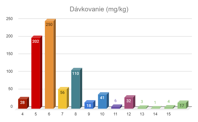
The starting dose for cats with wet or dry FIP and no signs of ocular or neurological disease is 4-6 mg/kg daily for 12 weeks, with younger and wet cases tending towards the lower end and dry cases towards the higher end. Cats with eye lesions and no neurological signs are started at 8 mg/kg daily for 12 weeks. Cats with neurological signs are started at 10 mg/kg daily for 12 weeks. If cats with wet or dry FIP initially develop ocular or neurological signs, they are switched to the appropriate ocular or neurological doses. The dose of GS is adjusted weekly to account for weight gain. Weight gain can be huge in many of these cats, either because they are in poor condition to begin with or because their growth has been stunted. If the cat does not gain weight during treatment, this is considered a bad sign. The initial dosage is not changed unless there are serious reasons for this, such as ineffectiveness of treatment or improvement in blood test values, improvement is very slow, low activity level, initial clinical symptoms have not resolved, or the disease form has changed with the appearance of ocular or neurological symptoms. If there are good reasons to increase the dosage, it should always be from +2 to +5 mg/kg per day and for at least 4 weeks. If these 4 weeks exceed the original 12-week treatment time, the treatment time is extended. A positive response to any increase in dosage can be expected, and if you don't see an improvement, it means that the dosage is still not high enough, drug resistance is emerging, the GS mark is not what it should be, the cat does not have FIP, or there are other diseases that confuse the treatment.
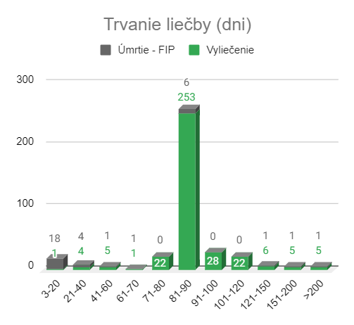
One of the most difficult decisions is determining when to stop treatment. Although some cats, often younger with wet FIP, can be cured as early as 8 weeks and possibly earlier, the usual treatment period is 12 weeks. Some cats may even require dose adjustments and even longer treatment periods. Critical blood levels such as hematocrit, total protein, albumin and globulin levels and absolute lymphocyte counts usually return to normal in curing cats after 8 to 10 weeks, when there is often an unexpected increase in activity levels. We believe, but there is no evidence yet, that after 8-10 weeks, the cat will have its own immune response against the infection. This is a situation that occurs in the treatment of people with hepatitis C, which is also a chronic RNA virus infection that often requires antiviral treatment for up to 12 weeks or more.
Cats with ocular disease and no neurological impairment show a rapid response to GS, and complete recovery of vision with minimal or no residual damage is expected in as little as two weeks. Cats that develop neurological abnormalities, develop neurological disease during the treatment of other forms of FIP, or develop neurological symptoms during the 12-week post-treatment observation period also improve rapidly, but the dose is much higher, the duration of treatment often longer and the cure rate slightly lower. Treatment failures in cats with neurological FIP are due to either insufficient dose or the development of drug resistance.
Unfortunately, there is no simple blood test that can determine when a cat with neurological impairment has fully recovered. Many cats with neurologic FIP show minimal blood abnormalities, especially those with primary neurologic FIP, and the abnormalities often disappear by the end of treatment, even though residual sites of inflammation remain in the brain or spinal cord. In addition, some cats that recover from the infection will have mild to moderate neurological deficits that are residual effects of the previous illness. These facts make it difficult to use blood test results or residual neurological deficits as indicators of cure or undertreatment. Although a thorough eye examination can clearly rule out active signs of disease, the true state of the disease in the brain and spinal cord can only be determined by an MRI, ideally together with an analysis of the cerebrospinal fluid. These procedures are expensive, not available to everyone, and may not provide definitive proof that the infection in the CNS has been cleared.
Fear of relapses means that many people involved in GS treatment are too cautious about a single blood parameter that is slightly abnormal (eg, slightly high globulin or slightly low A: G ratio), or final ultrasound results suggesting suspiciously enlarged abdominals. lymph nodes, small amounts of abdominal fluid or blurred irregularities in organs such as the kidneys, spleen, pancreas or intestines. It should be borne in mind that the normal range of blood values applies to most animals, but it is a bell-shaped curve, and that there are a few non-standard patients who will have values at the edge of these curves. Ultrasonographers must consider the degree of pathology that can occur in the FIP of the affected abdomen and how scars and other permanent consequences can change the normal appearance of successfully treated cats. In situations where such questions arise, it is better to focus in more detail on the overall picture and not just on one small part. The most important outcome of treatment is a return to normal health, which has two components - external signs of health and internal signs of health. External signs of health include a return to normal activity levels, an appetite, adequate weight gain or growth, and coat quality. The latter is one of the best criteria for cat health. Internal health symptoms include the return of certain critical values to normal based on periodic complete blood count (CBC) monitoring and serum chemical profiles. The most important values in CBC are hematocrit and relative and absolute total white blood cell, neutrophil and lymphocyte counts. The most important serum values for chemical analysis (or serum electrophoresis) are total protein, globulin, albumin and A: G ratio levels. Bilirubin is often elevated in cats with effusive FIP and may be useful in monitoring the severity and duration of inflammation. There are many other values in the CBC panels and serum, and it is not uncommon for some of them to be slightly higher or lower than normal, and it is better to ignore these values unless they are significantly elevated and associated with clinical signs. For example, high BUN and creatinine, which is also associated with increased water consumption, excessive urination, and urinary abnormalities. The number of machine-counted platelets in cats is notoriously low due to the trauma of blood collection and platelet aggregation and should always be verified by manual examination of blood smears. The final decision to discontinue or extend treatment when faced with unclear doubts about different testing procedures should always be based on external manifestations of health than on any single test result.
(This paragraph comes from the original article from 1/4/2021.)
Relapses usually refer to infections that have escaped into the central nervous system (brain, spine, eyes) during treatment for wet or dry FIP that are not accompanied by neurological or ocular symptoms. Doses of GS-441524 used to treat these forms of FIP are often insufficient to effectively cross the blood-brain or blood-ocular barrier. The blood-brain barrier is even more efficient than the blood-ocular barrier, which explains why eye lesions are easier to heal than brain and/or spinal cord infections. Post-treatment relapses involving the eyes, brain, or spine are usually treated for at least 8 weeks at an initial daily dose at least 5 mg/kg higher than the dose used during primary treatment (eg, 10, 12, 15 mg/kg per day). Cats that fail to clear the infection at doses up to 15 mg/kg per day are likely to have developed varying degrees of resistance to GS-441524. Partial resistance may allow suppression of disease symptoms but not cure, while complete resistance is manifested by varying severity of clinical symptoms during treatment.
Different groups focused on the treatment of FIP have made various modifications in the treatment protocols. Some groups will treat with an extremely high dose of GS from the beginning and not increase the dose when indicated, or will recommend discontinuing or extending the high dose for the last two weeks in the hope that this will reduce the risk of relapse. In addition to GS, systemic prednisolone is often prescribed, but should only be used temporarily to stabilize serious illness. Systemic steroids reduce inflammation but tend to mask the beneficial effects of GS, and if used for an unreasonably long time and in high doses, can interfere with the development of immunity to FIP. Restoration of immunity to FIP is thought to be an important part of successful GS treatment. Therefore, some people advocate the use of interferon omega or non-specific immunostimulants to further stimulate the immune system, and some come up with other modifications. There is no evidence that using an extremely high dose will improve the cure rate. Also, interferon omega and non-specific immunostimulants have not been shown to have beneficial effects on FIP, whether given as a single treatment or as an adjunct to GS. The practice of adding another antiviral drug, the viral protease inhibitor GC376, to the treatment of GS in cats that develop resistance to GS is also emerging, but this still requires further research. Finally, it is common for owners, treatment groups and veterinarians to add many supplements, tonics or injections (eg B12) to increase hematopoiesis or to prevent liver or kidney disease. However, such supplements are rarely necessary in cats with pure FIP.
Molnupiravir (EIDD-2801) – Molnupiravir is very similar to GS-441524, but is a cytidine rather than an adenine nucleoside analog. It is widely used as an oral treatment for early cases of COVID-19 in humans, but in the last 1-2 years it has been increasingly used to treat cats with FIP. Due to the toxicity observed in cats at higher doses and as yet unknown chronic side effects, it is most often recommended for cats that developed resistance to GS-441524 during primary treatment or relapsed with neurological/ocular signs after treatment with high doses of GS- 441524. Fortunately, molnupiravir has a different resistance profile than GS-441524.
The safe and effective dosing of molnupiravir in cats with FIP has not been established in properly controlled and monitored field studies such as those performed for GC376 and GS-441524. However, the estimated starting dose of molnupiravir in cats with FIP was derived from published EIDD-1931 and EIDD-2801 in vitro cell culture studies and other laboratory and experimental animal studies. Molnupiravir (EIDD-2801) has an EC50 of 0.4 µM/µL against FIPV in cell culture, while the EC50 of GS-441524 is approximately 1.0 µM/µL. Molnupiravir begins to show cellular cytotoxicity at concentrations of 400 µM or higher, while GS-441524 is non-toxic at 400 µM. Both have similar oral absorption of around 40-50 %. The current recommended starting dose of molnupiravir for neurologic and ocular FIP is 8–10 mg/kg orally every 12 hours for 84 days. Depending on the response to treatment, it may be necessary to increase it to a maximum of 15 mg/kg orally every 12 hours. At higher doses, molnupiravir toxicity is likely to occur as indicated by changes in the complete blood count.
Causes of treatment failure
Incorrect dosage adjustments - It is important to start treatment with the appropriate dosage and to monitor it closely with regular checks on temperature, weight and external signs of improvement. The CBC and serum chemical analysis panel, which contains baseline protein values (total protein, albumin, globulin (TP - albumin = globulin) and A: G), should be performed at least once a month. with GS-441524 Expensive serum protein electrophoresis does not provide much more valuable information.
Low quality GS-441524 - GS-441524 is not approved for marketing in any country and is sourced from a small number of Chinese chemical companies which sell it to distributors as pure powder. Vendors dilute it into injectable solutions or prepare oral forms for sale under their trade names. There is no independent mechanism to ensure the quality of the final product sold to cat owners. Nevertheless, the main providers of dilute forms for injectable solutions and / or oral preparations are surprisingly honest, and some even offer limited guarantees if treatment with some of their products does not cure the disease. However, the batches sold by some providers appear to be counterfeit and some are not in the specified concentration. There may also be differences between batches, probably due to occasional problems with the supply of raw GS by retailers or problems with meeting the needs and expectations of the cat owner. Various groups of FIP Warriors have good information about the most reliable brands.
Drug resistance - resistance to GS-441524 may already exist at the time of diagnosis, but this is unusual. It occurs more frequently during treatment and is initially only partial and requires only higher doses. In some cats, it may become complete. Resistance is the biggest problem in cats with neurological disease, or they develop brain infections during treatment or within a few days or weeks after stopping treatment. Many cats with partial drug resistance may be "treated" for their symptoms, but they relapse as soon as treatment is stopped, as is the case with HIV treatment, for example. There are cats that have been able to partially or completely treat the symptoms of FIP for more than a year, but without a cure. Resistance eventually worsens and the symptoms of the disease worsen, treatment difficulties become unbearable for the owner or the owner's financial resources run out.
GS side effects
GS-441524 treatment is incredibly free of systemic side effects. It can cause mild kidney damage in cats without significant kidney damage, but does not lead to latent disease or kidney failure. Systemic drug reactions such as vasculitis have been observed in several cats and can be confused with injection site reactions. However, these drug reactions are in places where injections are not given, and often stop on their own or respond well to short-term low-dose steroids. The main side effect of GS treatment is pain at the injection sites, which varies from cat to cat and according to the abilities of the person giving the injections (usually the owner). Swelling or ulcers at the injection site sometimes occur in owners who do not change the application site often enough (do not stay between the shoulder blades) and do not inject into the muscular and nervous layers under the skin. I recommend choosing places starting one inch behind the shoulder blades, down from the back to 1 to 2 inches in front of the root of the tail and one third to half way down to the chest and abdomen. Many people use gabapentin to relieve pain before injections. Swollen spots and ulcers at the injection site should be stripped of surrounding hair and gently cleaned 4 or more times a day with sterile cotton swabs soaked in homemade hydrogen peroxide diluted 1: 5. They usually do not require any more complicated treatment and will heal in about 2 weeks.
Prognosis of treatment with GS441524
Exact cure rate data with GS-441524 are not yet available, but it seems possible to cure more than 80% cats with confirmed FIP. Treatment failure is due to misdiagnosis of FIP, inadequate treatment monitoring and dose adjustment, complicating diseases, poor GS, resistance to GS, or economic difficulties. The cure rate is slightly lower in cats with neurological forms of FIP and in older cats. Older cats are more susceptible to other chronic diseases, which either predispose cats to FIP or complicate overall health.
Cats with neurological FIP may suffer permanent residual symptoms of the disease. This is especially true for cats with spinal involvement and urinary and/or fecal incontinence or hind paralysis. Hydrocephalus and syringomyelia are common complications of neurological FIP and often persist to some extent after the infection has cleared. Fortunately, most cats with neurologic FIP recover normal or near-normal function despite persistent traces of hydrocephalus and syringomyelia.
Legal treatment for FIP?
We hope that the legal form GS-441524 will be available soon. The drug, called Remdesivir, is the greatest hope of the present because Remdsivir breaks down into GS when given intravenously to humans, mice, primates and cats. Remdesivir (Veklury®) has been fully approved by the US FDA and similar approval is likely to follow in other countries. If so, it can be prescribed by any licensed human physician as well as veterinarians. However, the use of Remdesivir in the United States was initially limited to a specific subset of patients with Covid-19 and only under controlled conditions and with ongoing data collection. Until all restrictions are lifted, it will not be easily accessible for human use. We have no experience in treating cats with Remdesivir instead of GS-441524. The molar basis of Remdesivir is theoretically the same as GS-441524. GS-441524 has a molecular weight of 291.3 g / M, while Remdesivir is 442.3 g / M. Therefore, 442.3 / 291.3 = 1.5 mg of Remdesivir would be required to obtain 1 mg of GS-441524. The diluent for Remdesivir is significantly different from the diluent used for GS-441524 and intended for intravenous use in humans. How diluted Remdesivir will behave when given by subcutaneous injection over 12 weeks or more is not known. Mild signs of hepatic and renal toxicity were observed in humans. GS-441524 causes mild and progressive renal toxicity in cats, but without apparent hepatic toxicity. It is uncertain whether renal toxicity observed in humans receiving Remdesivir is due to its active substance (ie GS-441524) or to chemical agents designed to increase antiviral activity.
The GC376 approval process for cats (and humans) is ongoing at Anivive, but will take two or more years. GC376 is a viral protease inhibitor and, unlike GS-441524, which inhibits the initial stage of viral RNA replication, GC376 prevents viral replication in the final stage of its replication process. Therefore, it is unlikely to have a significant synergistic viral inhibitory effect and its use in combination will be much more important in inhibiting drug resistance (e.g. in combination antiviral therapy for HIV / AIDS).
Improper use of GS-441524
Some veterinarians, in collaboration with major Chinese supplier GS-441524, have advocated its use to eliminate feline enteric coronavirus (FECV) infection. The reason is to prevent the occurrence of a mutant virus causing FIP (FIPV) and thus prevent FIP. This approach was supported by limited and highly controversial studies with shelter cats, which were naturally exposed to the FECV. Although this approach is attractive at first glance, it is a very incorrect use of GS-441524 in cats. FECV infection originally occurs in kittens and is not associated with any significant symptoms of the disease. Elimination lasts for weeks, months, and in some cases indefinitely, but in most cats, it eventually stops when immunity develops. Most cats over the age of three will no longer shed the virus. GS-441524 treatment is highly unlikely to result in more permanent immunity than is observed in nature and to eliminate cycles of infection and reinfection in younger cats.
Although our current knowledge of FECV infection seriously challenges this approach, there are even more compelling reasons why we will not treat healthy cats GS-441525 or other antiviral agents in the future. We already know from published studies that some primary strains of FIPV are resistant to GS-441524 (and GC376). We also know that drug resistance has become a long-term problem in cats with long-term treatment for GS-441524, especially in neurological forms of FIP. Therefore, the use of drugs such as GS-441524 in a large population of healthy cats will undoubtedly lead to widespread resistance to enzootic FECV. This resistance will also manifest itself in FIP-causing FECV (FIPV) mutations from these populations, making it impossible to use GS-441524 in more and more FIP cats. Unfortunately, veterinary medicine does not have the means of human medicine, it is not stimulated by potential benefits, which would lead to the discovery, testing and approval of more and more antiviral drugs to circumvent either natural or acquired drug resistance, which is already the case in HIV / AIDS treatment. achieved (at least on time).
(This part comes from the original article from 1/4/2021.)
Niels C. Pedersen, DVM PhD Distinguished Professor Emeritus UC Davis, Center for Animal Health Companion January 4, 2021, updated February 10, 2023
I am pleased to announce that I have ended my advisory role with SOCKFIP and have officially become a member of the SOCKFIP Board of Directors. This reflects my transition from academic to private life, but it will not affect my commitment to FIP research. I hope that this more direct involvement will help SOCKFIP transition to a broader role in feline health issues beyond FIP. FIP research continues at the University of California, Davis, as well as at other institutions around the world. FIP research projects at UC Davis are summarized in “ Best regards SOCK FIP" of 2022. SOCKFIP continues to provide financial assistance for such studies through public donations, and I will provide scientific expertise whenever needed.
I wish there was a licensed antiviral treatment for FIP in cats, but even the efforts of many individuals and groups have not been able to change the current reality. Therefore, it is questionable whether legal antivirals for FIP will reach the market in the next 2 to 5 years, even if the obstacles are removed immediately. Fortunately, restrictions on the general use of closely related human medicines for COVID-19 are being eased worldwide, allowing them to be prescribed by all doctors and used more widely in the field. Full human approval allows their use in animals, provided the drug needed is derived directly from the actual human product. This would allow drugs made for humans, such as remdesivir and molnupiravir (EIDD-2801), to be used legally in animals, albeit at the cost of human drugs. The goal should still be for drugs specifically licensed for animals and available at a veterinary rather than a medical price.
More and more veterinarians are already helping owners with treatment. However, it still saddens me that some vets have not heard of effective treatments for FIP, believe that published treatment reports are bogus, or that obtaining drugs from unapproved markets is so scary that they can't even help with treatment once an owner buys it. I commend those veterinarians who accept the reality of treatment and work with owners and their cats with FIP.
The most significant discovery after GS-441524 is the use of molnupiravir (EIDD-2801) (Merck) as a second effective treatment for FIP. Molnupiravir is also extremely effective in treating cats that have developed resistance to GS-441524, which are the most common cats that develop neurologic FIP during or after treatment with GS-441524. Reports of its use in cats are just beginning to emerge and are being posted on the SOCK FIP website.
I believe that there are several areas of research that veterinary researchers should consider. One area concerns the safety and efficacy of EIDD-1931 (beta-d-N4-hydroxycytidine), which is the biologically active component of molnupiravir, just as GS-441524 is the active component of remdesivir. This orally administered drug has been the subject of research for almost half a century and should no longer be patent protected. Preliminary research at the University of California, Davis suggests that it may be even more effective and safer than molnupiravir. I also believe that the oral protease inhibitor (nirmatrelvir) component of Paxlovid (Pfizer) should be tested for non-ocular/non-neurological cases of FIP. Nirmatrelvir is broken down into a simple chemical modification of GC373, the active form of GC376. Paxlovid is widely available and can be easily prescribed by both pharmacists and doctors for general treatment of COVID-19. This should make it widely available for use by veterinarians. I also believe that further research should be pursued to find ways to limit FECV infection and to understand the factors that suppress the natural normal protective immunity against FECV mutants. At this point, it is clear that most healthy cats have strong natural and acquired immunity to FIP viruses. What is this immunity and how can this knowledge contribute to strengthening immunity against FIP?
Several issues often arise during FIP treatment. Before addressing these issues, it is important to mention the FIP treatment itself. Only antivirals that target specific viral proteins and inhibit FIP replication have been shown to have therapeutic effects. Currently, these include nucleoside analogs and RNA replication inhibitors GS-441524 (and a related prodrug Remdesivir), Molnupiravir (EIDD-2801) and the viral protease inhibitor GC376. Proper administration of these drugs has resulted in the cure of all forms of FIP in more than 90 % with minimal side effects. Most treatments are completed without complications. However, certain issues that are the subject of this article often arise.
I pointed out the problems associated with unwanted sexual behavior in intact females and males treated with specific antivirals. The questions often come from countries where castration is either postponed or not common practice. They fear that the stress of castration and vaccines may affect the outcome of antiviral treatment. I believe that such concerns are exaggerated. If a cat is in treatment and in remission or is considered cured, it is okay to sterilize or neuter it, but preferably in the least stressful way possible. Cats can be neutered and sterilized quickly and efficiently and returned to their homes on the same day (castration) or within one day (sterilization) with minimal preoperative, operative and postoperative drug treatment and restrictions (eg cages, E-collars). Such operations will be less stressful for cats (and owners, which will then be reflected in their cats) than their sexual behavior.
I am also not in favor of hormonal treatment to prevent unwanted sexual behavior in males or females, and I feel that effective castration and sterilization will be less stressful in the long run than such preventive measures. Therefore, if it is necessary to permanently change this behavior, surgical castration is more appropriate than chemical.
Some owners seem to want to keep cured cats intact so that they can be used for breeding later. We know that FIP has both genetic and environmental components, which has led to the recommendation that purebred cats that breed FIP kittens should not be used for breeding. This should be even more true for cats that have been cured of the FIP.
As far as vaccines are concerned, many already know that I am not a big fan of adult vaccines and the first annual booster vaccines because I feel that immunity is long-lasting. I also think that rabies vaccines cannot be used routinely in cats, whether in terms of public health or cats. Nevertheless, I accept that these views are not generally accepted and that the laws in several states require rabies to be vaccinated against rabbits, in some vaccination is not required and in others it is recommended but not required. I have not noticed the consequences of routine vaccinations in any of our cured cats. However, it is not something I would recommend for cats undergoing treatment. The immune system of these cats is responsible for other things than responding to vaccines.
What are the indications for drugs other than specific antivirals for the treatment of FIP? During the initial illness, supportive (symptomatic) treatment may be required to keep the cats alive long enough for the antivirals to take effect. Drugs often used in this early stage usually include antibiotics (doxycycline / clindamycin), analgesics (opioids, gabapentin), anti-inflammatory drugs (corticosteroids, NSAIDS), immunostimulants (interferons, non-specific immunostimulants), and drugs. I have tried to avoid excessive use of these drugs except for temporary use and only if it is strongly justified, especially in severely ill cats during the first days. The most important goal of FIP treatment is to stop the replication of the virus in macrophages, which immediately stops the production of the numerous inflammatory and immunosuppressive cytokines that cause the symptoms of FIP. Although some drugs, such as corticosteroids (prednisolone) or NSAIDs (meloxicam), may inhibit inflammatory cytokines and cause clinical improvement, they are not curative. They can also mask the beneficial effects of GS treatment, which are often monitored to assess the effect and course of treatment. The response to antiviral treatment is also used for diagnostic purposes. The only drugs that completely suppress these harmful cytokines and cure FIP are antivirals such as GS-441524, molnupiravir or GC376, and related compounds. These antivirals cause a dramatic improvement in fever, activity, appetite, etc. within 24-48 hours. This improvement will be much greater than any improvement made with other drugs. Therefore, if the use of other drugs is not warranted, they should be discontinued as soon as the symptoms of FIP have steadily improved.
I also do not believe in many supplements that are said to treat or prevent problems with the liver, kidneys, immune system or other organs. These supplements are expensive and have not been shown to be effective in what they claim. B12 injections only treat B12 deficiency, which is rare, and not anemia in FIP. The same goes for other vitamins. This also applies to a wide range of nutritional supplements and special diets for cats of many types. There is no essential ingredient in any of these supplements that could be provided by well-tested commercial cat food brands. There is also a possibility that some supplements interfere with the absorption of oral antivirals.
How should cats be monitored after treatment and during the post-treatment observation period? From a technical point of view, no further blood tests are needed, especially if routine health assessments such as weight, appetite and temperature are continued during this period. Blood tests during this period do not change the outcome and can only increase the cost of treatment and increase the owner's stress. However, it is common for successfully treated cats to routinely test for blood during a 12-week post-treatment observation, usually every 4 weeks, but sometimes more frequently. In some cases, routine blood testing is continued for 12 weeks after treatment, even out of fear of possible relapse or recurrence. Relapses or new infections after a 12-week observation period are rare and are preceded by external signs of the disease, such as weight loss, lethargy, anorexia, poor coat and fever, which would be the best indicators for a blood test. Blood test panels also contain many values, and it is not uncommon for one or more values to be abnormal in healthy cats. Care must be taken not to over-interpret such abnormalities and to lead to excessive concern or additional testing in order to determine their significance. For example, a mild to moderate increase in one in three liver enzymes in a healthy cat is much less important than in another cat with symptoms of the disease.
This article discusses the development of knowledge about feline infectious peritonitis (FIP) from its recognition in 1963 to the present and has been prepared to inform veterinarians, cat rescuers and carers, shelter staff and cat lovers. The causative agent of the feline coronavirus and its relationship to the ubiquitous and minimally pathogenic feline intestinal coronavirus, epizootology, pathogenesis, pathology, clinical signs and diagnostics are briefly mentioned. The main emphasis is placed on the risk factors influencing the incidence of FIP and the role of modern antivirals in successful treatment.
Introduction
Figure 1. Photo of the author and Dr. Jean Holzworthová (1915-2007) from 1991. Dr. Holzworth was the best feline veterinarian the author knew and was responsible for the first report of FIP as a specific disease. She spent her entire career at Angell Memorial Animal Hospital in Boston.
Feline infectious peritonitis (FIP) was described as a specific disease in 1963 by veterinarians at Angell Memorial Animal Hospital in Boston (Holzworth 1963) (Fig. 1). Pathology records from this institution and Ohio State University failed to identify earlier cases (Wolfe and Griesemer 1966), although identical cases were soon recognized worldwide. The initial pathological descriptions were of diffuse inflammation of the tissues lining the abdominal cavity and abdominal organs with extensive effusion of inflammatory fluid, after which the disease was eventually named (Wolfe and Griesemer 1966, 1971) (Figs. 2, 3). A second and less common clinical form of FIP, which presents with less diffuse and more widespread granulomatous lesions involving organ parenchyma, was first described in 1972 (Montali and Strandberg 1972) (Figs. 3,4). The presence of inflammatory effusions in the body cavity in the common form and the absence of effusions in the less common form led to the names wet (effusion, non-parenchymatous) and dry (non-effusion, parenchymatous) FIP.
The prevalence of FIP appears to have increased during the panzootic disease caused by feline leukemia virus (FeLV) in the 1960s–1980s, when many cases of FIP were found to be associated with FeLV (Cotter et al., 1973; Pedersen 1976a). The subsequent management of FeLV infection in domestic cats through rapid testing and vaccination resulted in an increase in the number of FIP cases. However, recent interest in breeding/rescue along with effective treatment has led to increased awareness of the disease and its diagnosis.
Figure 2. Gross necroptic appearance of the abdominal cavity of a cat with acute onset wet FIP. The abdomen is filled with several hundred ml of yellow viscous fluid, the omentum is reddened, edematous and contracted, and fibrin deposits (arrows) are visible on the surface of the spleen and the edges of the liver. A fiber of fibrin can be seen on the spleen
Figure 3. Appearance of an open abdomen at autopsy of a cat that died of a chronic form of effusive FIP. The abdomen is filled with a viscous, yellow-colored exudate, and the omentum is thickened and contracted. The main lesions are in the liver with numerous plaque-like structures (pyogranulomas) on the cover. More circumscribed lesions (granulomas), also oriented on the serous surface, look more fleshy and are elevated above the surface. These lesions also involve the underlying liver parenchyma and are more typical of dry FIP. This is an example of a case of FIP that transitions between the wet and dry forms (arrow).
Figure 4A – Gross section of kidneys of two cats with dry form of FIP. The lesions are superficial and extend into the underlying parenchyma.
Figure 4B - lesions of the dry form of FIP in organs such as the kidneys, cecum, colon and intestinal lymph nodes (Fig. 5) were grossly confused with renal lymphoma.
Figure 5. Gross enlargement of ileo-cecal-colic lymph nodes in a cat with dry FIP.
Etiological factor
The first attempts did not allow identifying the causative agent of FIP, but confirmed its infectious nature (Wolfe and Griesemer 1966). A viral etiology was established in 1968 using ultrafiltrates of infectious material (Zook et al., 1968). The causative virus was subsequently identified as a coronavirus (Ward 1970), which is closely related to enteric coronaviruses of dogs and pigs (Pedersen et al., 1978).
Confusion arose when feline enteric coronavirus (FECV) was isolated from the feces of healthy cats and proved to be indistinguishable from feline infectious peritonitis virus (FIPV) (Pedersen et al., 1981). Unlike FIPV, which readily induced FIP in laboratory cats, experimental infections with FECV were largely asymptomatic. The relationship between the two viruses became clear when FIPVs were found to be FECV mutants that arise in the body of every cat with FIP (Vennema et al., 1995; Poland et al., 1996).
FECV is ubiquitous in feline populations worldwide and is first shed in faeces from approximately 9–10 weeks of age, coinciding with the loss of maternal immunity (Pedersen et al., 2008 ;). The infection takes place via the faecal-oral route and targets the intestinal epithelium, and the primary signs of enteritis are mild or inconspicuous (Pedersen et al., 2008; Vogel et al., 2010). Subsequent faecal excretion occurs from the colon and usually stops after several weeks or months (Herrewegh et al., 1997; Pedersen et al., 2008; Vogel et al., 2010). Immunity is short-lived and repeated infections are common (Pedersen et al., 2008; Pearson et al., 2016). Over time, stronger immunity eventually develops and cats older than 3 years are less likely to shed the infection in their faeces (Addie et al., 2003). FECV is constantly subject to genetic drift into locally and regionally identifiable clades (Herrewegh et al., 1997; Pedersen et al., 2009).
FECV and FIPV are classified as biotypes of the feline coronavirus (FCoV) subspecies. The genomes of FECV and FIPV biotypes are related at >98 %, but with unique host cell tropism and pathogenicity (Chang et al., 2012; Pedersen et al., 2009). FECVs infect the mature intestinal epithelium, whereas FIPVs lose intestinal tropism and acquire the ability to replicate in monocytes/macrophages. The published names FECV or FIPV will be used here when discussing aspects of the disease specific to each biotype, while the term FCoV will be used when discussing features common to both biotypes.
Three types of mutations are involved in the biotype change of FECV to FIPV. The first type, which is unique to each cat with FIP (Poland et al., 1996), consists of an accumulation of missense and nonsense mutations in the c-terminus of the auxiliary 3c gene, often resulting in truncated 3c gene products (Pedersen et al., 2012 ; Vennema et al., 1995). The second type of mutation consists of two specific single nucleotide polymorphisms in the fusion peptide of the S gene, one or the other form being common to >95 % FIPV and absent in FECV (Chang et al., 2012). A third type of mutation, unique to each FIPV isolate and not found in FECV, occurs in and around the furin cleavage motif between the receptor binding domain (S1) and the fusion domain (S2) of the spike gene (S) (Licitra et al., 2013). These mutations have different effects on furin cleavage activity. Together and in an as yet undetermined manner, they are responsible for the shift of the tropism of the host cell from the enterocyte to the macrophage and for the profound change in the form of the disease.
FCoV, and therefore FECV and FIPV, exist in two serotypes identified by antibodies against the viral neutralizing epitope on the S gene (Herrewegh et al., 1998; Terada et al., 2014). Serotype I FCoVs are identified in cat sera and are prevalent in most countries. Serotype II FCoVs result from recombination with the S part of the canine coronavirus gene (Herrewegh et al., 1998; Terada et al., 2014) and are identified by canine coronavirus antibodies. Serotype II FIPVs are easily cultured in tissue culture, whereas serotype I FIPVs are difficult to adapt to growth in vitro. Serotype I and II FECVs were not grown in conventional cell cultures (Tekes et al., 2020).
FIPVs are found exclusively in activated monocytes and macrophages in affected tissues and effusions and are not secreted into the environment. Therefore, cat-to-cat (horizontal) transmission of FIPV is not the main mode of spread. Rather, FIP follows the pattern of an underlying enzootic FECV infection, with sporadic cases and occasional small outbreaks of disease (Foley et al., 1997). These clusters of cases can be mistaken for epizootics. The only report of an epizootic occurrence of FIP was associated with a single serotype II virus that appeared to develop in a shelter housing both dogs and cats (Wang et al., 2013). Horizontal transmission in this case followed an epizootic rather than an enzootic disease model, with infection spreading rapidly to cats of all ages and in close contact with the index case (Wang et al., 2013).
The low incidence of FIP cases in the population suggests that FIPV mutations arise infrequently. However, studies involving FECV infection in immunocompromised cats infected with FIV and FeLV suggest that FIP mutants may be common but only cause disease under certain circumstances. Nineteen cats infected with feline immunodeficiency virus (FIV) for 6 years and a control group of 20 littermates not infected with FIV were orally challenged with FECV (Poland et al., 1996). Cats in both groups remained asymptomatic for two months when two cats in the FIV-infected group developed FIP. In a second study, 26 young cats with enzootic FECV infection from a breeding colony with no history of FIP were contact-exposed to FeLV carriers (Pedersen et al., 1977). Two kittens in the group subsequently developed FIP 2–10 weeks after becoming FeLV viremic. The question remains, how long can FIPV viruses survive in the body before they are eliminated? According to one of the theories, they persist in the body for a certain time and become pathological only if immunity against them is impaired (Healey et al., 2022). This theory is supported by the way immunity to FeLV develops. Most cats resist FeLV by kitten age and develop robust and permanent immunity, but this occurs within a few weeks, during which the virus persists in a subclinical or latent state (Pedersen et al., 1982; Rojko et al., 1982). . Methylprednisolone given during this period, but not after, will abolish developing immunity and lead to a state of persistent viremia.
Epizootology
Epizootiology is the study of the occurrence, spread and possible control of animal diseases and the influence of environmental, host and agent factors. FIP is considered one of the most important infectious causes of death in cats, although there are no precise data on prevalence. It is estimated that 0.3–1.4 % deaths of cats presented to veterinary institutions are related to FIP (Rohrbach et al., 2001; Pesteanu-Somogyi et al., 2006; Riemer et al., 2016) and in some shelters and breeding stations up to 3.6–7.8 % (Cave et al., 2002). FIP is also described as an environmental disease with a higher incidence of multiple cats. Three-quarters of the FIP cases in the currently ongoing treatment study came from the field through foster carers/rescues and cat shelters, 14 % from kennels, and only 11 % from households.1
Studies based on cases observed in academic institutions have demonstrated the influence of age and gender on the incidence of FIP (Rohrbach et al., 2001; Pesteanu-Somogyi et al., 2006; Pedersen 1976a; Worthing et al., 2012; Riemer et al., 2016) . Three-quarters of the cases in these cohorts occurred in cats younger than 3 years of age, and few cases occurred after 7 years of age. This was also confirmed by a current and ongoing field study from the Czech Republic and Slovakia, in which it was found that more than 80 % cases of FIP occurred in cats under 3 years of age and only 5 % in cats older than 7 years (Fig. 6) .1 Earlier institutional studies differed on the effect of sex, but indications were that male cats were slightly more susceptible to FIP than female cats. This was also confirmed by current data from the field, which show a ratio of males to females of 1.3:1.1. It is unclear whether castration affects the incidence of FIP, with some reports suggesting that it may increase susceptibility (Riemer et al., 2016), while others do not report such a clear effect.1
Figure 6. Age of more than 607 cats from the Czech Republic and Slovakia at the time of diagnosis and treatment of FIP.1 Thirty percent of infections were in cats six months of age or younger, 50 % at one year of age, and 85 % at three years of age or younger.
Other environmental and viral risk factors have been implicated in the increased incidence of FIP, but their significance requires knowledge of disease occurrence in their absence. A possible baseline may have been provided by a study of enzootic FECV infection, which had been unrecognized for many years in a well-managed specific pathogen-free breeding colony (Hickman et al., 1995). This colony was kept in strict quarantine free of other infections and the standard of nutrition and husbandry was high. This colony produced hundreds of kittens each year before the first case of FIP was diagnosed. Such observations suggest that FIP may be a rare phenomenon in the absence of risk factors.
The importance of moving to a new home as a risk factor for FIP is only now being appreciated. Breeders, many of whom have not experienced any cases of FIP in their litters, are most concerned about the announcement that one of their kittens has developed FIP shortly after going to a new home. A recent study found that more than half of cats with FIP had experienced a change in environment, shelter or capture in the weeks before the illness.1 Cats are known to hide external signs of stress even when they are suffering from serious internal ill effects. Even simple procedures such as changing the cage will suppress immunity and reactivate latent herpes virus shedding and disease symptoms in cats (Gaskell and Povey, 1977). Stressful situations, even those that appear to be minor, can cause a decrease in lymphocyte levels and “sick behavior” (Stella et al., 2013).
Differences in the genetic make-up of enzootic FCoV strains may also contribute to the prevalence of FIP in the population. Serotype II FIPVs are thought to be more virulent than serotype I and more likely to be transmitted from cat to cat (Lin et al., 2009; Wang et al., 2013). It is also possible that certain FECV clades are more susceptible to mutation to FIPV, which should be studied. The author also observed a disproportionately high proportion of cats with neurologic FIP in some regions, suggesting that genetic determinants in certain FCoV strains may be more neurotropic.
Immunodeficiencies associated with retroviruses are associated with susceptibility to FIP. Up to half of FIP cases during the peak of FeLV panzootic disease were persistently infected with FeLV (Cotter et al., 1973; Pedersen 1976a; Hardy 1981). FeLV infection causes suppression of T-cell immunity, which may inhibit the protective immune response to FIP. The importance of FeLV infection for the incidence of FIP has declined significantly since the 1980s, when carrier elimination and vaccination pushed FeLV back into the wild, where exposures are less severe and immunity is the usual outcome. Chronic feline immunodeficiency virus (FIV) infection has also been shown to be a risk factor for FIP in FECV-infected cats under experimental conditions (Poland et al., 1996). In one recent field study, FeLV infection was recognized in 2 % and FIV in 1 % cats treated for FIP.1
The incidence of FIP in purebred cats is reported to be higher than in random breeding cats, with some breeds appearing to be more susceptible than others (Pesteanu-Somogyi et al., 2006; Worthing et al., Genetic predisposition to FIP has been investigated in several Persian cat breeds and is estimated to account for half the risk of the disease (Foley et al., 1997). Some breeds, such as the Birman, are more susceptible to developing dry than wet FIP (Golovko et al., 2013). Attempts to identify specific genes associated with susceptibility for FIP in Burmese cats included several immune-related genes, but none reached the desired significance (Golovko et al., 2013).The largest study of genetic susceptibility to FIP showed that it is extremely polymorphic and reported consanguinity as a major risk factor. breeding (Pedersen et al., 2016).Specific polymorphisms in several genes have also been associated with high levels of FECV shedding among several breeding cat breeds (Bubeniko and et al., 2020).
In females, FIP, usually the wet form, may develop during pregnancy or in the perinatal period. This phenomenon resembles the suppression of immunity in pregnant women and the predisposition to certain infections (Mor and Cardenas 2010). It is not clear whether subclinical FIP is activated by pregnancy or by increased susceptibility to new infection. Maternal infection early in pregnancy results in fetal death and resorption, while later infections often result in abortion (Fig. 7). Kittens can be born healthy, but develop disease in the perinatal period and die. Some babies are born uninfected thanks to the effectiveness of the placental barrier between mother and fetus or thanks to the help of antiviral treatment (Fig. 8).
Figure 7. Aborted kittens from a dam that developed wet FIP late in pregnancy. Miscarriage was the first symptom of FIP, quickly followed by the classic symptoms of abdominal wet FIP. The mother was successfully cured of FIP with the antiviral GS-441524.
Figure 8. This mother developed symptoms of wet abdominal FIP 3 weeks after the onset of pregnancy and was successfully treated with GS-441524. Subsequently, she gave birth to a litter of four kittens by caesarean section, one of which died and three survived and grew up healthy. Treatment was given during the remaining 6 weeks of gestation and continued for 6 weeks during which the kittens were successfully nursed. GS-441524 had no apparent side effects on the mother or the kittens.
A possible increase in the number of cases of FIP was observed in cats older than 10 years in studies conducted 50 years ago (Pedersen 1976a). Slightly more than 3 % cases of FIP in a recent study occurred in cats 10 years of age and older and 1.5 % in cats 12 years of age and older (Fig. 6).1 The occurrence of FIP in the elderly often involves two different scenarios. The first scenario also involves exposure to FECV faecal excretion, but in a unique way. It is common for old cats to mate as kittens and live together in relative isolation unexposed to FECV for many years. One cat in the pair dies, is left alone, and a much younger companion obtained from a rescue organization, shelter, or kennel is brought into the household that has a high probability of excreting FECV. Older cats are also susceptible to the same FIP risk factors as younger cats, as well as other factors associated with aging. The first of these is the impact of aging on the immune system, with the most consequential being the deterioration of cellular immune function (Day 2010). Other risk factors associated with old cats include the debilitating and potentially immunosuppressive effects of diseases such as cancer and chronic diseases of the kidneys, liver, oral cavity and intestines. Some diseases in old cats can be mistaken for FIP or complicate the treatment of FIP if they are present at the same time.
Other risk factors that need further investigation include loss of maternal systemic immunity by separation at birth, early weaning and loss of lactogenic immunity, malnutrition, common kitten infectious diseases, early neutering, vaccination, congenital heart defects, and even a shelter fire (Drechsler et al.), 2011; Healey et al., 2022; Pedersen 2009, Pedersen et al. 2019).1 However, the most important positive risk factor remains the presence of FECV in the population (Addie et al., 1995). The prevalence of FIP in several Persian cat breeds was also related in one study to the proportion of cats that shed FECV at a given time and to the proportion of these cats that are chronic shedders (Foley et al., 1997). The importance of exposure to FECV supports the need to find ways to either prevent infection or reduce its severity. One of the first steps is a better understanding of FECV immunity (Pearson et al., 2019).
Pathogenesis
The first interface between FECV and the immune system is the lymphatic tissues of the intestine (Malbon et al., 2019, 2020). Although the downstream events leading to FIP are not fully understood, it is possible to speculate based on what is already known about FECV and FIPV infections, other macrophage-tropic infections, and viral immunity in general. During intestinal infection, FECV particles and proteins reach the local lymphatic tissues and are processed by phagocytic cells first into peptides and finally into amino acids. Some of these peptides will be recognized as foreign when arrayed on the cell surface, triggering innate (innate or non-specific) and adaptive (acquired or specific) immune responses (Pearson et al., 2016). FECVs also mutate to FIPV at the same time and in the same cell type. Some of these mutations will allow the virus to replicate in these or closely related cells of a specific monocyte/macrophage lineage.
The host cell for FIPV appears to be a specific class of activated monocytes found around venules on the surface of intestinal and thoracic organs, mesentery, omentum, uveal tract, meninges, choroid and ependyma of the brain and spinal cord, and freely in effusions. These cells belong to the activated (M1) class (Watanabe et al., 2018) and resemble a subpopulation of small peritoneal macrophages described in mice (Cassado et al., 2015). This type of cell arises from circulating bone marrow-derived monocytes that are rapidly mobilized from the blood in response to infectious or inflammatory stimuli. A similar-looking population of activated monocytes has been described around blood vessels in the retina affected by FIP (Ziolkowska et al., 2017). These cells stained for calprotectin, indicating their blood origin. Although FIPV infection occurs initially in smaller activated monocytes, viral replication is most intense in large, vacuolated, terminally differentiated macrophages (Watanabe et al., 2018). The virus released from these cells rapidly infects activated monocytes produced in the bone marrow and drawn to the site from the bloodstream.
The cellular receptor used by FECVs to infect intestinal epithelial cells has not yet been determined. The cellular receptor that FIPVs use to infect activated monocytes is also unknown. RNAs for conventional coronavirus receptors such as aminopeptidase N (APN), angiotensin converting enzyme 2 (ACE2) and CD209L (L-SIGN) were not upregulated in infected peritoneal cells of cats with experimental FIP, and CD209 (DC-SIGN) was significantly underexpressed (Watanabe et al., 2018). An alternative route of infection of activated monocytes may involve immune complexation of the virus and entry into cells by phagocytosis (Dewerchin et al., 2008, 2014; Van Hamme et al., 2008). Activated monocytes in lesions stain strongly positive for FIPV antigen, IgG and complement (Pedersen, 2009) and mRNA for FcγRIIIA (CD16A/ADCC receptor) is markedly increased in infected cells (Watanabe et al., 2018), supporting infection through immune complexation and alternative receptors related to phagocytosis.
Macrophage pathogens are intracellular and elimination of infected cells occurs through lymphocyte-mediated killing. The first line of defense is non-specific lymphocytes, and if they fail, an adaptive immune response to FIPV follows through specific T-lymphocytes. If infected activated monocytes and macrophages fail to be contained and eliminated, they may disseminate locally in the abdominal cavity, possibly from lymph nodes in the lower intestinal region and the site of FECV replication. Spread locally and to distant sites via the bloodstream is by infected monocyte cells (Kipar et al., 2005).
FIP occurs in two basic forms, wet (effusive, nonparenchymatous) (Figures 2 and 3) or dry (noneffusive, parenchymatous) (Figures 4 and 5), with wet FIP accounting for 80 % cases.1 The term “wet” refers to the characteristic effusion of fluid in the abdomen or chest (Wolfe and Griesemer 1966, 1971). Wet FIP lesions are dominated by inflammation resembling immediate- or Arthus-type hypersensitivity (Pedersen and Boyle, 1980), whereas dry FIP lesions resemble delayed-type hypersensitivity reactions (Montali and Strandberg 1972; Pedersen 2009). Wet and dry forms of FIP therefore reflect competing influences of antibody- and cell-mediated immunity and associated cytokine pathways (Malbon et al., 2020, Pedersen 2009). Immunity to FIPV-infected cells, which is the norm, is thought to involve strong cell-mediated responses (Kamal et al. 2019). Dry FIP is thought to occur when cell-mediated immunity is partially effective in suppressing infection, while wet FIP occurs when cell-mediated immunity is ineffective and humoral immune responses predominate.
FIP is considered unique among macrophage infections because it is viral, but the dry form shares many clinical and pathogenic features with feline diseases caused by systemic mycobacterial (Gunn-Moore et al., 2012) and fungal infections (Lloret et al., 2013). . Similarities in pathogenesis also exist between wet FIP and antibody-enhanced viral infections such as dengue fever and dengue hemorrhagic shock syndrome (Pedersen and Boyle 1980; Rothman et al., 1999; Weiss and Scott 1981).
Host responses are thought to solely determine the outcome of FIPV infection and the resulting forms of disease. However, macrophage-tropic pathogens have evolved their own unique defense mechanisms against the host (Leseigneur et al., 2020). One of the mechanisms is the delay of programmed cell death (apoptosis). Delayed apoptosis allows sustained microbial replication and eventual release of more infectious agents, as has also been described in FIPV-infected macrophages (Watanabe et al., 2018). FIPV can also control the recognition and killing of infected activated monocytes by specific or non-specific T-cells. The cell surface targets for T-cells that kill infected cells are likely FIPV proteins (antigens) expressed on major histocompatibility complex class I (MHC-I) receptors. However, surface expression of viral antigens by MHC-I receptors was not detected on FIPV-positive cells collected from FIP tissues or effusions (Cornelissen et al., 2007). DC-Sign has been proposed as a receptor for FIPV (Regan and Whitaker, 2008), but RNA for DC-Sign is markedly underexpressed by infected peritoneal cells, whereas RNA for Fc (MHC-II) receptors is markedly overexpressed and RNA for MHC -I is reduced (Watanabe et al., 2018). This suggests that the normal mode of infection of host cells may be altered by FIPV to favor infection by phagocytosis instead of binding to specific viral receptors on the cell surface, fusion with the cell membrane, and internalization.
Pathology
Detailed descriptions of the gross and microscopic lesions in the wet form of FIP were first described by Wolfe and Griesemer (1966, 1971). The disease is characterized by vasculitis involving venules in the tissues lining the abdominal or thoracic cavity, organ surfaces, and supporting tissues such as the mesentery, omentum, and mediastinum. The inflammatory process leads to effusions in the abdominal or chest cavity up to a volume of one liter or more (Fig. 2, 3). The underlying lesion is a pyogranuloma, which consists of a focal accumulation of activated monocytic cells in various stages of differentiation, interspersed with non-degenerate neutrophils and sparse numbers of lymphocytes. Pyogranulomas are superficially oriented and appear grossly and microscopically as single and coalescent plaques (Fig. 2).
FIPV antigen is immunohistochemically (IHC) observed only in activated monocytes in lesions and effusions (Litster et al., 2013). Large vacuolated terminally differentiated macrophages are particularly rich in virus (Watanabe et al., 2018), reminiscent of the lepromatous form of leprosy (deSousa et al., 2017). Lymph nodes located near the sites of inflammation are hyperplastic and enlarged.
The relationship between dry and wet FIP was first described in 1972 in a report of cases of unknown etiology with similar pathology (Montali and Strandberg 1972). As the authors state, “this pathological syndrome was characterized by granulomatous inflammation in various organs, but mainly affected the kidneys, visceral lymph nodes, lungs, liver, eyes, and leptomeninges.” Tissue extracts from these lesions induced wet FIP in laboratory cats, confirming that wet and dry FIP are caused by the same agent.
The gross and microscopic pathology of dry FIP resembles that of other macrophage-tropic infections such as feline systemic blastomycosis, histoplasmosis, coccidioidomycosis (Lloret et al., 2013), tuberculosis and leprosy (Gunn-Moore et al., 2012). Lesions of dry FIP mainly involve the abdominal organs (Figs. 5, 6) and are rare in the thoracic cavity (Montali and Strandberg 1972; Pedersen 2009). Lesions are less widespread and focal than in wet FIP, with a tendency to extend from the serous surfaces into the parenchyma of the underlying organs (Figs. 5, 6). The target of the host immune response are small aggregates of infected monocytic cells associated with venules, similar to pyogranulomas in wet FIP, but surrounded by dense accumulations of lymphocytes and plasma cells and variable fibrosis. The florid hyperemia, edema, and microhemorrhage associated with wet FIP are mostly absent, therefore significant effusions in the body cavities are absent. The host response to foci of infection gives the lesions a gross tumor-like appearance (Figs. 5, 6). Infected activated monocytes in the central focus of infection are less dense and contain lower levels of virus than in the wet form (Pedersen 2009;), a feature of the tuberculoid form of leprosy (de Sousa et al., 2017). Lesions in some places, for example on the wall of the large intestine, can cause a dense surrounding zone of fibrosis, which resembles classic tuberculosis granulomas. Transitional forms also exist between wet and dry forms in a small number of cases and are mostly recognizable at autopsy (Fig. 3).
Ocular and neurological FIP are classified as forms of dry FIP (Montali and Strandberg 1972). However, pathology in the uveal tract and retina and in the ependyma and meninges of the brain and spinal cord is intermediate between wet and dry FIP (Fankhauser and Fatzer 1977; Peiffer and Wilcock 1991). This can be explained by the effect of the blood-ocular and blood-brain barrier in protecting these areas from systemic immune reactions.
Clinical characteristics of FIP
The five most common symptoms in cats with FIP, regardless of clinical form and frequency of occurrence, are lethargy, loss of appetite, enlarged abdominal lymph nodes, weight loss, fever, and deteriorating coat.1 These symptoms can appear quickly, within a week, or they can exist for many weeks or even months before a diagnosis is made. The course of the disease tends to be more rapid in cats with wet FIP than with dry FIP, and growth retardation is common in young cats, especially those with more chronic disease. 20 % cats with fever as the main symptom are eventually diagnosed with FIP (Spencer et al., 2017).
The wet form of FIP occurs in approximately 80 % cases, more often in younger cats, and tends to be more severe and more rapidly progressive than the dry form. Abdominal effusion (ascites) is four times more common than pleural effusion, with abdominal distension (Fig. 9) and dyspnea being common symptoms. Pyrexia and jaundice are more common symptoms in cats with wet than dry FIP (Tasker, 2018).
Figure 9. Adult longhair cat with chronic abdominal wet FIP. The cat was in acceptable health except for mild weight loss, lethargy, poor coat quality, and occasional low-grade fever. Abdominal distension was not noted for some time, and the abdominal fluid contained relatively low protein and white blood cell counts.
Figure 9. A young cat who presented with rapid onset of high fever, loss of appetite, abdominal distension and abdominal fluid with a high protein and white blood cell content.
Most cats with dry FIP present with disease symptoms limited to the abdomen and/or chest. The most common clinical signs of dry FIP are palpable or ultrasound-identifiable masses in the kidney (Fig. 4), cecum, colon, liver, and associated lymph nodes (Fig. 5). Lesions of dry FIP usually spare the thoracic cavity and rarely occur in the skin, nasal passages, pericardium, and testes as part of a wider systemic disease.
Neurological and ocular disease are the sole or secondary features of 10 % of all FIP cases and are 10 times more often associated with dry than wet FIP (Pedersen 2009). The neurological and ocular forms of FIP have been classified as forms of dry FIP, but it may be more appropriate to classify them as distinct forms of FIP resulting from the modifying effects of the blood-ocular and blood-brain barriers behind which they occur. These barriers have a strong impact on the nature of eye and central nervous system (CNS) disease and response to antiviral therapy.
Clinical signs of neurologic FIP involve both the brain and spinal cord and include posterior weakness and ataxia, generalized incoordination, seizures, mental dullness, anisocoria, and varying degrees of fecal and/or urinary incontinence (Foley et al., 1998; Dickinson et al., 2020) ( Fig. 10). Extreme intracranial pressure can lead to sudden herniation of the cerebellum and brainstem into the spinal canal and spinal shock syndrome. Prodromal symptoms include compulsive wall or floor licking, litter eating, involuntary muscle twitching, and reluctance or inability to jump to high places. Eye involvement may precede or accompany neurological disease. Neurological FIP is a common phenomenon with antiviral therapy, either occurring during treatment of non-CNS forms of FIP or as a manifestation of disease relapse after treatment cessation (Pedersen et al., 2018, 2019; Dickinson et al., 2020).
Figure 10. A young cat with dry FIP and neurological impairment. The cat is lethargic, emaciated and with poor fur. The fur in the perineal area is wet and stained from urinary incontinence.
Figure 11. This cat's right iris staining was the first sign of FIP-associated uveitis. There is a slight haze in the anterior chamber, and there are fibrin deposits rich in red blood cells on the inside of the cornea. The pupils are also unequal (anisocoria).
Figure 12. A young cat with ocular FIP presenting as anterior uveitis in the right eye with secondary glaucoma causing globe enlargement. The iris has changed color due to inflammation, the vessels at the base of the iris are congested, and there is turbidity of aqueous humor and inflammatory products on the back of the cornea. Intraocular pressure is usually low in uncomplicated uveitis but elevated in cats with glaucoma.
Figure 13. This young cat had anterior uveitis, but her FIP therapy with GS-441524 was delayed, allowing glaucoma to develop in both eyes. Treatment cleared the underlying uveitis and greatly improved external health, but secondary glaucoma and blindness persisted.
Eye involvement is usually obvious and is confirmed by ophthalmoscopic examination of the anterior and posterior chambers. Ocular FIP affects the iris, ciliary bodies, retina, and optic disc to varying degrees (Peiffer and Wilcock, 1991; Ziółkowska et al., 2017; Andrew, 2000). The earliest symptom is often a unilateral change in the color of the iris (Fig. 11). The anterior chamber may appear cloudy and may show high protein levels and water turbidity on refraction. Inflammatory products in the form of activated macrophages, red blood cells, fibrin markers and small blood clots are washed into the anterior chamber. This material often adheres to the back of the cornea as keratic precipitates (Fig. 12). The disease can also affect the retina in tapetal and non-tapetal areas and lead to retinal detachment. Intraocular pressure is usually low, except in cases complicated by involvement of the ciliary body and glaucoma (Fig. 12, 13).
FIP Diagnosis
Signaling, environmental history, clinical signs, and physical examination findings often point to FIP (Tasker, 2018). A thorough physical examination should include body weight and temperature, coat and body condition, manual palpation of the abdomen and abdominal organs, gross assessment of cardiac and pulmonary function, and a cursory examination of the eyes and neurological system. Strong suspicion of an effusion in the abdominal or thoracic cavity may warrant confirmatory aspiration and even in-house fluid analysis as part of the initial examination.
Abnormalities in the complete blood count (CBC) and basic serum biochemical panel are important factors in the diagnosis of FIP (Tasker, 2018; Felten and Hartmann, 2019) and monitoring of antiviral therapy (Pedersen et al., 2018, 2019; Jones et al., 2021). ; Krentz et al., 2021) (Fig. 14). Total leukocyte counts are most likely high in cats with wet FIP, but low counts can occur with severe inflammation. A high leukocyte count is often associated with neutrophilia, lymphopenia, and eosinopenia. Mild to moderate non-regenerative anemia is also frequently seen in both wet and dry FIP. Total protein is usually elevated due to elevated globulin levels, while albumin values tend to be low (Fig. 14). This results in an A:G ratio that is often lower than 0.5-0.6 and is considered one of the most consistent indicators of FIP. However, a low A:G ratio can occur in situations where both albumin and globulin are within the reference range or in other diseases. Therefore, the A:G ratio should not be the only FIP indicator and should always be evaluated in the context of other FIP indicators (Tasker, 2018; Felten and Hartmann, 2019). Serum protein values obtained from most serum chemistry panels are usually adequate. Serum protein electrophoresis can provide additional information, especially if protein values from serum chemistry are questionable (Stranieri et al., 2017).
Figure 14. Complete blood count (CBC) (a) of a young cat with acute wet abdominal FIP. Although the leukocyte count was not elevated, relative but not absolute neutrophilia, relative and absolute lymphopenia, relative and absolute eosinopenia, and unresponsive anemia were noted, indicated by low red blood cells, hematocrit, and hemoglobin with a normal reticulocyte count.
Figure 14. Serum biochemical examination (b) of a young cat with acute wet abdominal FIP. Relevant values in the serum chemistry panel were elevated total protein, low albumin, high globulin, low albumin/globulin (A:G) ratio, and elevated total and direct bilirubin. Liver enzymes were normal except for mildly elevated AST and BUN and creatinine were normal, indicating the absence of significant liver or kidney disease. Globulin values are not always given, but a reasonable estimate can be calculated by subtracting the albumin level from the total protein.
Overreliance on CBC and serum biochemistry abnormalities can lead to diagnostic uncertainty when absent, despite the fact that no test value is consistently abnormal in all cases of FIP (Tasker, 2018)1. The biggest differences are between the clinical form of the disease, with leukocytosis and lymphopenia being more common in cats with wet than with dry FIP (Riemer et al., 2016). Hyperbilirubinemia is common in cats with FIP, but especially in cats with wet FIP (Tasker, 2018). The author also found that many cats with primary neurological FIP show minor or no blood abnormalities. Blood test values for FIP also vary from study to study (Tasker, 2018).
A complete analysis of the effusion is important to diagnose wet FIP and to rule out other potential causes of fluid accumulation (Dempsey and Ewing, 2011). It includes color (clear or yellow), viscosity (thin or viscous), presence of precipitates, ability to form a partial clot on standing, protein content, leukocyte count, and differential. The nature of the fluid may vary depending on the duration of the disease and its severity. Effusions in cats with more severe disease usually have protein values close to serum values, are more viscous, contain more leukocytes, are more yellow in color, and have a greater ability to form partial clots on standing. Chronic effusions tend to be less inflammatory in nature, with lower protein and leukocyte counts, less viscous and clearer. These values can be determined on the spot in most clinics. The clotting factor is determined by comparing the fluid collected in the serum and in the anticoagulant tubes after standing. Color and viscosity can be approximated and protein levels can be estimated using a handheld refractometer to determine total solids. Cells are pelleted from the fluid and analyzed on a fast-stained slide using light microscopy, and the leukocyte count and differential are estimated. Cells include nonseptic neutrophils, small and medium-sized mononuclear cells, and large vacuolated macrophages (Fig. 15). It is important to note that effusions can occur in a variety of conditions, such as heart failure, cancer, hypoproteinemia, and bacterial infections. Effusions in these other diseases usually have different identifying features.
Figure 15. Stained smear of peritoneal cells centrifuged from the abdominal fluid of a cat with wet FIP and examined on a fast-stained slide by light microscopy. The predominant cells are large strongly vacuolated macrophages, smaller differentiated activated monocytes and neutrophils. The greatest concentration of viral particles is in the intracytoplasmic vacuoles of macrophages (arrows).
Figure 16. Positive result of the Rivalt test. A small sample of abdominal or thoracic fluid is carefully dropped into a small beaker filled with diluted acetic acid (8 ml of distilled water and 1 drop of concentrated acetic acid). Inflammatory proteins almost immediately precipitate and sink to the bottom (positive). Less inflammatory fluids will form diffuse precipitates (questionable) or diffuse freely in solution (negative).
A positive Rivalt test on abdominal or chest fluid is often used to diagnose FIP as a cause of effusion, and a negative test tends to rule it out (Fischer et al., 2010) (Fig. 16). However, the test may be positive in inflammatory effusions of another cause and negative in some cats with FIP. Therefore, Rivalt's test is most helpful in combination with other clinical findings of FIP and should not replace a thorough fluid analysis (Felten and Hartmann, 2019).
Serum total and direct bilirubin levels are often elevated, especially in cats with wet FIP (Fig. 14), and may be associated with jaundice and bilirubinuria. Hyperbilirubinemia in FIP is not caused by liver disease (Tasker, 2018), but rather by vasculitis, microhemorrhage, hemolysis, and destruction of damaged red blood cells by macrophages locally and in the liver. The released hemoglobin is finally metabolized to bilirubin, which is then conjugated in the hepatocytes and excreted in the urine. Glucuronidation is essential for bilirubin excretion, and genetic disorders affecting glucuronidation in humans prevent its excretion (Kalakonda et al., 2021). Cats as a species are deficient in the enzymes required for glucuronidation, making it difficult to excrete substances such as bilirubin (Court and Greenblatt 2000).
Although FIP can affect the kidneys and liver, it is not severe enough to cause significant loss of kidney or liver function. However, serum tests for blood urea nitrogen (BUN) and creatinine as indicators of kidney disease and alanine aminotransferase (ALT), alkaline phosphatase (ALP), and gamma glutamyltransferase (GGT) as indicators of liver disease are often mildly elevated in cats with FIP, especially with a more acute and serious disease (Fig. 14). Therefore, slightly abnormal test values should not be interpreted excessively if other clinical signs of liver or kidney disease are not present, while their significant increase should point to the possibility of concurrent and possibly predisposing diseases of these organs.
Serum can also be tested for other markers of systemic inflammation, such as increased levels of alpha-1-acid glycoprotein (AGP) (Paltrinieri et al., 2007) and feline serum amyloid A (fSAA) (Yuki et al., 2020). They may also prove useful in monitoring response to antiviral therapy (Krentz et al., 2021).
Radiography can be helpful in identifying chest and abdominal effusions. Abdominal ultrasound can reveal a smaller amount of effusion, identify enlarged mesenteric and ileo-cecal-colic lymph nodes, thickening of the colonic wall and lesions in organs such as the kidneys, liver and spleen (Lewis and O'Brien 2010). It may also be useful in examining the chest for lesions and assisting with needle aspiration or biopsy.
Antibody titers against FCoV have decreased since the first report nearly 50 years ago (Pedersen 1976b). The reference antibody test uses indirect fluorescent antibody staining (IFA) IFA titers ≥ 1:3200 in FIP cats are higher than most FECV-exposed cats (1:25–1:400). Newer tests often use ELISA procedures for rapid in-house or laboratory testing, but are qualitative rather than quantitative. IFA antibody titers decrease during successful antiviral treatment in many cats, but remain high in others (Dickinson et al., 2020; Krentz et al., 2021). Sequential titers can show a gradual increase in titers during the development of FIP (Pedersen et al., 1977), but previous serum samples are rarely available for comparison. Like most tests, FCoV antibody levels should not be used as the sole criterion to diagnose or rule out FIP (Felten and Hartmann, 2019) or to assess treatment success (Krentz et al., 2021).
Reverse transcriptase polymerase chain reaction (RT-PCR) is the primary means of identifying FCoV RNA in inflammatory effusions, fluids, or affected tissues (Felten and Hartmann, 2019). Accessory gene 7b RNA is present at the highest levels in tissues, fluids or exudates infected with FECV or FIPV, making it the most sensitive target for detecting low levels of virus (Gut et al., 1999). RT-PCR for FIPV S gene mutations is often used in samples that are positive for 7b RNA to be specific for FIPV (Felten et al., 2017). Other studies suggest that RT-PCR assays for FIPV-specific S gene mutations have similar specificity for FIP, but at the cost of a significant loss of sensitivity (Barker et al., 2017). A decrease in sensitivity is associated with an increase in the number of false negative results. False-negative RT-PCR tests also occur in samples that do not contain sufficient numbers of infected macrophages or in cats with very low levels of virus. False-negative results are especially common when testing whole blood.
Immunohistochemistry (IHC) detects feline coronavirus nucleocapsid protein in formalin-fixed tissues with high sensitivity and specificity, but is not as popular as RT-PCR (Litster et al., 2013; Ziółkowska et al., 2019). Specimens for IHC must contain intact infected macrophages (Fig. 17), which requires careful separation of cells from effusions and mounting them on slides, or formalin-fixed, paraffin-embedded diseased tissues that show lesions compatible with FIP. The coronavirus antigen in macrophages within a typical FIP lesion or fluid is seen only in FIP, giving IHC a high level of specificity.
Figure 17. Histological section from the thickened colon of a cat with the intestinal form of FIP. The thickened wall contained foci of macrophages (square area), which stained positive (brown-red) for FIPV nucleocapsid protein by immunoperoxidase.
A thorough ophthalmological examination is necessary to diagnose the characteristic changes of FIP (Pfeiffer and Wilcock 1991; Andrew, 2000). A sample of aqueous humor from the anterior chamber of an inflamed eye may also be useful for cytology, PCR and IHC.
Neurological FIP is often diagnosed using contrast-enhanced magnetic resonance imaging (MRI) and is often associated with cerebrospinal fluid (CSF) analysis (Crawford et al., 2017; Tasker, 2018; Dickinson et al., 2020). However, these are expensive procedures that are not always available and carry a certain risk for the cat. MRI lesions include obstructive hydrocephalus, syringomyelia, and herniation of the foramen magnum with contrast enhancement of the meninges of the brain and spinal cord and ependyma of the third ventricle, mesencephalic aqueduct, and brainstem. CSF shows an increased number of proteins and cells (neutrophils, lymphocytes, monocytes/macrophages) and, if present, can be reliable material for PCR or IHC examination.
Neurologic and/or ocular forms of FIP are often confused with systemic feline toxoplasmosis, and many cats with FIP are empirically treated for toxoplasmosis before a diagnosis of FIP is made. Fortunately, the availability of effective treatment for FIP has curtailed this practice. Systemic toxoplasmosis is much less prevalent than FIP, and fewer than 1 % cats with FIP were serologically positive in one field study.1 Therefore, testing or treatment for toxoplasmosis should only be considered once FIP has been adequately diagnosed.
Antiviral treatment as a diagnostic tool
Figure 18. A cat with FIP at the start of treatment with GS-441524 (a) and after 1 week (b). The answer is quick, the fever will disappear within 24-48 hours and the general state of health will improve significantly within 1-2 weeks. This type of response is often used to confirm a diagnosis of FIP.
Situations commonly occur where clinical findings point to FIP but doubts remain. Then there is a choice of performing several diagnostic tests, which may not lead to a more definitive diagnosis. An alternative diagnostic approach is treatment with a suitable antiviral for 1-2 weeks in the correct dose for the suspected form of FIP.2 Treatment often produces clinical improvement in as little as 24-48 hours and this rapidly progresses over the next 2 weeks and the total duration of treatment (Fig. 18). No response to test treatment and/or deterioration in health would indicate the need for further investigation of the cause(s) of ill health.
FIP Treatment
Before 2017, there was no cure for FIP, and treatment was mainly aimed at alleviating the symptoms of the disease (Izes et al., 2020). Such supportive treatment was aimed at maintaining good nutrition, controlling inflammation (corticosteroids), changing immune responses (interferons, cyclophosphamide, chlorambucil) and inhibiting key cytokine responses (pentoxifylline and other TNF-alpha inhibitors). Nutritional supplements that were supposed to help specific organ functions were also commonly used, such as one (Polyprenyl Immunostimulant) that was supposed to improve immunity and prolong survival in cats with dry but not wet FIP (Legendre et al., 2017). The effect of good supportive care on survival could not be determined because most cats were euthanized after diagnosis or within days or weeks. The survival rate for even the mildest forms of dry FIP and the most permanent treatment in one study was only 13 % at 200 days and 6 % at 300 days (Legendre et al., 2017).
Many commercially available drugs and compounds inhibit FIPV infection or replication in vitro, some of which are drugs known to inhibit specific HIV or hepatitis C proteins, while others work by inhibiting normal cellular processes that the virus usurps for its own life cycle (Hsieh et al., 2010; Izes et al., 2020; Delaplace et al., 2021). These various drugs and agents include cyclosporine and related immunophilins, several nucleoside and protease inhibitors, vioporin inhibitors, pyridine N-oxide derivatives, chloroquine and related compounds, ivermectin, several plant lectins, ubiquitin inhibitors, itraconazole, and several antibiotics. However, the concentrations required to inhibit viral replication in vitro often approach toxic values for cells. It has also been difficult to transfer favorable in vitro conclusions to animals, and studies in sick cats have rarely followed. Ribavarin inhibits FIPV replication in vitro, but was not effective as a treatment for experimental FIP (Weiss et al., 1993). The efficacy of chloroquine was tested in laboratory cats infected with FIPV, but clinical outcomes in treated cats were only slightly better than untreated ones and hepatotoxicity was demonstrated (Takano et al., 2013). A 3-month-old kitten with chest wet FIP treated with itraconazole and prednisolone developed neurological FIP and was euthanized after 38 days of treatment (Kameshima et al., 2020). Mefloquine also inhibited FIPV replication at low concentrations in cultured feline cells without cytotoxic effects, and preliminary pharmacokinetic studies in cats appeared favorable (Yu et al., 2020), but evidence of its safety and efficacy in clinical trials in cats with FIP has yet to be established. published.
A breakthrough in the treatment of FIP occurred in 2016-2019 when antiviral drugs were reported that target specific FIPV proteins essential for replication. The first of these drugs was GC376, a major protease inhibitor (Mpro ) FIPV (Kim et al., 2016; Pedersen et al., 2018). Protease inhibitors prevent the formation of individual viral proteins by inhibiting their cleavage from polyprotein precursors. GC376 was able to cure all experimentally infected cats and 7 of 21 cats with naturally occurring wet and dry FIP, but was less effective for cats with ocular or neurological signs (Pedersen et al., 2018). The second of these drugs was GS-441514, the active part of the prodrug remdesivir (Gilead Sciences; Murphy et al., 2018; Pedersen et al., 2019). GS-441524 is an adenosine nucleoside analog that blocks FIPV replication by inserting a nonsense adenosine into the developing viral RNA. GS-441524 was also able to cure all experimentally infected cats (Murphy et al., 2018) and 25/31 cats with naturally occurring wet and dry FIP (Pedersen et al., 2019). It has also been shown to be effective at higher doses in several cats with ocular and neurological FIP (Pedersen et al., 2019) and is now the drug of first choice for cats with neurological FIP (Dickinson et al., 2020). GS-441524 has cured thousands of FIP cats from around the world over the past three years, with an overall cure rate of just over 90 % (Jones et al., 2021).1
Although the ability of GC376 and GS-441524 to treat cats has been known for several years, neither is currently legally available in most countries. The rights to GC376 have been purchased by Anivive, but it has not yet been launched.3 Potential conflicts with the development of remdesivir for the treatment of COVID-19 in humans led Gilead Sciences to withhold rights to GS-441524 for animal use, prompting the creation of an unapproved source for GS-441524 from China (Jones et al, 2021).1,2,4 Remdesivir is rapidly metabolized in the body to GS-441524 and has been approved for the treatment of FIP in some countries.2 GS-441524 can also be administered orally in higher doses and is currently commonly used in practice (Krentz et al., 2021).1
The efficacy of drugs such as GC376 and GS-441524 on FIP cats, the use of which preceded the COVID-19 pandemic, has been recognized by researchers investigating related SARS-CoV 2 inhibitors (Yan et al., 2020; Vuong et al., 2021). Remdesivir, an injectable drug called glaucoma (Gilead), has been used worldwide to reduce mortality from COVID-19 (Beigel et al., 2020). GC373, the active form of the prodrug GC376, has undergone simple modifications to increase efficacy and oral bioavailability (Vuong et al., 2021). The GC373-related drug, nirmatrelvir, has been successfully tested against early COVID-19 infections and has been approved for the treatment of early COVID-19 and marketed as paxlovid (Pfizer). Paxlovid consists of two medicines, nirmatrevir and the HIV protease inhibitor ritonavir. Ritonavir is not a significant inhibitor of SARS-CoV 2, but is reported to prolong the half-life of Mprowhen used in combination (Vuong et al., 2020). Nirmatrelvir and paxlovid have not been tested in cats with FIP at present, but based on experience with the closely related drug GC376, oral treatment of some forms of FIP may be important in the future.
Two other nucleoside analogs, EIDD-1931 and EIDD-2801 (Painter et al., 2021), have been investigated for the treatment of multiple RNA virus infections in humans and animals. EIDD-1931 is the experimental designation for beta-D-N4-hydroxycytidine, a compound widely studied since the 1970's. Beta-D-N4-hydroxycytidine is metabolized to a ribonucleoside analog, which is incorporated into RNA instead of cytidine and leads to fatal mutations in the viral RNA strand. The compound is an inhibitor of a wide variety of human and animal RNA viruses, including all known coronaviruses. EIDD-1931 was modified to increase oral absorption and was termed EIDD-2801 (molnupiravir) (Painter et al., 2021). Molnupiravir is deesterified in the body to its active ingredient, beta-D-N4-hydroxycytidine. Therefore, EIDD-1931 and molnupiravir are analogous to GS-441524 and remdesivir. Molnupiravir is marketed for the home treatment of primary COVID-19 under the names Lagevrio (Merck, USA) or Molnulup (Lupine, India).
Both EIDD-1931 and EIDD-2801 have been shown to be effective in inhibiting FIPV in tissue culture (Cook et al., 2021), and EIDD-2801 is currently used to treat some cases of FIP in the field.5,7 The effective concentration of 50 % (EC50) for EIDD-1931 against FIPV is 0.09 µM, EIDD-2801 0.4 µM and GS-441524 0.66 µM (Cook et al., 2021). The percentage cytotoxicity at 100 μM for these compounds is 2.8, 3.8 and 0.0. Thus, EIDD-1931 and -2801 are slightly more inhibitory to viruses, but more cytotoxic than GS-441524. Resistance to GS-441524 has been reported in some cases of FIP (Pedersen et al., 2019) and to remdesivir in patients with COVID-19 (Painter et al., 2021), but these isolates remain sensitive to molnupiravir (Sheahan et al., 2020). This may prove useful in combating resistance to GS-441524 in cats and humans and in developing multidrug therapy to prevent the development of resistance.
What will full approval of medicines like molnupiravir and paxlovid mean for cats? Full human approval should allow veterinarians in most countries to legally procure medicinal products authorized for human consumption for direct use in animals, provided that the guidelines for use in non-food producing animals are followed.6 This requires a reformulation of a medicine made for humans and purchased at a price for humans. Hopefully, antivirals similar or identical to those approved for humans will be licensed exclusively for animals and sold at a much lower price, but this is likely to take years.
Commercial and policy issues that limit the current use of antivirals such as GS-441524 in animal diseases such as FIP are for current cat owners and feline support groups who have already bypassed the current drug approval system and its emphasis on overriding human needs, irrelevant (Jones et al., 2021; Krentz et al., 2021). Advocates of FIP treatment are currently found around the world and often associate under the expanded FIP Warrior brand. Members of these groups often act as intermediaries between owners, veterinarians and antiviral suppliers and often provide advice to those who are unable to obtain veterinary treatment assistance. Some of these groups, such as FIP Warriors Czech Republic / Slovakia7, have placed their experience with FIP treatment on the Internet, where they provide much-needed information about current antiviral treatment.
Current situation of FIP treatment
The current drug of choice for the treatment of FIP is the adenosine nucleoside analog GS-441524, which was first published in the scientific literature under experimental conditions (Murphy et al., 2018) and later against naturally occurring disease (Pedersen et al., 2019). Although initial experimental and field studies of GS-441524 were conducted in collaboration between researchers at Gilead Sciences and the University of California, Davis, the relationship between Remdesivir and GS-441524 and the onset of the COVID-19 pandemic in 2019 led Gilead Sciences to eventually did not grant rights to use GS-441524 to animals on the grounds that it could interfere with the development of Remdesivir for human use.4 Objections to this decision have been raised directly by the company and in several internet forums.4 Subsequent pressure from cat owners, cat rescue groups and cat lovers, along with opportunistic Chinese drug manufacturers, quickly created an alternative unapproved source of GS-441524, its market and treatment network.4 This network has largely bypassed veterinarians, most of whom have decided to wait for the drug to be legalized (Jones et al., 2021). The result of this relationship was an almost seamless transition of FIP treatment with GS-441524 from the laboratory to a rapidly expanding worldwide network of groups, under the umbrella of FIP Warriors (Jones et al., 2021).4,7
The sale and use of GS-441524 in practice for the treatment of FIP began almost immediately with the first publication of the results of field trials (Pedersen et al., 2019) (Fig. 19).
Figure 19. Graph of the monthly development of treatment of cats from the Czech Republic and Slovakia since August 2019. This graph comes from the FIP Warrior CZ / SK website.1 These figures reflect the experiences of other FIP Warrior groups around the world. Since 2019, when the first field study GS-441524 was published (Pedersen et al. 2019), thousands of cats have been successfully treated for FIP worldwide. The winter peaks of the disease reflect a late spring and summer increase in the number of kittens born and a high incidence of FIP, which usually begins at 3 to 6 months of age (Fig. 6). This chart is from the FIP Warrior CZ / SK website.1
Figure 20. The main participants in the GS-441524 treatment. This chart is from the FIP Warriors CZ / SK website.1
The fact that GS-441524 is not legally approved for use in animals has prevented many veterinarians from recognizing or participating in this treatment. Only 25 % cats in the CZ / SK treated group received veterinary support during treatment (Fig. 20), although more veterinarians may have been involved in the diagnosis of the disease. Interestingly, this number was higher than the 8.7 % treated cats in the United States that received veterinary care (Jones et al., 2021). However, participants in CZ / SK studies and similar groups around the world are not without medical experience, as many of them are engaged in temporary care / rescue and have had considerable direct and indirect veterinary experience with cat diseases and their treatment and castration programs.
From the first laboratory studies and research of Chinese manufacturers, it was known that GS-441524 can be absorbed orally, although with less efficiency (Kim et al. 2016).9 The first sellers of GS-441524 further investigated this fact and found that effective blood levels could be achieved by increasing the amount administered orally compared to injection.8 Supplements have often been added to GS-441524 oral capsules or tablets, claiming that they increase absorption or have additive therapeutic benefits (Krentz et al., 2011). Most major retailers of GS-441524 now offer oral versions, and oral therapy is becoming increasingly popular either as a single treatment or in combination with GS-441524 (Figure 21). The success of GS-441524 oral therapy did not differ significantly from GS-441524 injection therapy (Figure 22).
Figure 21. Comparison of the use of oral (tablets or capsules) and injectable (subcutaneous) forms of GS-441524 for the treatment of FIP in cats from the Czech Republic and Slovakia. This chart is from the FIP Warriors CZ / SK website.1
Figure 22. There is no significant difference in treatment success with oral administration of GS-441524 compared to subcutaneously administered GS, but the actual amount (mg) of drug administered orally in each dose is up to twice the amount contained in the same dose of GS injection. This chart is from the FIP Warriors CZ / SK.1 website
The recommended dosing schedule for GS-441524 based on published data from field studies (Pedersen et al., 2019) was 4 mg / kg, subcutaneous (SC), daily (q24h), ie 4 mg / kg, SC, q24h. This recommended starting dose for cats with wet or dry FIP without ocular or neurological symptoms tended to increase to 6 mg / kg SC q24h over time (Fig. 23). 8 mg / kg SC q24h is the current recommended dose for cats with ocular symptoms and 10 or 12 mg / kg SC q24h for cats with neurological symptoms.
Figure 23. Daily dose of GS-441524, which was used to treat FIP in cats from the Czech Republic and Slovakia. The usual starting dose was 6 mg / day, with some cats requiring higher doses based on response to treatment, form of the disease and recurrence after treatment appeared to be successful. GS-441524 oral formulations are usually labeled to match the dosage used for the injectable drug, but contain up to twice the labeled amount. This chart is from the FIP Warrior CZ / SK website.1
The optimal duration of treatment, as determined in the initial clinical study, is 84 days (Pedersen et al., 2019). In some cases of acute wet FIP in younger cats, healing has been achieved in 6-8 weeks, but some cats need more than 84 days. As shown in Figure 24.72 % cats were treated for 81-90 days, 19 % longer and only 9 % were treated shorter. Unfortunately, there is no simple and accurate test to determine the moment of cure, and the decision to stop treatment is based on a complete return to health and normal blood test values. Cats treated for much longer than 100 days were usually those requiring a GS dose higher than 12 mg / kg per day by injection or equivalent oral dose, cats that relapsed during the 12-week post-treatment observation period, cats with neurological disease or cats that have become resistant to GS-441524.
Figure 24. Duration of treatment with GS-141524 in 352 cats successfully treated for all forms of FIP. This chart is from the FIP Warriors CZ / SK website.1
Figure 25. Initial treatment was successful in 88.1 % cats and 6.2 % cats died or were sacrificed either due to insufficient response to treatment, financial reasons or treatment side effects. Another 5.7 % cats relapsed after initial treatment and approximately the same number of cats either recovered or died after further treatment. This chart is from the FIP Warriors CZ / SK.1 website
The treatment success rate for all forms of FIP in cats from the Czech Republic and Slovakia is 88.1 % in the first treatment, but when cats that relapsed after the first treatment and recovered after the second treatment (3.1 %) were included, the overall success rate was more as 91 % (Fig. 25). This cure rate is identical to the cure rate of other groups of FIP fighters (Jones et al., 2021). Treatment success did not differ between cats with wet or dry FIP and without ocular or neurological impairment (Fig. 26). However, the cure rate in cats with ocular and neurological impairments was lower, at 80 % compared to 92 % in all other forms of FIP (Fig. 26).
Figure 26. Cure rate of cats with wet or dry FIP without ocular or neurological symptoms and cats with ocular or neurological disease as the main feature of their disease. This chart is from the FIP Warriors CZ / SK website.1
Figure 27. The condition of the cats one year after the successful completion of treatment with GS-441524. This chart is from the FIP Warriors CZ / SK website.1
Cats that have been successfully treated for FIP have been followed for 4 to 5 years, including cases reported in the first field studies. There have been no recurrences or recurrent cases of FIP in this group of first field trials. Data on annual survival are available from a much larger population of the CZ / SK study, which shows that 90.5 % cats are still healthy one year after the end of treatment (Fig. 27). Only 1.3 % of these cats died from causes other than FIP and 8.2 % cohort is currently in an unknown medical condition. The low proportion of cats that died of unknown causes within a year of treatment and their positive response to treatment suggest that FIP has been diagnosed correctly.
EIDD-2801 (molnupiravir) is currently being used in the field for the main treatment and for the treatment of GS-441524-resistant cats.5,7,9 EIDD-1931, the active form of EIDD-2081, needs to be further researched because it is no longer covered by patent protection and is thus easily approved for use in animals if it is found to be truly safe and effective.5 Nirmatrelvir, an oral form of GC373 and a closely related GC376, still needs to be studied for the treatment of FIP.
Acknowledgement
I am indebted to Ladislav Mihok and his collaborators at “FIP Warriors Czech Republic/Slovakia” for allowing me to share data from their website. This website contains the most significant, thorough and organized collection of data on the treatment of FIP with antivirals to date. The website also contains useful information and advice on initiating, managing and monitoring current treatment. The collection of cats and their data is continuously and regularly updated and at the time of writing included over 600 cats with FIP.
References
Addie DD, Toth S, Murray GD, Jarrett O, 1995. Risk of feline infectious peritonitis in cats naturally infected with feline coronavirus. American Journal of Veterinary Research, 56, 429-34.
Addie DD, Schaap IA, Nicolson L, Jarrett O, 2003. Persistence and transmission of natural type I feline coronavirus infection. Journal of General Virology 84, 2735–2744.
Andrew SE, 2000. Feline infectious peritonitis. Veterinary Clinics of North America and Small Animal Practice 30, 987-1000.
Barker EN, Stranieri A, Helps CR, Porter EL, Davison AD, Day MJ, Knowles T, Kipar A, Tasker S, 2017. Limitations of using feline coronavirus spike protein gene mutations to diagnose feline infectious peritonitis. Veterinary Research 48, 60.
Beigel JH, Tomashek KM, Dodd LE, Mehta EK, Zingman BS, et al., 2020. Remdesivir for the Treatment of Covid-19 - Final Report. New England Journal of Medicine, 383, 1813-1826,
Bubenikova J, Vrabelova J, Stejskalova K, Futas J, Plasil M, Cerna P, Oppelt J, Lobova D, Molinkova D, Horin P, 2020. Candidate gene markers associated with fecal shedding of the feline enteric coronavirus (FECV). Pathogens 9, 958.
Cassado Ados A, D'Império Lima, Bortoluci KR., 2015. Revisiting mouse peritoneal macrophages: heterogeneity, development, and function. Frontiers in Immunology 6, 225.
Cave TA, Thompson H, Reid SW, Hodgson DR, Addie DD, 2002. Kitten mortality in the United Kingdom: a retrospective analysis of 274 histopathological examinations (1986 to 2000). Veterinary Record 151, 497–501.
Cook SE, Vogel H, Castillo D, Olsen M, Pedersen N, Murphy BG, 2021. Investigation of monotherapy and combined anticoronaviral therapies against feline coronavirus serotype II in vitro. Journal of Feline Medicine and Surgery. doi: 10.1177 / 1098612X211048647. Epub ahead of print. PMID: 34676775.
Cornelissen E, Dewerchin HL, Van Hamme E, Nauwynck HJ, 2007. Absence of surface expression of feline infectious peritonitis virus (FIPV) antigens on infected cells isolated from cats with FIP. Veterinary Microbiology. 121, 131-137,
Cotter SM, Gilmore CE, Rollins C. 1973, Multiple cases of feline leukemia and feline infectious peritonitis in a household. Journal of the American Veterinary Medical Association 162, 1054–1058.
Court MH., Greenblatt DJ. 2000, Molecular genetic basis for deficient acetaminophen glucuronidation by cats: UGT1A6 is a pseudogene, and evidence for reduced diversity of expressed hepatic UGT1A isoforms Pharmacogenetics, 10, 355-369
Crawford AH, Stoll AL, Sanchez-Masian D, Shea A, Michaels J, Fraser AR, Beltran E, 2017. clinicopathologic features and magnetic resonance imaging findings in 24 cats with histopathologically confirmed neurologic feline infectious peritonitis. Journal of Veterinary Internal Medicine 31, 1477–1486.
Day MJ, 2010. Aging, immunosenescence and inflammageing in the dog and cat. Journal of Comparative Pathology 142 Suppl 1, S60-69.
Delaplace M, Huet H, Gambino A, Le Poder S, 2021. Feline coronavirus antivirals: A review. Pathogens 10, 1150. doi: 10.3390 / pathogens10091150.
Dempsey SM, Ewing PJ, 2011. A Review of the Pathophysiology, Classification, and Analysis of Canine and Feline Cavitary Effusions. Journal of the American Animal Hospital Association 47, 1–11.
de Sousa JR, Sotto MN, Simões Quaresma JA, 2017. Leprosy as a complex infection: Breakdown of the Th1 and Th2 immune paradigm in the immunopathogenesis of the disease. Frontiers in Immunology 8,1635.
Dewerchin HL, Cornelissen E, Van Hamme E, Smits K, Verhasselt B, Nauwynck HJ, 2008. Surface-expressed viral proteins in feline infectious peritonitis virus-infected monocytes are internalized through a clathrin- and caveolae-independent pathway. Journal of General Virology 89, 2731–2740
Dewerchin HL, Desmarets LM, Noppe Y, Nauwynck HJ, 2014. Myosins 1 and 6, myosin light chain kinase, actin and microtubules cooperate during antibody-mediated internalization and trafficking of membrane-expressed viral antigens in feline infectious peritonitis virus infected monocytes. Veterinary Research 45, 17.
Dickinson PJ, Bannasch M, Thomasy SM, Murthy VD, Vernau KM, Liepnieks M, Montgomery E, Knickelbein KE, Murphy B, Pedersen NC, 2020. Antiviral treatment using the adenosine nucleoside analogue GS ‐ 441524 in cats with clinically diagnosed neurological feline infectious peritonitis. Journal of Veterinary Internal Medicine 34, 1587–1593.
Drechsler Y, Alcaraz A, Bossong FJ, Collisson EW, Diniz PP, 2011. Feline coronavirus in multicat environments. Veterinary Clinics North America and Small Animal Practice41, 1133-1169.
Fankauser R, Fatzer R, 1997. Meningitis and chorioependymitis granulomatosa der Katze. Possible conditions for infectious peritonitis (FIP). Client Practice 22, 19–22.
Felten S, Leutenegger CM, Balzer HJ, Pantchev N, Matiasek K, Wess G, Egberink H, Hartmann K, 2017. Sensitivity and specificity of a real-time reverse transcriptase polymerase chain reaction detecting feline coronavirus mutations in effusion and serum / plasma of cats to diagnose feline infectious peritonitis. BMC Veterinary Research 13, 228.
Felten S, Hartmann K, 2019. Diagnosis of Feline Infectious Peritonitis: A Review of the Current Literature. Viruses 11, 1068.
Fischer Y, Sauter-Louis C, Hartmann K, 2012. Diagnostic accuracy of the Rivalta test for feline infectious peritonitis. Veterinary Clinical Pathology 41, 558–67.
Foley JE, Poland A, Carlson J, Pedersen NC, 1997. Risk factors for feline infectious peritonitis among cats in multiple-cat environments with endemic feline enteric coronavirus. Journal of the American Veterinary Medicine Association 210, 1313-1318.
Foley JE, Lapointe JM, Koblik P, Poland A, Pedersen NC, 1998. Diagnostic features of clinical neurologic feline infectious peritonitis. Journal of Veterinary Internal 12, 415–423.
Gaskell RM, Povey RC, 1977. Experimental induction of feline viral rhinotracheitis virus re-excretion in FVR-recovered cats. Veterinary Record 100, 128–133.
Golovko L, Lyons LA, Liu H, Sørensen A, Wehnert S, Pedersen NC, 2013. Genetic susceptibility to feline infectious peritonitis in Birman cats. Virus Research 175, 58–63.
Gunn-Moore DA, Gaunt C, Shaw DJ, 2012. Incidence of mycobacterial infections in cats in great britain: estimate from feline tissue samples submitted to diagnostic laboratories. Transboundary and Emerging Diseases. 60, 338-344.
Gut, M, Leutenegger, CM, Huder, JB, Pedersen NC, H, 1999. One-tube fluorogenic reverse transcription-polymerase chain reaction for the quantitation of feline coronaviruses. Journal of Virological Methods 77, 37–46.
Hardy WD Jr., 1981. Feline leukemia virus non-neoplastic diseases. Journal of the American Animal Hospital Association 17, 941-949.
Healey EA, Andre NM, Miller AD, Whitaker GR, Berliner EA, 2022. Outbreak of feline infectious peritonitis (FIP) in shelter-housed cats: Molecular analysis of the feline coronavirus S1/S2 cleavage site consistent with a 'circulating virulent-avirulent theory' of FIP pathogenesis. Journal of Feline Medicine and Surgery Open Reports 8, 20551169221074226.
Herrewegh AAPM, Mähler M, Hedrich HJ, Haagmans BL, Egberink HF, Horzinek MC, Rottier PJM, de Groot RJ, 1997. Persistence and evolution of feline coronavirus in a closed cat-breeding colony. Virology 234, 349–363.
Herrewegh AA, Smeenk I, Horzinek MC, Rottier PJ, de Groot RJ, 1998. Feline coronavirus type II strains 79-1683 and 79-1146 originate from a double recombination between feline coronavirus type I and canine coronavirus. Journal of Virology 72, 4508–4514.
Hickman MA, Morris JG, Rogers QR, Pedersen NC, 1995. Elimination of feline coronavirus infection from a large experimental specific pathogen-free cat breeding colony by serologic testing and isolation, Feline Practice 23, 96–102.
Hsieh LE, Lin CN, Su BL, Jan TR, Chen CM, Wang CH, Lin DS, Lin CT, Chueh LL. 2010. Synergistic antiviral effect of Galanthus nivalis agglutinin and nelfinavir against feline coronavirus. Antiviral Research 88, 25–30.
Holzworth J, 1963. Some important disorders of cats. Cornell Veterinarian 53, 157–160.
Izes AM, Yu J, Norris JM, Govendir M, 2020. Current status on treatment options for feline infectious peritonitis and SARS-CoV-2 positive cats. Veterinary Quarterly 40, 322–330.
Jones S, Novicoff W, Nadeau J, Evans S, 2021. Unlicensed GS-441524-like antiviral therapy can be effective for at-home treatment of feline infectious peritonitis. Animals 11, 2257.
Kalakonda A, Jenkins BA, John S. Physiology, Bilirubin. [Updated 2021 Sep 16]. In: StatPearls [Internet]. Treasure Island (FL): StatPearls Publishing; 2022 Jan-. Available from: https://www.ncbi.nlm.nih.gov/books/NBK470290/
Mustaffa-Kamal F, Liu H, Pedersen NC, Sparger EE, 2019. Characterization of antiviral T cell responses during primary and secondary challenge of laboratory cats with feline infectious peritonitis virus (FIPV). BMC Veterinary Research 15,165.
Kameshima S, Kimura Y, Doki T, Takano T, Park CH, Itoh N, 2020. Clinical efficacy of combination therapy of itraconazole and prednisolone for treating effusive feline infectious peritonitis. Journal of Veterinary Medical Science 82, 1492–1496.
Kim Y, Liu H, Galasiti Kankanamalage AC, Weerasekara S, Hua DH, Groutas WC, Chang KO, Pedersen NC, 2016. Reversal of the progression of fatal coronavirus Infection in cats by a broad-spectrum coronavirus protease inhibitor. PLoS Pathogens 12: e1005531.
Kipar A, May H, Menger S, Weber M, Leukert W, Reinacher M, 2005. Morphologic features, and development of granulomatous vasculitis in feline infectious peritonitis. Veterinary Pathology 42, 321–330.
Krentz D., Zenger K., Alberer M., Felten S., Bergmann M, Dorsch R., Matiasek, K., Kolberg, L., Hofmann-Lehmann, R., Meli, ML, et al., 2021. Curing cats with feline infectious peritonitis with an oral multi-component drug containing GS-441524. Viruses 13, 2228.
Legendre AM, Kuritz T, Galyon G, Baylor VM, Heidel RE, 2017. Polyprenyl immunostimulant treatment of cats with presumptive non-effusive feline infectious peritonitis in a field study. Frontiers in Veterinary Science 4, 7.
Teacher C, Lê-Bury P, Pizarro-Cerdá J, Dussurget O, 2020. Emerging Evasion Mechanisms of Macrophage Defenses by Pathogenic Bacteria. Frontiers in Cellular and Infection Microbiology, 10, 538.
Lewis KM, O'Brien RT, 2010. Abdominal ultrasonographic findings associated with feline infectious peritonitis: a retrospective review of 16 cases. Journal of the American Animal Hospital Association. 46, 152-60.
Licitra BN, Millet JK, Regan AD, Hamilton BS, Rinaldi VD, Duhamel GE, Whittaker GR, 2013. Mutation in spike protein cleavage site and pathogenesis of feline coronavirus. Emerging Infectious Diseases 19, 1066–1073.
Lin CN, Su BL, Wang CH, Hsieh MW, Chueh TJ, Chueh LL, 2009. Genetic diversity and correlation with feline infectious peritonitis of feline coronavirus type I and II: A 5-year study in Taiwan. Veterinary Microbiology 136, 233–239.
Litster AL. Pogranichniy R, Lin TL, 2013. Diagnostic utility of a direct immunofluorescence test to detect feline coronavirus antigen in macrophages in effusive feline infectious peritonitis. Veterinary Journal 198, 362–366.
Lloret A, Hartmann K, Pennisi MG, Ferrer L, Addie D, Belák S, Boucraut-Baralon C, Egberink H, Frymus T, Gruffydd-Jones T, et al., 2013. Rare systemic mycoses in cats: blastomycosis, histoplasmosis and coccidioidomycosis: ABCD guidelines on prevention and management. Journal of Feline Medicine and Surgery 15, 624–627.
Longstaff L, Porter E, Crossley VJ, Hayhow SE, Helps CR, Tasker S, 2017. Feline coronavirus quantitative reverse transcriptase polymerase chain reaction on effusion samples in cats with and without feline infectious peritonitis. Journal of Feline Medicine and Surgery 19, 240–245.
Mahase E. 2021. Covid-19: Molnupiravir reduces risk of hospital admission or death by 50% in patients at risk, MSD reports. BMJ 375, n2422.
Malbon AJ, Meli ML, Barker EN, Davidson AD, Tasker S, Kipar A, 2019. inflammatory mediators in the mesenteric lymph nodes, site of a possible intermediate phase in the immune response to feline coronavirus and the pathogenesis of feline infectious peritonitis? Journal of Comparative Pathology 166, 69–86.
Malbon AJ, Russo G, Burgener C, Barker EN, Meli ML, Tasker S, Kipar A, 2020. the effect of natural feline coronavirus infection on the host immune response: A whole-transcriptome analysis of the mesenteric lymph nodes in cats with and without feline infectious peritonitis. Pathogens 7, 524.
Mor G, Cardenas I, 2010. The immune system in pregnancy: A unique complexity. American Journal of Reproductive Immunology 63, 425–433.
Murphy BG, Perron M, Murakami E, Bauer K, Park Y, Eckstrand C, Liepnieks M, Pedersen NC, 2018. The nucleoside analog GS-441524 strongly inhibits feline infectious peritonitis (FIP) virus in tissue culture and experimental cat infection studies. Veterinary Microbiology 219, 226–233.
Painter WP, Holman W, Bush JA, Almazedi F, Malik H, Eraut NCJE, Morin MJ, Szewczyk LJ, Painter GR, 2021. Human safety, tolerability, and pharmacokinetics of molnupiravir, a novel broad-spectrum oral antiviral agent with activity against SARS-CoV-2. Antimicrobial Agents and Chemotherapeutics 65: e02428-20.
Paltrinieri S, Giordano A, Tranquillo V, Guazzetti S, 2007. Critical assessment of the diagnostic value of feline α1-acid glycoprotein for feline infectious peritonitis using the likelihood ratios approach. Journal of Veterinary Diagnostic Investigation. 19, 266-272.
Pearson M, LaVoy A, Evans S, Vilander A, Webb C, Graham B, Musselman E, LeCureux J, VandeWoude S, Dean GA, 2019. Mucosal Immune Response to Feline Enteric Coronavirus Infection. Viruses 11, 906.
Pedersen NC, 1976b. Serologic Studies of Naturally Occurring Feline Infectious
peritonitis. American Journal of Veterinary Research 37, 1447–1453.
Pedersen NC, 2009. A review of feline infectious peritonitis virus infection: 1963-2008. Journal of Feline Medicine and Surgery 11, 225–258.
Pedersen NC, Boyle J, 1980. Immunologic Phenomena in the Effusive Form of Feline Infectious Peritonitis. American Journal of Veterinary Research 41: 868–876.
Pedersen NC, Ward J, Mengeling WL, 1978. Antigenic relationship of the feline infectious peritonitis virus to coronaviruses of other species. Archives of Virology58, 45–53.
Pedersen NC, Allen CE, Lyons LA, 2008. Pathogenesis of feline enteric coronavirus infection. Journal of Feline Medicine and Surgery 10, 529–541.
Pedersen NC, Theilen G, Keane MA, Fairbanks L, Mason T, Orser B, Che CH, Allison C, 1977. Studies of naturally transmitted feline leukemia virus infection. American Journal of Veterinary Research 38, 1523–1531.
Pedersen NC, Boyle JF, Floyd K, Fudge A, Barker J, 1981. An enteric coronavirus infection of cats and its relationship to feline infectious peritonitis. American Journal of Veterinary Research 42, 368-377.
Pedersen NC, Meric SM, Hoe E, Johnson L. Plucker S, Theilen GH, 1982. The clinical significance of latent feline leukemia virus infection. Feline Practice 14, 32–48.
Pedersen NC, Black JW, Boyle JF, Evermann JF, McKeirnan AJ, Ott RL, 1984. Pathogenic differences between various feline coronavirus isolates. Advances in Experimental Medicine and Biology 173, 365–380.
Pedersen NC, Liu H, Dodd KA, Pesavento PA, 2009. Significance of coronavirus mutants in feces and diseased tissues of cats suffering from feline infectious peritonitis. Viruses1, 166-184.
Pedersen NC, Liu H, Durden M, Lyons LA, 2016. Natural resistance to experimental feline infectious peritonitis virus infection is decreased rather than increased by positive genetic selection. Veterinary Immunology and Immunopathology 171, 17–20.
Pedersen NC, Liu H, Scarlett J, Leutenegger CM, Golovko L, Kennedy H, Kamal FM, 2012. Feline infectious peritonitis: role of the feline coronavirus 3c gene in intestinal tropism and pathogenicity based upon isolates from resident and adopted shelter cats. Virus Research 165,17-28
Pedersen NC, Kim Y, Liu H, Galasiti Kankanamalage AC, Eckstrand C, Groutas WC, Bannasch M, Meadows JM, Chang KO, 2018. Efficacy of a 3C-like protease inhibitor in treating various forms of acquired feline infectious peritonitis. Journal of Feline Medicine and Surgery 20, 378–392.
Pedersen NC, Perron M, Bannasch M, Montgomery E, Murakami E, Liepnieks M, Liu H, 2019. Efficacy and safety of the nucleoside analog GS-441524 for the treatment of cats with naturally occurring feline infectious peritonitis. Journal of Feline Medicine and Surgery 21, 271–281.
Peiffer RL Jr, Wilcock BP, 1991. Histopathologic study of uveitis in cats: 139 cases (1978-1988). Journal of the American Veterinary Medical Association 198, 135–138.
Pesteanu-Somogyi LD, Radzai C, Pressler BM, 2006. Prevalence of feline infectious peritonitis in specific cat breeds. Journal of Feline Medicine and Surgery 8, 1–5.
Poland AM, Vennema H, Foley JE, Pedersen NC, 1996. Two related strains of feline infectious peritonitis virus isolated from immunocompromised cats infected with the feline enteric coronavirus. Journal of Clinical Microbiology 34, 3180–3184.
Regan A, Whitaker G, 2008. Utilization of DC-SIGN for entry of feline coronaviruses into host cells. Journal of Virology 82, 11992-11996.
Riemer F, Kuehner KA, Ritz S, Sauter-Louis C, Hartmann K, 2016. Clinical and laboratory features of cats with feline infectious peritonitis – a retrospective study of 231 confirmed cases (2000-2010). Journal of Feline Medicine and Surgery 18, 348–356.
Rohrbach BW, Legendre AM, Baldwin CA, Lein DH, Reed WM, Wilson RB, 2001. Epidemiology of feline infectious peritonitis among cats examined at veterinary medical teaching hospitals. Journal of the American Veterinary Medical Association 218, 1111–1115.
Rojko J, Hoover E, Quackenbush, S. Olsen RG, 1982. Reactivation of latent feline leukemia virus infection. Nature 298, 385–388.
Sheahan TP, Sims AC, Zhou S, Graham RL, Pruijssers AJ, Aostini, ML, Leist, SR, Schäfer, A, Dinnon, KH 3rd., Stevens, LJ et al., 2020. An orally bioavailable broad-spectrum antiviral inhibitions SARS-CoV-2 in human airway epithelial cell cultures and multiple coronaviruses in mice. Science Translational Medicine. 12, eabb5883.
Spencer, SE, Knowles, T, Ramsey, IK. 2017. Pyrexia in cats. retrospective analysis of signalment, clinical investigations, diagnosis and influence of prior treatment in 106 referred cases. Journal of Feline Medicine and Surgery 19, 1123–1130.
Stella J, Croney C, Buffington T, 2013. Effects of stressors on the behavior and physiology of domestic cats. Applied Animal Behavior Science 143, 157-163.
Stranieri A, Giordano A, Bo S, Braghiroli C, Paltrnieri S, 2017. Frequency of electrophoretic changes consistent with feline infectious peritonitis in two different time periods (2004–2009 vs 2013–2014). Journal of Feline Medicine and Surgery 19, 880–887.
Takano T, Katoh Y, Doki T, Hohdatsu T, 2013. Effect of chloroquine on feline infectious peritonitis virus infection in vitro and in vivo. Antiviral Research. 99, 100–107.
Tasker S, 2018. Diagnosis of feline infectious peritonitis: Update on evidence supporting available tests. Journal of Feline Medicine and Surgery 20, 228–243.
Tekes G, Ehmann R, Boulant S, Stanifer ML, 2020. Development of feline ileum- and colon-derived organoids and their potential use to support feline coronavirus infection. Cells 9, 2085.
Terada Y, Matsui N, Noguchi K, Kuwata R, Shimoda H, Soma T, Mochizuki M, Maeda K, 2014. Emergence of pathogenic coronaviruses in cats by homologous recombination between feline and canine coronaviruses. PLoS One 9, e106534.
Van Hamme E, Dewerchin HL, Cornelissen E, Verhasselt B, Nauwynck HJ, 2008. Clathrin- and caveolae-independent entry of feline infectious peritonitis virus in monocytes depends on dynamin. Journal of General Virology 89, 2147–2156.
Vennema H, Poland A, Foley J, Pedersen NC, 1995. Feline infectious peritonitis viruses arise by mutation from endemic feline enteric coronaviruses. Virology 243, 150–157.
Vogel L, Van der Lubben M,, Te Lintelo EG, Bekker CPJ, Geerts T, Schuif LS, Grinwis GCM, Egberink HF, Rottier PJM, 2010. Pathogenic characteristics of persistent feline enteric coronavirus infection in cats. Veterinary Research 41, 71.
Vuong, W, Fischer C, Khan MB, van Belkum MJ, Lamer T, Willoughby, KD, Lu, J, Arutyenova, E, Joyce, MA, Saffran, HA et al., 2021. Improved SARS-CoV-2 Mpro inhibitors based on feline antiviral drug GC376: Structural enhancements, increased solubility, and micellar studies. European Journal of Medicinal Chemistry, 222, 113584.
Wang YT, Su BL, Hsieh LE, Chueh LL, 2013. An outbreak of feline infectious peritonitis in a Taiwanese shelter: Epidemiologic and molecular evidence for horizontal transmission of a novel type II feline coronavirus. Veterinary Research, 44, 57.
Ward JM, 1970. Morphogenesis of a virus in cats with experimental feline infectious peritonitis. Virology 41, 191–194.
Watanabe R, Eckstrand C, Liu H, Pedersen NC, 2018. Characterization of peritoneal cells from cats with experimentally-induced feline infectious peritonitis (FIP) using RNA-seq. Veterinary Research 49, 81.
Weiss RC, Scott FW, 1981. Antibody-mediated enhancement of disease in feline infectious peritonitis: comparisons with dengue hemorrhagic fever. Comparative Immunology, Microbiology and Infectious Diseases 4, 175-189.
Weiss RC, Cox NR, Martinez ML, 1993. Evaluation of free or liposome-encapsulated ribavirin for antiviral therapy of experimentally induced feline infectious peritonitis. Research in Veterinary Science 55, 162e72.
Wolfe, LG, Griesemer, RA, 1971. Feline infectious peritonitis: a review of gross and histopathologic lesions. Journal of the American Veterinary Medical Association 158, 987–993.
Worthing KA, Wigney DI, Dhand NK, Fawcett A, McDonagh P, Malik R, Norris JM, 2012. Risk factors for feline infectious peritonitis in Australian cats. Journal of Feline Medicine and Surgery 14, 405–412.
Yan VC, Muller FL, 2020. Advantages of the Parent Nucleoside GS-441524 over Remdesivir for Covid-19 Treatment. ACS Medicinal Chemistry Letters 11, 1361–1366
Yu J, Kimble B, Norris JM, Govendir M, 2020. Pharmacokinetic profile of oral administration of mefloquine to clinically normal cats: A preliminary in-vivo study of a potential treatment for feline infectious peritonitis (FIP). Animals 10, 1000.
Yuki M, Aoyama R, Nakagawa M, Hirano T, Naitoh E, Kainuma D, 2020. A Clinical Investigation on serum amyloid A concentration in client-owned healthy and diseased cats in a primary care animal hospital. Veterinary Sciences, 7, 45.
Ziółkowska N, Paździor-Czapula K, Lewczuk B, Mikulska-Skupień E, Przybylska-Gornowicz B, Kwiecińska K, Ziółkowski H, 2017. Feline infectious peritonitis: immunohistochemical features of ocular inflammation and the distribution of viral antigens in the structures of the eye. Veterinary Pathology, 54, 933-940.
Zook BC, King NW, Robinson RL, McCombs HL, 1968. Ultrastructural evidence for the viral etiology of feline infectious peritonitis. Veterinary Pathology 5, 91–95.
Hughes D, Howard G, Malik R, 2021. Treatment of FIP in cats with Remdesivir. Clinical review, 2021. The Veterinarian. https://www.turramurravet.com.au/wp-content/uploads/2021/07/FIP-Article_The-Veterinarian.pdf (Accessed 5 March 2022).
Anonymous. Thanks to Cats, One Promising Coronavirus Treatment is Already in Development-The GC376 story. 2021, https://anivive.com/coronavirus (Accessed 4 April 2022)
Those who have followed my career know that I have many interests in addition to infectious diseases of cats. However, I am best known for feline medicine and diseases that plague multi-cat environments. This interest in infectious diseases started in 1965 as a second-year veterinary student but evolved after I joined the faculty of the UC Davis School of Veterinary Medicine in 1972. My first appointment was to help win President Nixon’s war on cancer. This war emphasized potential viral causes of cancer, in particular retroviruses and human leukemias. This was my entry back into the world of feline leukemia virus (FeLV). Of course, my interest was more on FeLV infection as it applied to cats than any application to human cancers. It became rapidly apparent that FeLV infection was a serious panzootic (pandemic) of cats that had unknowingly spread from feral to pet cats in the preceding decades and would account for one-third of mortality in cats in the 1960s and 70s. Cat lovers quickly mobilized once the virus was discovered and started raising money to support FeLV research. The original SOCK was created by a group of amazing cat lovers led by Vince, Connie and Dorothy Campanile and friends. SOCK it to leukemia became the rallying cry of the group and I was privileged to join forces with them from their beginning to end. Thereafter, donations from cat lovers and not federal research funds provided the bulk of our research into FeLV infection at UC Davis. This research led to an understanding of how FeLV became a pandemic of pet cats, how it caused a wide range of diseases, and how it could be controlled. FeLV infection of pet cats was brought under control in the 1970’s and 1980’s through rapid diagnostic tests and vaccination. The conquest of FeLV infection was one of the highlights of veterinary research of the period, and perhaps one of the most important contributions of modern feline medicine in the 20th century. SOCK it to leukemia had ultimately worked itself out of existence with over $1M dollars raised towards the ultimate conquest of FeLV infection. FeLV infection still exists in nature, where it remains a problem for a small number of younger cats coming into foster/rescues and shelters from the field.
At the same time, another very deadly disease emerged. In 1963, veterinarians at Angell Memorial Animal Hospital in Boston first recorded feline infectious peritonitis (FIP). It was later found to be closely related to FeLV infection, and it was hoped that with FeLV control, it would largely disappear. This was not confirmed, and FIP soon replaced FeLV as the leading infectious cause of death in cats. As a result, the torch was passed from “SOCK it to leukemia” to “SOCK it to FIP.” This was also a natural progression for my research. FIP had been my first “love” since I helped investigate the first cases of FIP as a veterinary student at the University of California, Davis in 1965. My interest in FIP briefly took a back seat in the 1980s when I was working on HIV/AIDS and subsequently discovered feline immunodeficiency virus (FIV). FIP has become the main focus of my research for the past three decades.
I am pleased to have had the support of SOCK FIP over these later years. One of our greatest discoveries at UC Davis was how an innocuous and ubiquitous feline enteric coronavirus (FECV) ends up causing such a highly fatal disease as FIP. Our theory that the virus of FIP arose as an internal mutation of FECV was first met with great skepticism but is now universally accepted. The internal mutation theory has led to a much better understanding of the conditions under which FIP occurs and how the FIP virus causes disease. Unfortunately, no one, including us, was able to find a successful vaccine for FIP. This failure led to my interest in curing rather than preventing FIP using modern antiviral drugs, which I became familiar with during the HIV/AIDS pandemic. The capstone of my almost 50-year experience with FIP was the discovery of two antiviral drugs that could cure FIP. Thousands of cats from round the world have been cured of FIP with antiviral drugs researched at UC Davis over the last 3 years. Our discoveries at UC Davis could have been impossible without the significant long-term financial and moral support of SOCK FIP and cat owners who have donated money.
The discovery of a cure for FIP has once again brought SOCK FIP to a logical ending, just as the conquest of FeLV infection ended the need for the original SOCK. Although I am retired, I continue to work with cat owners and caregivers on how to use antiviral drugs to treat FIP and will maintain my relationship with SOCK FIP as a consultant on FIP treatment and a lifelong member. Admittedly, there is still research to be done with FIP, mainly in the areas of disease prevention. Hopefully, others will take up this and other areas of FIP research. The question now is how SOCK can best improve the health of our cats and kittens. SOCK FIP is in the process of evaluating a broader mission than just FIP. This mission may or may not involve fund raising for research and could be more informational. We welcome suggestions on how the long history of SOCK’s can be used to improve the health of our cats and kittens.
Origin of FIP exudates. Sweat in wet FIP comes from small vessels (venules) that line the surface of the abdominal and thoracic organs (visceral) and walls (parietal), mesentery / mediastinum, and omentum. The spaces around these vessels contain a specific type of macrophages that come from monocyte progenitors that constantly recirculate between the bloodstream, the interstitial spaces around the venules, the afferent lymph, the regional lymph nodes, and back into the bloodstream. Other sites of this recirculation are located in the meninges, brain ependyma, and uveal eye tract. A small proportion of these monocytes develop into immature macrophages (monocyte / macrophage) and eventually into resident macrophages. Macrophages are constantly looking for infections.
FIPV is caused by a mutation in feline enteric coronavirus (FECV) present in lymphoid tissues and lymph nodes in the lower intestine. The mutation changes FECV cell tropism from enterocytes to peritoneal-type macrophages. Monocytes / macrophages appear to be the first cell type to be infected. This infection causes more monocytes to leave the bloodstream and begin to turn into macrophages, which continue the cycle of infection. [2]. Monocytes / macrophages do not undergo programmed cell death as usually expected, but continue to mature into large virus-loaded macrophages. These large macrophages eventually undergo programmed cell death (apoptosis) and release large amounts of virus, which then infects new monocytes / macrophages. [1]. Infected monocytes / macrophages and macrophages produce several substances (cytokines) that mediate the intensity of inflammation (disease) and immunity (resistance). [1,2].
Inflammation associated with FIP leads to three types of changes in the venules. The first is loss of vascular wall integrity, micro-bleeding, and leakage of plasma protein rich in activated complement clotting and activation factors and other inflammatory proteins. The second type of damage involves thrombosis and blocking blood flow. The third injury occurs in more chronic cases and involves fibrosis (scarring) around the blood vessels. Variations in these three events determine the amount and composition of exudates according to the four Starling forces that determine the movement of fluids between the bloodstream and interstitial spaces. [3].
The classic effusion in wet FIP is mainly due to acute damage to the vessel walls and leakage of plasma into the interstitial spaces and finally into the body cavities. Protein that escapes into the interstitial spaces attracts additional fluids, which can be exacerbated by blocking venous blood flow and increasing capillary pressure. This type of effusion, known as exudate, also contains high levels of protein, which is involved in inflammation, immune responses and blood clotting.
This fluid also contains a large number of neutrophils, macrophages / monocytes, macrophages, eosinophils and a lower number of lymphocytes and red blood cells. This classic type of fluid has the consistency of egg white and forms weak clots containing a high amount of bilirubin. Bilirubin does not originate from liver disease, but rather from the destruction of red blood cells that escape into interstitial tissue cells and are taken up by monocytes / macrophages and macrophages. Red blood cells break down and hemoglobin is broken down into heme and globin. Globin is further metabolized to biliverdin (greenish color) and finally to bilirubin (yellowish color), which is then excreted by the liver. However, cats lack the enzymes used for conjugation and are therefore ineffective in removing bilirubin from the body. [4]. This leads to the accumulation of bilirubin in the bloodstream and gives the effusion a yellow tinge. The darker the yellow tint, the more bilirubin is in the effusion, the more severe the initiating inflammatory response and the more severe the resulting bilirubinemia, bilirubinuria and jaundice.
At the opposite extreme of the classic and more acute effusion in FIP are effusions that arise predominantly from chronic infections and blockage of venous blood flow and subsequent elevation of capillary pressure. High capillary pressure results in an effusion that more closely resembles interstitial fluid than plasma, has a lower protein content, is watery rather than viscous, is clear or slightly yellow in color, is not prone to clotting, and has a lower number of acute inflammatory cells such as neutrophils. There are also FIP effusions that fall between these extremes, depending on the relative degree of acute inflammation and chronic fibrosis. These transitional types of fluid are commonly referred to in the veterinary literature as modified transudate, but this is a misnomer. Modified transudate begins as a transudate and changes as it persists and causes mild inflammation. The low-protein, low-cell effusions of FIP arise as exudates rather than transudates and do not fit this description. A more accurate term is “modified exudate” or “variant exudative effusion.”
How long do sweats usually last in cats treated with GS-441524 or GC376? The presence of abdominal effusions often leads to a large dilation of the abdomen and is confirmed by palpation, hollow needle aspiration, X-ray or ultrasound. Cats with thoracic effusions are most often presented with severe shortness of breath and are confirmed by radiological examination and aspiration. Chest effusions are almost always removed to relieve shortness of breath and recur slowly compared to abdominal effusions. Therefore, abdominal effusions are usually not removed unless they are massive and do not interfere with respiration, as they are quickly replaced. Repeated drainage of abdominal effusions can also deplete proteins and cause harmful changes in fluid and electrolyte balance in severely ill cats.
Chest effusions disappear faster with GS-441524 treatment, with improved breathing within 24-72 hours and usually disappearing in less than 7 days. Abdominal effusions usually decrease significantly within 7-14 days and disappear within 21-28 days. The detection of exudates that persist after this time depends on their amount and method of detection. Small amounts of persistent fluid can only be detected by ultrasound.
Persistence of exudates during or after antiviral treatment. There are three basic reasons for the persistence of exudates. The first is the persistence of the infection and the resulting inflammation at a certain level, which can be caused by inappropriate treatment, poor medication or drug resistance. Inadequate treatment may be the result of incorrect dosing of the wrong drug or the acquisition of virus resistance to the drug. The second reason for fluid persistence is chronic venous damage and increased capillary pressure. This may be due to a low-grade infection or residual fibrosis from an infection that has been removed. The third reason for persistence is the existence of other diseases, which can also manifest as exudates. These include congenital heart disease, in particular cardiomyopathy, chronic liver disease (acquired or congenital), hypoproteinemia (acquired or congenital) and cancer. Congenital diseases causing effusions are more common in young cats, while acquired causes and cancer are more commonly diagnosed in older cats.
Diagnosis and treatment of persistent effusions. A thorough examination of the fluid, as described above, is a prerequisite for diagnosis and treatment. If the fluid is inflammatory or semi-inflammatory and the cell pellet is positive by PCR or IHC, the reason for the persistence of the infection must be determined. Was the antiviral treatment performed correctly, was the antiviral drug active and its concentration correct, was there evidence of acquired drug resistance? If the fluid is inflammatory and PCR and IHC are negative, what other diseases are possible? Low protein and non-inflammatory fluids that are negative for PCR and IHC indicate a diagnosis of residual small vessel fibrosis and / or other contributing causes such as heart disease, chronic liver disease, hypoproteinemia (bowel disease or kidneys). Some of the disorders causing this type of effusion may require an exploratory laparotomy with a thorough examination of the abdominal organs and a selective biopsy to determine the origin of the fluid. The treatment of persistent effusions will vary greatly depending on the end cause. Persistent effusions caused by residual small vessel fibrosis in cats cured of the infection often resolve after many weeks or months. Persistent discharges caused in whole or in part by other diseases require treatment for these diseases.
Identification and characteristics of persistent effusions. The presence of fluid after 4 weeks of GS treatment is unpleasant and is usually detected in several ways depending on the amount of fluid and its location. Large amounts of fluid are usually determined by the degree of abdominal dilation, palpation, X-ray and abdominal aspiration, while smaller amounts of fluid are best detected by ultrasound. Persistent pleural effusion is usually detected by X-rays or ultrasound. Overall, ultrasound is the most accurate means of detecting and semiquantitatively determining thoracic and abdominal effusions. Ultrasound can also be used in combination with thin needle aspiration to collect small and localized amounts of fluid.
The second step in examining persistent effusions is to analyze them based on color, protein content, white and red blood cell counts, and the types of white blood cells present. Fluids generated primarily by inflammation will have protein levels close to or equal to plasma and a large number of white blood cells (neutrophils, lymphocytes, monocytes / macrophages and large vacuolated macrophages). Fluids produced by increased capillary pressure are more similar to interstitial fluid with proteins closer to 2.0 g / dl and cell counts <200. The Rivalt test is often used to diagnose FIP-related effusions. However, this is not a specific test for FIP, but rather for inflammatory effusions. It is usually positive for FIP effusions that are high in protein and cells, but is often negative for very low protein and cell effusions. The effluents that are between these two types of effusions will be tested either positively or negatively, depending on where they are in the spectrum.
The third step is the analysis of exudates for the presence of FIP virus. This usually requires 5 to 25 ml or more of fluid. For fluids with a higher protein and cell count, a smaller amount may suffice, while for fluids with a low protein and cell count, a larger amount is required. The freshly collected sample should be centrifuged and the cell pellet analyzed for the presence of viral RNA by PCR or cytocentrifuged for immunohistochemistry (IHC). The PCR test should be for FIPV 7b RNA and not for specific FIPV mutations, as the mutation test does not have sufficient sensitivity and does not provide any diagnostic benefits [5]. Samples that are positive by PCR or IHC provide definitive evidence of FIP. However, up to 30 % samples from known cases of FIP may have a false negative test either due to an inappropriate sample and its preparation, or because the RNA level of the FIP virus is below the level of detection. It is also true that the less inflammatory the fluid, the lower the virus levels. Therefore, effusions with lower protein and white blood cell levels are more likely to be tested negative because viral RNA is below the detection limit of the test.
References
[1] Watanabe R, Eckstrand C, Liu H, Pedersen NC. Characterization of peritoneal cells from cats with experimentally-induced feline infectious peritonitis (FIP) using RNA-seq. Vet Res. 2018 49 (1): 81. doi: 10.1186 / s13567-018-0578-y.
[2]. Kipar A, Meli ML, Failing K, Euler T, Gomes-Keller MA, Schwartz D, Lutz H, Reinacher M. Natural feline coronavirus infection: differences in cytokine patterns in association with the outcome of infection. Vet Immunol Immunopathol. 2006 Aug 15; 112 (3-4): 141-55. doi: 10.1016 / j.vetimm.2006.02.004. Epub
[4]. Court MH. Feline drug metabolism and disposition: pharmacokinetic evidence for species differences and molecular mechanisms. Vet Clin North Am Small Anim Pract. 2013; 43 (5): 10391054. doi: 10.1016 / j.cvsm.2013.05.002
[5]. Barker, EN, Stranieri, A, Helps, CR. Limitations of using feline coronavirus spike protein gene mutations to diagnose feline infectious peritonitis. Vet Res 2017; 48: 60.