Infectious feline peritonitis (FIP) is a life-threatening infectious disease of cats caused by virulent feline coronavirus (FIP: FIPV). Several effective antivirals have recently emerged for the treatment of FIP, but many are not available for practical use. 5-Aminolevulinic acid (5-ALA) is a low molecular weight amino acid synthesized in plant and animal cells. 5-ALA can be synthesized in large quantities and is widely used in the medical and agricultural industries. We hypothesized that 5-ALA inhibits FIPV infection. Therefore, we evaluated its antiviral activity against FIPV in whole felis catus fetal 4 cells and in primary feline macrophages. FIPV infection was significantly inhibited by 250 μM 5-ALA. Our study suggested that 5-ALA is useful for the treatment and prevention of FIPV infection.
Feline Infectious Peritonitis (FIP) is a life-threatening infectious disease caused by feline coronavirus (FCoV) in domestic and wild Felidae species. FCoV is highly prevalent in cats throughout the world. FCoV is an enveloped single-stranded positive RNA virus. This virus belongs to the genus Alphacoronavirus of the subfamily Orthocoronavirinae of the family Coronaviridae (1). FCoV is divided into two serotypes according to the amino acid sequence of the spike protein (S), FCoV serotype I and FCoV serotype II (2). Serological and genetic studies have revealed that type I FCoV is dominant worldwide (3-5). FCoV is spread mainly by faecal-oral transmission (6). Most FCoV-infected cats are subclinical. However, several mutations have occurred in the VS protein that have led to the development of a virulent type called feline infectious peritonitis virus (FIPV) (7, 8). Serum in the peritoneal and pleural cavities and pyogranulomatous lesions in several organs are characteristic pathological findings of FIP in cats (9).
FIP is an immune-mediated and difficult-to-treat viral infection. Several effective antivirals for the treatment of FIP have recently been published (10, 11), but many are not available for practical use. Some anti-FCoV drugs, such as itraconazole, are available in animal kinetics, but their therapeutic effects are limited (12). Because FIP is a chronic and systemic disease, clinical remission is difficult to achieve. Therefore, it is desirable that therapeutic drugs for FIP have the following properties: (1) Few side effects for cats, (2) low cost, and (3) low pathogen mutagenesis.
5-Aminolevulinic acid (5-ALA) is a low molecular weight amino acid synthesized in plant and animal cells (13, 14). It is an intermediate in tetrapyrrole biosynthesis. Because 5-ALA is highly soluble in water and has low cytotoxicity, it is widely used in the medical and agricultural industries (15). Several studies have been published on the effects of 5-ALA on infectious diseases. Suzuki et al. stated that when 5-ALA and iron ion were administered orally to mice infected with a malaria parasite (Plasmodium yoelii), the mice survived (16). On the other hand, its effects on viral infection are unclear.
In veterinary medicine, photodynamic therapy (PDT) using 5-ALA has been studied in the treatment of tumors in dogs (17), but to our knowledge the effect of 5-ALA on infectious animal diseases has not been studied. We investigated whether 5-ALA could be administered as an anti-FCoV drug in vitro.
Materials and methods
Cell cultures, animals and viruses
Feline fetal 4-cell Felis catus (fcwf)-4 (kindly provided by Dr. MC Horzinke from Utrecht University) were grown in Eagles’ MEM containing 50% of Leibovitz’s L-15 medium, 5% of fetal calf serum (FCS), 100 U/ml penicillin and 100 μg/ml streptomycin. The maintenance medium had the same composition as the growth medium except for the FCS concentration (2%). Feline primary macrophages were selected for primary macrophages. Feline alveolar macrophages were obtained from four specific pathogen-free (SPF) cats aged 3–5 years by bronchoalveolar lavage with Hank’s balanced salt solution. Feline primary macrophages were maintained in D-MEM supplemented with 10% FCS, 100 U/ml penicillin, and 100 μg/ml streptomycin. SPF cats were maintained in an isolated, temperature-controlled facility. The animal experiment was approved by the President of Kitasato University based on the decision of the Institutional Animal Care and Use Committee of Kitasato University (18-050) and performed in accordance with the Guidelines for Animal Experiments of Kitasato University. Sample sizes were determined based on our previous study and a minimum number of cats were used. The FCoV KU-2 type I strain (FIPV-I KU-2) was isolated in our laboratory. FCoV-II 79-1146 was kindly provided by Dr. MC Horzinek from Utrecht University. These viruses were cultured in fcwf-4 cells at 37°C.
Compounds
5-ALA and sodium ferrous citrate (SFC) were provided by Neopharma Japan (Tokyo, Japan). 5-ALA and SFC were dissolved in culture medium at 200 mM and 50 mM. The SFC solution was used as the 5-ALA solvent. On the day of the experiments, these compounds were diluted to the desired concentrations in the culture medium.
Cytotoxic effects of compounds
Fcwf-4 cells were seeded in 96-well plates. Compounds were loaded into wells in triplicate. After incubation for 96 hours, the culture supernatants were removed, WST-8 solution (Kishida Chemical, Osaka, Japan) was added, and the cells were returned to the incubator for 1 hour. The absorbance of the formazan produced was measured at 450 nm using a 96-well spectrophotometric plate reader, according to the manufacturer's recommendations. Percent cell viability was calculated using the following formula: Cell viability (%) = [(OD of untreated compounds - cells treated with compound) / (OD of cells not treated with compound)] x100. 50% cytotoxicity concentration (CC50 ) was defined as the cytotoxic concentration of each compound that reduced the absorbance of treated cells to 50% compared to the absorption of untreated cells.
Antiviral effects of 5-ALA
Confluent monolayers of fcwf-4 cells were cultured in or without compound media at the indicated concentrations in 24-well multi-plate plates at 37 ° C for 24 or 48 hours. The cells were washed and the virus (MOI 0.01) was adsorbed into the cells at 37 ° C for 1 hour. After washing, cells were cultured in 1.5% carboxymethylcellulose (CMC) -MEM or MEM with or without compounds. For cells cultured in CMC-MEM, cell monolayers were incubated at 37 ° C for 48 hours, fixed and stained with 1% crystal violet solution containing 10% buffered formalin, and then the resulting plaques were counted. The percentage of decrease or increase in plaques was calculated using the following formula: Percent reduction of plaques (%) = [(number of plaques of compounds treated with compound) / (number of plaques of cells not treated with compound)] x100. EC50 was defined as the effective concentration of compounds that reduced the virus titer in the culture supernatant of infected cells to 50% compared to the virus control titer. For cells cultured in MEM, culture supernatants were collected 48 hours after infection and virus titers were measured by TCID assay.50.
Primary feline macrophages were cultured in medium with or without compounds at the indicated concentrations in 24-well multiple plates at 37 ° C for 48 hours. After washing with PBS, FIPV 79-1146 (1 x 10⁴ TCID50) adsorbed into cells at 37 ° C with 5% CO2 for 1 hour. After washing with PBS, the cells were cultured in medium and the supernatants were collected. Virus titers were measured using the TCID assay50.
Statistical analysis
Data from only two groups were analyzed using Student's t-test (Welch t-test) and data from multiple groups were analyzed by one-way ANOVA followed by Tukey's test. A P value <0.05 was considered significant.
The results
Cytotoxic and antiviral effects of 5-ALA
A cytotoxicity assay was performed to determine the non-toxic concentration of 5-ALA against fcwf-4 cells (Figure 1). More than 75% fcwf-4 cells survived in the presence of 1,000 μM 5-ALA (maximum concentration in this experiment). The vehicle does not show any cytotoxic effects on fcwf-4 cells.
Figure 1.
Cytotoxic effects of 5-ALA on fcwf-4 cells. The viability of fcwf-4 cells was measured using the WST-8 assay. Black ring: 5-ALA. White circle: Vehicle (SFC). The vehicle (control solvent) was the same as the vehicle in the 5-ALA solution at each serial dilution. Results are presented as mean ± SE. The data represent three independent experiments (n = 3).
Effects of 5-ALA on FIPV infection in a feline cell line
The antiviral effects of 5-ALA against FIPV were evaluated in a plaque inhibition assay in fcwf-4 cells. Cells were treated with 5-ALA as follows: 24-hour pre-treatment (pre-24h), 24-hour pre-treatment followed by 49-hour FIPV treatment (pre-24h and co-49h) and 48-hour pre-treatment, after which followed by a 49-hour FIPV treatment (pre-48h and co-49h). At pre-24h, the percent plaque inhibition increased significantly at 500 μM or higher (Figures 2A, B). At pre-24h and co-49h, the percentage of plaque inhibition increased significantly at 125μM or higher (Figures 2C, D). At pre-48h and co-49h, the percentage of plaque inhibition in FIPV type I reached 125 μM 5-ALA 75% (Figures 2E, F). The control vehicle, SFC, showed no plaque inhibitory effects on FCoV under any conditions. According to the titration assay, FIPV type I and type II production was significantly reduced at 250 and 500 μM 5-ALA (Figure 3).
Figure 2. FIPV plaque inhibition assay in 5-ALA-treated fcwf-4 cells. (A, B) Effects of 24-hour pretreatment on 5-ALA antiviral activity. Plaque inhibition rate of FIPV-infected fcwf-4 cells pretreated with 5-ALA for 24 hours. (C, D) Effects of 24-hour pre-treatment and 48-hour post-treatment on 5-ALA antiviral activity. Plaque inhibition rate of FIPV-infected fcwf-4 cells pre-treated for 24 hours and post-treated for 48 hours with 5-ALA. (E, F) Effects of 48-hour pre-treatment and 48-hour post-treatment on 5-ALA antiviral activity. Plaque inhibition rate of FIPV-infected fcwf-4 cells pretreated for 48 hours followed by 5-ALA for 48 hours. (A, C, E) type I FIPV. (B, D, F) type II FIPV. Black column: 5-ALA. White column: Vehicle (control solvent). Results are presented as mean ± SE. The data represent four independent experiments (n = 4). ** p <0.01 (* p <0.05) vs. vehicle.
Figure 3. Inhibition of FIPV 5-ALA infection in fcwf-4 cells. (A, C) Effects of 48-hour pre-treatment and 48-hour post-treatment on antiviral activity of 250 μM 5-ALA. (B, D) Effects of 48-hour pre-treatment and 48-hour post-treatment on the antiviral activity of 500 μM 5-ALA. (A, B) 250 μM 5-ALA. (A, B) type I FIPV. (C, D) type II FIPV. Black ring: 5-ALA. White circle: vehicle (control solvent). Results are presented as mean ± SE. The data represent four independent experiments (n = 4). ** p <0.01 (* p <0.05) vs. vehicle.
Effects of 5-ALA on FIPV infection in a feline cell line
FIPV-infected macrophages are involved in the progression of FIP symptoms to severe. We investigated whether 5-ALA inhibits FIPV proliferation in macrophages. In this experiment, FIPV 79-1146 type II with a high ability to reproduce in primary cat macrophages was used. Virus production in FIPV-infected macrophages was reduced at 250 μM 5-ALA in three of the four cats (Figure 4).
Figure 4. Inhibition of FIPV infection in macrophages. Effects on 5-ALA antiviral activity (250μM) in primary cat macrophages. Black column: 5-ALA. White column: Vehicle (control solvent). The data represent three independent experiments (n = 3).
Discussion
5-ALA is an intermediate in the synthesis of tetrapyrrole in animals, plants and microorganisms (13-15). The potential for 5-ALA efficacy in plants was reported in the 1980s (18), but it was difficult to produce 5-ALA in sufficient quantities for practical use because only a small amount is produced in microorganisms. After Nishikawa et al. introduced the method of mass production of 5-ALA using bacteria (19), the effectiveness of 5-ALA has been confirmed not only in agriculture but also in the medical and biological fields. 5-ALA is an inexpensive drug and is practically used as a supplement to improve animal performance and immune response in veterinary medicine (14, 20).
5-ALA inhibited FIPV growth in fcwf-4 cells. The metal complexes of the 5-ALA metabolite, protoporphyrin IX (PpIX), were found to have antiviral activity (21-23). The PpIX metal complex, heme, inhibits the multiplication of dengue virus (21). On the other hand, the multiplication of Zika virus is not inhibited by heme (23). It is not clear whether heme inhibits FCoV proliferation. Generally, an increase in intracellular heme stimulates the production of hemeoxygenase-1 (HO-1), a heme degrading enzyme. HO-1 has been reported to induce antiviral activity (24, 25). However, in a preliminary experiment, we confirmed that the expression level of HO-1 mRNA in cells treated with 250μM 5-ALA remained unchanged (data not shown). Based on this finding, 5-ALA-induced inhibition of FIPV infection occurs due to a factor other than heme and HO-1.
There are already many studies on therapeutic drugs in FIP. Many drugs effective in FIP have been identified in vitro, and some have been shown to have therapeutic effects when administered to cats with FIP (10-12). However, the effects of all drugs were weak in cases with neurological manifestations. Poor transmission of these drugs to the central nervous system was thought to be the cause; therefore, there is a need for a drug that has antiviral effects against FIPV capable of entering brain tissue. 5-ALA is an amino acid with low molecular weight and possible good penetration ability into brain tissue (26). It is further reported that the diffusion of 5-ALA from the blood into normal brain tissue is very low (27), suggesting that it has fewer adverse effects. FIP can only be unambiguously diagnosed by detecting the FCoV antigen in the lesion (28). However, when treatment is not started until a clear diagnosis has been made, the symptoms have meanwhile progressed to a state where, in many cases, there is no longer a response to treatment. Therefore, if there is a drug that can be given prophylactically before the diagnosis of FIP, it is possible to prevent the progression of symptoms, for which 5-ALA may be an ideal means. However, in our experiment with FIPV target cells, macrophages, the antiviral effects of 5-ALA were not observed in some cats. Therefore, when 5-ALA is used as a therapeutic drug for FIP, anti-FCoV drugs such as GS-441524 (29), GC-376 (30), U18666A (31) and itraconazole (32) or anti-inflammatory drugs such as anti-TNF-alpha antibody (33) should be used concomitantly.
FECV transmission mainly occurs in cats in the field, while horizontal FIPV infection between cats is considered rare (34). FIPV has been found to be generated by a genetic mutation of FECV. Thus, if there are means to prevent FECV infection on a daily basis, the development of FIP can be prevented. No vaccine has been developed to prevent FECV infection. Addie et al. reported that viral gene secretion in faeces ceased in FECV-infected cats treated with a synthetic adenosine analog (35). Therefore, elimination of FECV infecting the gut is expected by administering FECV antivirals to infected cats. However, synthetic adenosine analogs can induce coronavirus gene mutations (36). In addition, although the level of the FECV gene in feces has fallen below the limit of detection in cats treated with a synthetic adenosine analog, it is possible that FECV has latently infected the gut or other tissues. Long-term administration of a synthetic adenosine analog is required to prevent FECV gene mutation and reliably eliminate FECV infection, but this is not realistic due to its high cost. On the other hand, 5-ALA is practically used as a dietary supplement. 5-ALA has low toxicity to animals and plants, which strongly suggests the possibility of long-term administration of 5-ALA to cats. In the future, it is necessary to examine whether 5-ALA is useful as an adjunct to prevent the development of FIP in cats infected with FECV.
In this study, we confirmed the possibility that 5-ALA inhibits FIPV proliferation and TNF-alpha production. Because 5-ALA is an amino acid present in the body, immediate administration is possible. However, it is necessary to administer 5-ALA to cats with FIP and to monitor whether therapeutic effects can be achieved. It is further investigated whether long-term administration of 5-ALA eliminates the virus and inhibits FIP development in FECV-infected cats.
Motokawa K, Hohdatsu T, Aizawa C, Koyama H, Hashimoto H. Molecular cloning and sequence determination of the peplomer protein gene of feline infectious peritonitis virus type I. Arch Virol. (1995) 140: 469–80. 10.1007 / BF01718424 [PMC free article] [PubMed] [CrossRef] [Google Scholar]
Hohdatsu T, Okada S, Ishizuka Y, Yamada H, Koyama H. The prevalence of types I and II feline coronavirus infections in cats. J Vet Med Sci. (1992) 54: 557–62. 10.1292 / jvms.54.557 [PubMed] [CrossRef] [Google Scholar]
Kummrow M, Meli ML, Haessig M, Goenczi E, Poland A, Pedersen NC, et al. . (2005). Feline coronavirus serotypes 1 and 2: seroprevalence and association with disease in Switzerland. Clin Diagn Lab Immunol. 12: 1209–15. 10.1128 / CDLI.12.10.1209-1215.2005 [PMC free article] [PubMed] [CrossRef] [Google Scholar]
Wang YT, Chueh LL, Wan CH. An 8-year epidemiologic study based on baculovirus-expressed type-specific spike proteins for the differentiation of type I and II feline coronavirus infections. BMC Vet Res. (2014) 10: 186 10.1186 / s12917-014-0186-7 [PMC free article] [PubMed] [CrossRef] [Google Scholar]
Pedersen NC. An overview of feline enteric coronavirus and infectious peritonitis virus infections. Feline Pract. (1995) 23: 7-21. [Google Scholar]
Pedersen NC, Kim Y, Liu H, Galasiti Kankanamalage AC, Eckstrand C, Groutas WC, et al. . Efficacy of a 3C-like protease inhibitor in treating various forms of acquired feline infectious peritonitis. J Feline Med Surg. (2018) 20: 378–92. 10.1177 / 1098612X17729626 [PMC free article] [PubMed] [CrossRef] [Google Scholar]
Pedersen NC, Perron M, Bannasch M, Montgomery E, Murakami E, Liepnieks M, et al. . Efficacy and safety of the nucleoside analog GS-441524 for treatment of cats with naturally occurring feline infectious peritonitis. J Feline Med Surg. (2019) 21: 271–81. 10.1177 / 1098612X19825701 [PMC free article] [PubMed] [CrossRef] [Google Scholar]
Kameshima S, Kimura Y, Doki T, Takano T, Park CH, Itoh N. Clinical efficacy of combination therapy of itraconazole and prednisolone for treating effusive feline infectious peritonitis. J Vet Med Sci. (2020) 82: 1492–6. 10.1292 / jvms.20-0049 [PMC free article] [PubMed] [CrossRef] [Google Scholar]
Czarnecki O, Grimm B. New insights in the topology of the biosynthesis of 5-aminolevulinic acid. Plant Signal Behav. (2013) 8: e23124. 10.4161 / psb.23124 [PMC free article] [PubMed] [CrossRef] [Google Scholar]
Hendawy AO, Khattab MS, Sugimura S, Sato K. Effects of 5-aminolevulinic acid as a supplement on animal performance, iron status, and immune response in farm animals: a review. Animals. (2020) 10: 1352. 10.3390 / ani10081352 [PMC free article] [PubMed] [CrossRef] [Google Scholar]
Sasaki K, Watanabe M, Tanaka T, Tanaka T. Biosynthesis, biotechnological production and applications of 5-aminolevulinic acid. Appl Microbiol Biotechnol. (2002) 58: 23–9. 10.1007 / s00253-001-0858-7 [PubMed] [CrossRef] [Google Scholar]
Suzuki S, Hikosaka K, Balogun EO, Komatsuya K, Niikura M, Kobayashi F, et al. . In vivo curative and protective potential of orally administered 5-aminolevulinic acid plus ferrous ion against malaria. Antimicrob Agents Chemother. (2015) 59: 6960–7. 10.1128 / AAC.01910-15 [PMC free article] [PubMed] [CrossRef] [Google Scholar]
Osaki T, Yokoe I, Ogura S, Takahashi K, Murakami K, Inoue K, et al. . Photodynamic detection of canine mammary gland tumors after oral administration of 5-aminolevulinic acid. Vet Comp Oncol. (2017) 15: 731–9. 10.1111 / vco.12213 [PubMed] [CrossRef] [Google Scholar]
Rebeiz CA, Montazer-Zouhoor A, Hopen HJ, Wu SM. Photodynamic herbicides: 1. Concept and phenomenology. Enzyme Microb Technol. (1984) 6: 390-401. 10.1016 / 0141-0229 (84) 90012-7 [CrossRef] [Google Scholar]
Nishikawa S, Watanabe K, Tanaka T, Miyachi N, Hotta Y, Murooka Y. Rhodobacter sphaeroides mutants which accumulate 5-aminolevulinic acid under aerobics and dark conditions. J Biosci Bioeng. (1999) 87: 798–804. 10.1016 / S1389-1723 (99) 80156-X [PubMed] [CrossRef] [Google Scholar]
Sasaki A, Iwatani N, Harada K. Improvement effect of 5-aminolevulinic acid on hyperlipidemia in miniature schnauzer dogs: an open study in 5 cases of one pedigree. Yonago Acta Med. (2020) 63: 234–8. 10.33160 / yam.2020.08.006 [PMC free article] [PubMed] [CrossRef] [Google Scholar]
Assunção-Miranda I, Cruz-Oliveira C, Neris RLS, Figueiredo CM, Pereira LPS, Rodrigues D, et al. . Inactivation of dengue and yellow fever viruses by heme, cobalt-protoporphyrin IX and tin-protoporphyrin IX. J Appl Microbiol. (2016) 120: 790–804. 10.1111 / jam.13038 [PubMed] [CrossRef] [Google Scholar]
Cruz-Oliveira C, Almeida AF, Freire JM, Caruso MB, Morando MA, Ferreira VN, et al. . Mechanisms of vesicular stomatitis virus inactivation by protoporphyrin IX, zinc-protoporphyrin IX, and mesoporphyrin IX. Antimicrob Agents Chemother. (2017) 61: e00053-17. 10.1128 / AAC.00053-17 [PMC free article] [PubMed] [CrossRef] [Google Scholar]
Neris RL, Figueiredo CM, Higa LM, Araujo DF, Carvalho CA, Verçoza BR, et al. Co-protoporphyrin IX and Sn-protoporphyrin IX inactivate Zika, Chikungunya and other arboviruses by targeting the viral envelope. Sci Rep. (2018) 8: 9805 10.1038 / s41598-018-27855-7 [PMC free article] [PubMed] [CrossRef] [Google Scholar]
Espinoza JA, González PA, Kalergis AM. Modulation of antiviral immunity by heme oxygenase-1. Am J Pathol. (2017) 187: 487–93. 10.1016 / j.ajpath.2016.11.011 [PubMed] [CrossRef] [Google Scholar]
Tseng CK, Lin CK, Wu YH, Chen YH, Chen WC, Young KC, et al. . Human heme oxygenase 1 is a potential host cell factor against dengue virus replication. Sci Rep. (2016) 6: 32176. 10.1038 / srep32176 [PMC free article] [PubMed] [CrossRef] [Google Scholar]
Novotny A, Xiang J, Stummer W, Teuscher NS, Smith DE, Keep RF. Mechanisms of 5-aminolevulinic acid uptake at the choroid plexus. J Neurochem. (2000) 75: 321–8. 10.1046 / j.1471-4159.2000.0750321.x [PubMed] [CrossRef] [Google Scholar]
Ennis SR, Novotny A, Xiang J, Shakui P, Masada T, Stummer W, et al. . Transport of 5-aminolevulinic acid between blood and brain. Brain Res. (2003) 959: 226–34. 10.1016 / S0006-8993 (02) 03749-6 [PubMed] [CrossRef] [Google Scholar]
Tasker S. Diagnosis of feline infectious peritonitis: update on evidence supporting available tests. J Feline Med Surg. (2018) 20: 228–43. 10.1177 / 1098612X18758592 [PubMed] [CrossRef] [Google Scholar]
Murphy BG, Perron M, Murakami E, Bauer K, Park Y, Eckstrand C, et al. . The nucleoside analog GS-441524 strongly inhibits feline infectious peritonitis (FIP) virus in tissue culture and experimental cat infection studies. Vet Microbiol. (2018) 219: 226–33. 10.1016 / j.vetmic.2018.04.026 [PMC free article] [PubMed] [CrossRef] [Google Scholar]
Kim Y, Liu H, Galasiti Kankanamalage AC, Weerasekara S, Hua DH, WC Groutas, et al. . Reversal of the progression of fatal coronavirus infection in cats by a broad-spectrum coronavirus protease inhibitor. PLoS Pathog. (2016) 12: e1005531. 10.1371 / journal.ppat.1005531 [PMC free article] [PubMed] [CrossRef] [Google Scholar]
Takano T, Endoh M, Fukatsu H, Sakurada H, Doki T, Hohdatsu T. The cholesterol transport inhibitor U18666A inhibits type I feline coronavirus infection. Antiviral Res. (2017) 145: 96–102. 10.1016 / j.antiviral.2017.07.022 [PMC free article] [PubMed] [CrossRef] [Google Scholar]
Takano T, Akiyama M, Doki T, Hohdatsu T. Antiviral activity of itraconazole against type I feline coronavirus infection. Vet Res. (2019) 50: 5. 10.1186 / s13567-019-0625-3 [PMC free article] [PubMed] [CrossRef] [Google Scholar]
Doki T, Takano T, Kawagoe K, Kito A, Hohdatsu T. Therapeutic effect of anti-feline TNF-alpha monoclonal antibody for feline infectious peritonitis. Res Vet Sci. (2016) 104: 17–23. 10.1016 / j.rvsc.2015.11.005 [PMC free article] [PubMed] [CrossRef] [Google Scholar]
Pedersen NC. An update on feline infectious peritonitis: virology and immunopathogenesis. Vet J. (2014) 201: 123–32. 10.1016 / j.tvjl.2014.04.017 [PMC free article] [PubMed] [CrossRef] [Google Scholar]
Addie DD, Curran S, Bellini F, Crowe B, Sheehan E, Ukrainchuk L, et al. . Oral Mutian® X stopped faecal feline coronavirus shedding by naturally infected cats. Res Vet Sci. (2020) 130: 222–9. 10.1016 / j.rvsc.2020.02.012 [PMC free article] [PubMed] [CrossRef] [Google Scholar]
Pruijssers AJ, Denison MR. Nucleoside analogues for the treatment of coronavirus infections. Curr Opin Virol. (2019) 35: 57–62. 10.1016 / j.coviro.2019.04.002 [PMC free article] [PubMed] [CrossRef] [Google Scholar]
Pets may become infected with SARS-CoV-2, but based on the limited information available so far, it is not known whether the new UK variant B.1.1.7 can more easily infect certain animal species or increase the possibility of human-to-animal transmission. In this study, we report the first cases of infection of domestic cats and dogs by British variant B.1.1.7 SARS-CoV-2 diagnosed at a specialized veterinary hospital in the south-east of England. We further found that many owners and caregivers of these pets developed respiratory symptoms in Covid-19 3-6 weeks before their pets became ill, and also had a positive PCR test for Covid-19. Interestingly, all of these B.1.1.7 infected pets developed atypical clinical manifestations, including severe myocarditis-related cardiac abnormalities and severe deterioration of the patient's overall health, but without any primary respiratory symptoms. Our findings demonstrate for the first time the ability of pet animals to be infected with variant B.1.1.7 SARS-CoV-2 and raise questions about its pathogenicity in these animals. In addition, given the increased infectivity and transmissibility of variant B.1.1.7 for humans, these findings also highlight, more than ever, the risk that pets may potentially play a more important role in the dynamics of SARS-CoV-2 outbreaks than previously thought.
Introduction
The COVID-19 pandemic due to severe acute respiratory syndrome caused by type 2 coronavirus (SARS-CoV-2), characterized by a change in the amino acid spike protein (S) D614G (referred to as variant B. 1), has affected several million cases worldwide. This global situation has favored the occurrence of many genomic mutations, some of which have created variants with selective advantages. 1. At the end of autumn 2020, three notable variants emerged in several countries, which then spread rapidly around the world, including B.1.1.7 (also known as 20I / N501Y.V1), which was first identified in England. 2, B.1.351 (20J / N501Y.V2) first detected in South Africa and recently identified "Brazilian" variants of P.1 (20I / N501Y.V3). These three variants carry a constellation of genetic mutations, including those at the level of the protein S receptor binding domain (RDB), which is necessary for binding to the ACE-2 receptor of the host cell to facilitate virus entry.
It is estimated that variant B.1.1.7, also referred to as the dreaded variant (VOC) 202012/01 or 20I / 501Y.V1, appeared in September 2020 in Kent, a county in the south-east of England, and quickly surpassed other existing ones in England. variants due to increased portability and infectivity 2. Multiple evidence suggests that its increased portability is driven by the N501Y mutation and the Δ69 / 70 amino acid deletion in RDB 3. Subsequently, the incidence of B.1.1.7 increased during the national lockdown introduced by the UK Government from 5 November to 2 December 2020, despite severe restrictions, which led to an extraordinary increase in COVID-19 cases, which particularly affected the Greater London area. . As of February 7, 2021, VOC 202012/01 represented approximately 95% new SARS-CoV-2 infections in the United Kingdom and has currently been identified in at least 86 countries.
There have been several cases of SARS-CoV-2 infection in pets (especially cats and dogs) worldwide, and these animals are thought to have been infected by their owners or caregivers. Pet infections have mostly led to unnoticeable to mild digestive and respiratory symptoms such as cough, rhinitis, sneezing and conjunctivitis. 4-6. Interestingly, despite the uncontrolled increase in COVID-19 cases in the UK since November 2020, no SARS-CoV-2 infections have been reported in pets. More surprisingly, to the best of the authors' knowledge, no natural infection of any B.1.1.7 animal has ever been documented, either in England or anywhere else.
The results
We report a sudden increase in the number of domestic dogs and cats with myocarditis at The Ralph Veterinary Referral Center (RVRC) on the outskirts of London (UK) between December 2020 and February 2021, with an unexpected increase in incidence from 1.4% to 12.8% (8, 5% in cats and 4.3% in dogs). This sudden increase in cases appears to have followed the curve and timeline of the COVID-19 human pandemic in the UK as a result of option B.1.1.7, which began in mid-December 2020 and culminated in late January 2021 before returning to historical levels in mid-February. 2021
None of these myocarditis patients had a history of heart disease and their clinical picture was similar and characterized by acute onset of lethargy, anorexia, tachypnoea / dyspnoea (secondary to congestive heart failure) and in some cases syncopal events. Diagnostic examinations revealed the presence of elevated cardiac troponin-I (median 6.8; range 0.68 to 61.1 ng / ml [normal reference range 0.0-0.2 ng / ml]) accompanied by echocardiographic evidence of myocardial remodeling and / or signs of pleural effusion and / or pulmonary edema, often confirmed on chest radiographs, and / or severe ventricular arrhythmias on electrocardiography (see Additional Figure S1). All affected animals achieved remarkable improvement after cage rest, oxygen therapy, acute diuresis, and in some cases antiarrhythmic treatment with sotalol and fish oil before being discharged for oral treatment after several days of intensive care. Notably, most owners and caregivers of these pets with myocarditis developed respiratory symptoms in Covid-19 within 3-6 weeks before their pets became ill, and many of these owners had a positive PCR test for Covid-19. Due to this coincidence and the interesting simultaneous development of myocarditis in these pets and the outbreak of B1.1.7 COVID-19 in the UK, we decided to investigate SARS-CoV-2 infection in these animals. For this purpose, serum samples as well as oro / nasopharyngeal and rectal swabs were taken from seven animals (six cats and one dog) at the initial presentation at the RVRC between 22 January and 10 February 2021 (Table 1). During the same period, we took blood samples from four other pets (two cats and two dogs) during their recovery, 2-6 weeks after they developed symptoms of myocarditis. None of the 11 myocarditis animals developed any influenza-like symptoms and all improved clinically over several days of intensive care, although one cat (LL) had a relapse of her clinical symptoms characterized by severe lethargy and uncontrolled ventricular tachycardia one week after discharge. its owners to opt for euthanasia. All cats and dogs were neutered and aged from one to 12 years. After collection, all samples were stored at -20 ° C until transport on ice to the MIVEGEC laboratory in Montpellier, France for serological and virological examinations.
Kind of
Tribe
Age
Gender
Days
General symptoms
T ° C
Cardiac abnormalities
Troponin ng / ml
Covid-19 + contact
SN
SARS-CoV-2 ddPCR
Serology
?
DSH
9
M
2
Lethargy Anorexia
37.1
CHF
7.9
✔️
–
–
–
?
DSH
9
M
2
Lethargy Anorexia
37.0
CHF, VA
0.68
✔️
–
33 copies of RNA / µL
–
?
Manx
12
F
2
Lethargy
35.2
CHF, VA
6.8
❓
–
–
–
?
Sphynx
10
F
3
Syncope
37.9
CHF, VA
45.6
❓
–
12 copies RNA / µL
–
?
Labrador
9
F
4
Lethargy Anorexia Hemorrhagic diarrhea
37.6
VA
43.5
✔️
–
13 copies RNA / µL
–
?
DSH
9
M
8
Lethargy
37.8
CHF
1.31
❓
–
–
N protein
?
Scottish Fold
1
M
10
Lethargy Anorexia
37.3
CHF
12.1
❓
–
–
–
?
Mastiff
8
F
14
Syncope
38.0
CHF, VA
2.5
❓
–
ON THE
–
?
Siberian
1
F
28
Lethargy
38.5
CHF
4.92
✔️
–
ON THE
N protein RBD S trimmer
?
Dalmatian
8
M
37
Syncope
38.2
VA
61.1
✔️
+
ON THE
RBD S trimmer
?
Persian
1
M
64
Lethargy
37.5
CHF, VA
0.83
❓
–
ON THE
–
Table 1: Characteristics of dogs and cats diagnosed with myocarditis at the Ralph Veterinary Clinic between 22 January and 10 February 2021 Days: Number of days between onset of symptoms and consultation with a veterinarian, DSH: Domestic shorthair, CHF: congestive heart failure, VA: Ventricular arrhythmia, NA: Not applicable
Oro / nasopharyngeal and rectal swabs were tested by droplet digital RT-PCR (ddPCR) targeting one region specific for the SARS-CoV-2 N gene and two Spike protein regions specific for the three current predominant SARS-CoV-2 variants, namely 20I / N501Y.V1, 20J / N501Y.V2 and 20I / N501Y.V3. One target region containing the N501Y mutation is common to three variants and the other target region containing the Δ69-70 deletion is specific to variant B.1.1.7. Sera were tested for SARS-CoV-2 specific IgG using three microsphere immunoassays (MIA) that detected IgG binding to N protein, S1-RBD protein or S trimeric protein, as well as a retrovirus-based assay to detect SARS-CoV-2. 2 neutralizing antibodies (see Materials and Methods).
All oro / nasopharyngeal swabs were found to be negative for SARS-CoV-2 ddPCRs. However, positive ddPCR signals were obtained for three areas from rectal swabs from three of the seven animals (two cats and one dog), indicating infection with British variant B.1.1.7. The RNA concentration ranged from 12 to 33 copies / μL of sample, indicating a low viral load (Table 1). In addition, samples from one animal taken during the acute phase of the disease, which showed a negative ddPCR result, as well as from two of the four animals taken during the recovery period, were found to contain SARS-CoV-2 antibodies. Thus, in total, six of our 11 animals tested were shown to be SARS-CoV-2 positive, either ddPCR or serologically. More interestingly, considering only five animals whose owners or caregivers were laboratory confirmed for Covid-19, four showed SARS-CoV-2 positive (Table 1).
Figure 1: The ECG trace recorded in one of the dogs shows signs of acute myocarditis showing persistent monomorphic ventricular tachycardia at 320 bpm (sample obtained from a 5-minute continuous 6-lead ECG recording, 50 mm / s, 10 mm / mV).
Discussion
As far as we know, this is the first report of an infection of cats and dogs by the British variant SARS-CoV-2 B.1.1.7. Given the increased infectivity and transmissibility of human B.1.1.7, the discovery of B.1.1.7 infected cats and dogs emphasizes more than ever the risk that pets may potentially play a significant role in the dynamics of SARS-CoV-2 outbreaks, as previously thought. Therefore, further studies are urgently needed to examine the likelihood of transmission of animal-animal as well as animal-human variant B.1.1.7 and to demonstrate whether the N501Y mutation and the Δ69-70 deletion cause the virus to be more infectious to these animals.
Another remarkable and unexpected finding of our study is the development of unusual clinical manifestations in cats and dogs infected with B.1.1.7, including severe cardiac abnormalities secondary to myocarditis and severe deterioration of the patient's overall health, but without any primary respiratory symptoms. With the exception of the only cat in Spain who experienced cardiorespiratory failure leading to severe respiratory distress 7, both natural and experimental SARS-CoV-2 infections have been reported in cats and dogs, either asymptomatic or with mild upper respiratory disease. Although B.1.1.7 infection in humans appears to be associated with higher COVID-19 mortality or clinical severity, the association between myocarditis and B.1.1.7 infection in pets should be recognized and addressed. 8 In this context, it is important to emphasize the fact that myocarditis associated with multisystem inflammatory syndrome is also a well-known complication of COVID-19 in humans (both adults and children), probably due to an exaggerated immune response of the host. 9,10.
Our findings demonstrate the ability of pet animals to become infected with SARS-CoV-2 B.1.1.7 and raise questions about its pathogenicity in these animals. Therefore, there is an urgent need to significantly accelerate and strengthen the investigation and surveillance of animal infections with highly transmissible variants such as British B.1.1.7, South African B1.351 and Brazilian P.1 as part of the global response to the ongoing multi-species pandemic of COVID-19.
Materials and methods
RNA extraction
Rectal and oro / nasopharyngeal swabs were resuspended by vortexing in 300 μL PBS. Total RNA was extracted from 200 μL of rectal swab supernatant and 200 μL of nasopharyngeal swab virus transport medium. Extraction was performed on an IndiMag 48 extraction system (Indical Bioscience) using magnetic bead technology with the IndiMag Pathogen kit according to the manufacturer's instructions. The elution volume was 100 μL.
One-step dRT-PCR
The RT-dPCR procedure was performed according to the manufacturer's instructions using a QIAcuity 8.5 plexus (Qiagen, Germany), a QIAcuity One-Step Viral RT-PCR kit (cat. No. 1123145, Qiagen, Germany) and a 24-well 26K nanoplate ( Cat. No. 250001, Qiagen, Germany). The ddRT-PCR technique showed higher sensitivity and specificity for the diagnosis of COVID-19 compared to RT-qPCR. 11
Briefly, the RT-dPCR reaction mixture was assembled as follows: 4x one-step viral RT-PCR Master Mix 10 μl, 100x multiplex reverse transcription mixture 0.4 μl, 20x primer and probe set 0149, 0130, 0150 (ref IAGE) 2 μl x3 (6 μL), RNase-free water 22.6 μl and RNA template 1 μl, in a final volume of 40 μl. Target 0130 target 2019-nCoV_N2 NC_045512v2 fluorophore HEX, amplicon length 67 bp. Target region 0149: deletion of mutations 69-70, lines B1.1 and B1.258, fluorophore HEX, amplicon length 100 bp. Target region 0150 S: mutation N501Y, line B1.1.7, fluorophore Cy5, amplicon length 133 bp. The sequences are confidential and are stored under number EP20306715.2. The mixture was prepared on a pre-plate and then transferred to a 24-well 26K nanoplate. It was then introduced into the QIAcuity 8, a fully automated system. The procedure included i) a priming and rolling step to generate and isolate the ventricular compartments, ii) an amplification step according to the following cyclic protocol: 50 ° C for 40 minutes for reverse transcription, 95 ° C for 2 minutes for enzyme activation, 95 ° C after for 5 s for denaturation and 60 ° C for 30 s for annealing / extension for 40 cycles and iii) the imaging step was performed by reading in the following channels FAM, HEX and CY5. The entire workflow time for these three steps was approximately 2 hours. Experiments were performed using a negative control (no template control, NTC) and a positive control (the patient sample was confirmed as positive by RT-PCR using our routine diagnostic testing). All reactions had at least 25,400 compartments. Data were analyzed using QIAcuity Suite Software V1.1.3 (Qiagen, Germany) and expressed as copies / μl.
Microsphere immunoassay
Serum samples from cats and dogs were tested by multiplex microscopic immunoassay (MIA). 10 μg of three recombinant SARS-CoV-2 antigens were used to capture specific serum antibodies: nucleoprotein (N), receptor binding domain (RBD) and trimeric spike (tri-S). Separate MagPlex microsphere kits (Luminex Corp) were appropriately coupled to viral antigens using an amine coupling kit (Bio-Rad Laboratories) according to the manufacturer's instructions, while the recombinant human protein-coupled microsphere kit (O6-methylguanine DNA methyltransferase) was used as a control in the assay. . The MIA procedure was performed as described in 12. Briefly, microsphere mixtures were sequentially incubated protected from light on an orbital shaker with serum samples (1: 400), biotinylated protein A and biotinylated protein G (4 μg / ml each) (Thermo Fisher Scientific) and Streptavidin-R-phycoerythrin (4 μg / ml) (Life technologies). Measurements were performed using a Magpix instrument (Luminex). Relative fluorescence intensities (RFI) were calculated for each sample by dividing the MFI signal measured for the antigen-coated sets of microspheres by the MFI signal obtained for the set of control microspheres to take into account the non-specific binding of the antibodies to the beads. Specific seropositivity limits for each antigen were set to three standard deviations above the mean RFI value of serum samples from 29 dogs and 30 cats collected before 2019. Based on the pre-pandemic population, the MIA specificity for N protein was set at 96.6%. for dogs and cats, 96.6% for RBD for cats and 100% for dogs and 100% for tri-S for cats and 96.6% for dogs.
Measurement of neutralizing activity
An MLV-based pseudo-particle carrying a SARS-CoV2 tip pseudotyped (SARS-CoV-2pp) was used to measure neutralizing activity in cat and dog sera. Each SARS-CoV2 positive sample detected by MIA was treated by a neutralization procedure as described in 13. The level of infectivity was expressed as % GFP positive cells and compared to SARS-CoV-2pp infected cells incubated without serum. Pre-pandemic sera from France were used as negative controls and anti-SARS-CoV-2 RBD antibody was used as a positive control.
Declaration of conflict of interest
None of the authors in this study has any conflict of interest (financial or personal).
Financing
The study was funded by the French National Research Agency (ANR-RA-COVID-19; Geographical and Temporal Serological Study of SARS-CoV-2 Pet Infection during the Second COVID-19 Wave in France, CoVet) and the IDEXLYON project from the Université de Lyon as part of the "Investissements d'Avenir Program" (ANR-16-IDEX-0005) and the Institut de Recherche pour le Développement (IRD).
Acknowledgement
We are grateful to the owners of the pets for giving us consent to take samples from their animals. We also thank Dr. Laurent Locquet and Dr. Altin Cala for contributing to the clinical management of these patients. We also thank Franz Durandet (IAGE - Environmental Engineering and Analysis, Grabels, France), Alix de Mont-Marin (Innovative Diagnostics, France) and Afif M. Abdel Nour (Qiagen, France) for their help in molecular biology. We also thank François Loïco Cosset, Solène Denolly, Bertrand Boson of CIRI - Center for International Research in Infectiology, Team EVIR, Univ Lyon, Claude Bernard Lyon 1 University, Inserm, U111, UMR5308, ENS Lyon, for the development of seroneutralization technology. In conclusion, we thank Dr. Thierry Buronfosse for the kind gift of pre-pandemic sera.
References
Rambaut, A., et al. A dynamic nomenclature proposal for SARS-CoV-2 lineages to assist genomic epidemiology. Nature microbiology 5, 1403–1407 (2020).Google Scholar
Leung, K., Shum, MH, Leung, GM, Lam, TT & Wu, JT Early transmissibility assessment of the N501Y mutant strains of SARS-CoV-2 in the United Kingdom, October to November 2020. Euro surveillance: bulletin Europeen sur transmissible males = European communicable disease bulletin 26 (2021).Google Scholar
Barrs, V., et al. SARS-CoV-2 in Quarantined Domestic Cats from COVID-19 Households or Close Contacts, Hong Kong, China. Emerging Infectious Disease journal 26, 3071 (2020).Google Scholar
Hosie, MJ, et al. Respiratory disease in cats associated with human-to-cat transmission of SARS-CoV-2 in the UK. bioRxiv, 2020.2009.2023.309948 (2020).Google Scholar
Sit, THC, et al. Infection of dogs with SARS-CoV-2. Nature 586, 776–778 (2020).Google Scholar
Segalés, J., et al. Detection of SARS-CoV-2 in a cat owned by a COVID-19-affected patient in Spain. Proceedings of the National Academy of Sciences 117, 24790–24793 (2020).Abstract / FREE Full TextGoogle Scholar
Challen, R., et al. Risk of mortality in patients infected with SARS-CoV-2 variant of concern 202012/1: matched cohort study. BMJ 372, n579 (2021).Abstract / FREE Full TextGoogle Scholar
Morris, SB, et al. Case Series of Multisystem Inflammatory Syndrome in Adults Associated with SARS-CoV-2 Infection - United Kingdom and United States, March-August 2020. MMWR Morb Mortal Wkly Rep 69, 1450–1456 (2020).CrossRefPubMedGoogle Scholar
Valverde, I., et al. Acute Cardiovascular Manifestations in 286 Children With Multisystem Inflammatory Syndrome Associated With COVID-19 Infection in Europe. Circulation 143, 21–32 (2021).Google Scholar
Falzone, L., et al. Sensitivity assessment of droplet digital PCR for SARS-CoV-2 detection. Int J Mol Med 46, 957–964 (2020).CrossRefGoogle Scholar
Fritz, M., et al. High prevalence of SARS-CoV-2 antibodies in pets from COVID-19 + households. One Health 11, 100192 (2021).Google Scholar
Legros, V., et al. A longitudinal study of SARS-CoV-2-infected patients reveals a high correlation between neutralizing antibodies and COVID-19 severity. Cellular & Molecular Immunology 18, 318–327 (2021).Google Scholar
Feline infectious peritonitis (FIP) is a systemic, potentially fatal viral disease. The aim of this study was to investigate the clinical and laboratory characteristics and treatment of cats with highly suspected FIP in Wuhan, China. The clinical records of 127 cats with highly suspicious FIP were reviewed for history, clinical signs, physical findings, and diagnostic test results. Gender, castration status, breed, age and month of onset of the disease were compared with the characteristics of the clinical population. Age and castration status were significantly correlated with suspected FIP. Gender, breed and month of onset of symptoms were not related to FIP. Many more cases of suspected FIP have been reported in young cats or intact males. The effusion was observed in 85.8% cats suspected of having FIP. Elevations in serum amyloid A (SAA) and lymphopenia were common abnormalities in FIP cases. Furthermore, 91.7% cats with highly suspected FIP had an albumin / globulin (A / G) ratio <0.6, while 85.3% had an A / G ratio <0.5. Mortality for cats suspected of FIP was 67% and six cases were confirmed by FIP-specific immunohistochemistry. Of the 30 cats treated with GS-441524 and / or GC376, 29 were clinically cured. The study highlights the diversity of clinical manifestations in the diagnosis of this potentially fatal disease. The A / G ratio and SAA had a higher diagnostic value. GS-441524 and GC376 have been shown to be effective in treating cats suspected of having FIP.
Keywords: Zoology, Diagnosis of infectious diseases
Introduction
Infectious feline peritonitis (FIP) is a worldwide disease of domestic and feral cats. The pathogen of FIP is feline infectious peritonitis virus (FIPV), which was caused by a mutation in feline coronavirus (FCoV) 1,2. Although extensive FIP research has been conducted, FIP remains one of the most widespread and deadly infectious diseases in cats.
In addition to non-specific clinical symptoms such as malaise, anorexia, fluctuating fever, and weight loss, the clinical picture of FIP is complex and variable. Different clinical signs may be the result of variations in the virus and the nature of the individual host's immune response. FIP can be divided into three types - effusive, non-effusive and mixed - depending on the different clinical features that occur, these types may change over time for each individual cat. Effusive (wet) FIP manifests itself mainly as ascites and / or pleural effusion, while non-effusive (dry) FIP manifests itself mainly by the formation of granulomas in the central nervous system, eyes and abdominal organs and does not produce effusion in the body cavity.3. If the symptoms of effusive and non-effusive form appear simultaneously, it is a mixed FIP4. To our knowledge, there are few studies on the clinical and laboratory properties of FIP in China.
Diagnosing FIP before death is a controversial and complicated issue. It is more difficult to diagnose FIP in vivo in non-exuding cats because the clinical signs and laboratory characteristics are relatively unclear. Immunofluorescence staining of FCoV antigen in macrophages is highly specific, but is difficult to detect due to the low number of macrophages in the effusion. Immunohistochemistry (IHC) is currently the gold standard for FIP diagnostics to identify FCoV antigens in affected tissues. However, this invasive method has low operability and requires professional detection equipment and personnel. At the same time, a negative IHC result does not rule out the diagnosis of FIP, which poses another challenge for the diagnosis of this disease.5. FIP has an extremely high mortality rate and until recently was considered a fatal disease. In recent years, a 3C protease inhibitor (GC376) and a nucleoside analogue (GS-441524) have been shown to have some therapeutic effect on FIP.6-9. The purpose of this study is to determine the epidemiological properties and associated risk factors of FIP, to summarize common clinical signs and laboratory characteristics, and to evaluate the therapeutic effects of GS-441524 and GC376.
Materials and methods
Case selection
This study retrospectively evaluated medical records in a database of 12 veterinary hospitals in Wuchan, China, from January 1, 2019 to January 1, 2020. Due to the limitations of diagnostic facilities in veterinary hospitals and the reluctance of cat owners to consent to invasive diagnostic procedures such as biopsy, most cases were not confirmed by the IHC. Therefore, based on previously published literature in this area, the following inclusion criteria have been established for the diagnosis of suspected FIP cases.5-15. Highly suspected cases of FIP were defined as those that met at least four of the following characteristics: 1) Typical clinical signs of FIP effusion (pleural effusion and / or ascites) or typical clinical signs of non-fusive FIP (ocular and neurological symptoms); 2) Decreased albumin / globulin ratio (A / G <0.6); 3) Rival's test showing positive results in effusion; 4) Positive detection of FCoV RNA in reverse transcription polymerase chain reaction (RT-PCR) effusion; 5) Tissue samples taken at necropsy that show typical histological features of FIP, namely systemic vasculitis and pyogranulomatous lesions.
Of the 20,984 cats registered in 12 veterinary hospitals, 127 cases with a high suspicion of FIP were included according to the above criteria. Through the collection of background information, history, clinical signs, haematological and biochemical analyzes, diagnostic imaging, RT-PCR, Rivalt's test, exploratory laparotomy and histopathology, cases were comprehensively diagnosed and analyzed and detailed information and test results were recorded (see Supplemental Table S1 for FIP case record sheet). All information was collected with the consent and cooperation of veterinarians and cat owners. The protocol was approved by the Animal Management and Ethics Committee of the Center for Laboratory Animals at Huazhong Agricultural University (approval number: HZAUCA-2018-005).
Statistical analysis
Statistical analysis was performed using commercial software (SPSS version 21.0 [IBM]). Descriptive statistics were used for all variables evaluated. Categorical data were analyzed using Pearson's chi-square test (χ2). Fisher's exact two-tailed results were used in 2 × 2 contingency tables with expected cell values <5. The sex, castration, breed, age, and onset of symptoms of suspected FIP were compared to a feline clinical population (20,984 cats) presented from January 2019 to January 2020 from 12 veterinary hospitals to determine if these risk factors were associated with FIP.
The history, clinical signs, and laboratory results of FIP suspicions were retrospectively analyzed to summarize common clinical signs and laboratory abnormalities of the disease. Samples were randomly selected from cats undergoing a laparotomy and sent to LABOKLIN GMBH & CO. KG. KG (Bad Kissingen, Germany) to the IHC to verify that the inclusion criteria for FIP cases are correct and justified. In addition, the therapeutic effect of GS-441524 and GC376 was evaluated.
Ethical approval
All relevant international, national and institutional guidelines for the care and use of animals have been complied with. All procedures performed in animal studies were in accordance with the ethical standards of the institution in which the studies were performed.
The results
Risk factors
No significant correlation was found between FIP and gender (p = 0.083). However, FIP significantly correlated with castration status (p <0.001) and intact males were more susceptible to the disease. Regarding the breed, whether classified as crossbred or purebred (p = 0.069) or specific to each breed (p = 0.246), no correlation was found with the FIP. There was a significant correlation between age and FIP (p <0.001) and young cats were more susceptible. The age of cases of suspected FIP ranged from 1 month to 8 years and 2 months, with a mean age of 13.1 months and a median age of 8 months. In these cases, 40.2% was less than 6 months old, 67% was less than 1 year old, and 90.6% was less than 2 years old; in addition, the prevalence in older cats was significantly lower than the prevalence in young cats (Table 1). The correlation of the month of onset of the disease was studied for the first time and the number of cats suspected of having FIP and the clinic population were counted for each month. The results showed that the highest prevalence was 1% in May. The prevalence in January (0.95%), February (0.87%), September (0.66%) and November (0.77%) was higher than the overall prevalence and the lowest prevalence was 0.35% in July. However, in the statistical analysis, there was no correlation between FIP and month (p = 0.135; Table 1).
Table 1
Risk factors for cats with a high suspicion of FIP compared to the clinical population.
Risk factors
Suspected FIP (n= 127)
Clinical population (n= 20984)
χ2
p Value
OR
95% CI
Gender
n= 127 (%)
n= 17,494 (%)
3.009
0.083
Male
78 (61.4)
9397 (53.7)
1.372
0.958–1.963
Female
49 (38.6)
8097 (46.3)
0.729
0.509-1.043
Castration status
n= 127 (%)
n= 16041 (%)
45.684
<0.001 ***
Uncastrated male
69 (54.3)
4887 (30.5)
Ref
Castrated male
9 (7.1)
3791 (23.6)
0.168
0.084-0.337
Uncastrated female
39 (30.7)
4472 (27.9)
0.618
0.416-0.917
Castrated female
10 (7.9)
2891 (18.0)
0.245
0.126-0.476
Tribe
n= 127 (%)
n= 16469 (%)
3.307
0.069
Crossed
36 (28.3)
5953 (36.1)
0.699
0.474–1.029
Purebred
91 (71.7)
10,516 (63.9)
1.431
0.972–2.108
Breed (specific)
n= 127 (%)
n= 16469 (%)
16.752
0.053
Crossed
36 (28.3)
5953 (36.1)
Ref
British Shorthair
60 (47.2)
6558 (39.8)
1.513
0.999-2.290
American Shorthair
8 (6.3)
1275 (7.7)
1.038
0.481–2.237
Ragdoll
9 (7.1)
877 (5.3)
1.697
0.815–3.535
Siamese
6 (4.7)
430 (2.6)
2.307
0.967–5.506
Exotic cat
3 (2.4)
1019 (6.2)
0.487
0.150-1.584
Chinchilla (Persian)
2 (1.6)
258 (1.6)
1.282
0.307-5353
Scottish Fold
1 (0.8)
44 (0.3)
3.758
0.504-28281
American curl
1 (0.8)
28 (0.2)
5.906
0.782–44.581
Bengali
1 (0.8)
27 (0.2)
6.124
0.810–46.291
Age groups
n= 127 (%)
n= 19772 (%)
220.276
<0.001 ***
≤ 6 M
51 (40.2)
1530 (7.7)
Ref
6 M <A ≤ 1Y
34 (26.8)
3254 (16.5)
0.313
0.202-0.486
1Y <A ≤ 2Y
30 (23.6)
4660 (23.6)
0.193
0.123-0304
2Y <A ≤ 4Y
8 (6.3)
4027 (20.4)
0.060
0.028-0.126
4Y <A ≤ 7Y
3 (2.4)
3507 (17.7)
0.026
0.008-0.082
A> 7Y
1 (0.8)
2794 (14.1)
0.011
0.001-0.078
Starting months
n= 127 (%)
n= 20984 (%)
16.166
0.135
January
16 (12.6)
1685 (8.0)
Ref
February
13 (10.2)
1500 (7.1)
0.913
0.438–1.904
March
7 (5.5)
1768 (8.4)
0.417
0.171-1,016
April
7 (5.5)
1656 (7.9)
0.445
0.183-1.085
May
17 (13.4)
1689 (8.1)
1.054
0.531–2.094
June
10 (7.9)
2109 (10.1)
0.499
0.226–1.103
July
7 (5.5)
2022 (9.6)
0.365
0.150-0.888
August
9 (7.1)
1773 (8.4)
0.535
0.236–1.213
September
12 (9.4)
1812 (8.6)
0.697
0.329–1.479
October
8 (6.3)
1734 (8.3)
0.486
0.207–1.138
November
13 (10.2)
1698 (8.1)
0.806
0.387–1.681
December
8 (6.3)
1529 (7.3)
0.551
0.235–1.291
Data expressed as n (%); χ2= Chi-square; OR = Odds ratio; CI = Confidence Interval; Ref = Reference category; A = Age; Y = Years; ***: p<0.001, expresses a statistically significant difference.
Pre-diagnosis stress events were documented in 60/127 cats. Because some cats had more stressors, a total of 77 stressors were recorded. Of these events, environmental change was the most common factor leading to stress (40.3%), followed by new domestic pets that coexisted with other diseases, intimidation, and dietary changes (Fig. 1). In addition, information was collected on the density of domestic cats, of which 43 (51.8%) were single-cat households and 40 (48.2%) were single-cat households.
Figure 1 Frequency of documented stress situation of cats with high suspicion of FIP.
Clinical signs
Of the 127 cats with highly suspicious FIP, 12 (9.4%) developed dry FIP and 109 (85.8%) developed effusive FIP, of which 92 exhibited ascites (84.4%), 11 had pleural effusion (10.1%). and 6 with ascites and pleural effusion (5.5%). In addition, 6 cats (4.7%) had mixed FIP. Some first developed dry FIP, followed by ascites and the development of effusive FIP. Some cats initially showed effusive FIP and developed neurological symptoms several days before death.
The clinical signs of cats with a high suspicion of FIP are listed in Table 2. The most common non-specific signs were weight loss (93.8%), malaise (86.2%) and anorexia (86%). In more than half of the cases, jaundice and 59.4% and 52.9% fever, respectively, were present. Of the 85 cats with highly suspected FIP for whom the temperature was documented on physical examination, 52.9% had a temperature above 39.5 ℃, 36.5% had a temperature above 40 ℃ and 10.6% had a temperature above 40.5 ℃ (see Additional Figure S1.). As a result, the 41.3% cats had a palpable mass on abdominal palpation, which could be either an enlargement of the mesenteric lymph nodes or an increase in the intestinal wall. Diarrhea was less common and occurred in only 17.6%, while dyspnoea occurred in 35.2% and was most common in cats with pleural effusion. In addition, ocular and neurological symptoms generally occurred only in dry or mixed forms of FIP.
Table 2
Clinical signs of cats with high suspicion of FIP.
Clinical signs
Examined animals (n)
Number of cats with clinical signs (n)
Percentage of cats with clinical signs (%)
Weight loss
64
60
93.8
Illness
94
81
86.2
Anorexia
100
86
86
Jaundice
64
38
59.4
Fever
85
45
52.9
Abdominal mass
63
26
41.3
Dyspnoea
71
25
35.2
Diarrhea
85
15
17.6
Eye symptoms
103
9
8.7
Neurological symptoms
103
9
8.7
FIP tips
Dry
127
12
9.4
Ascites
127
92
Efusive pleural
127
11
85.8
Both
127
6
Mixed
127
6
4.7
Ocular symptoms included uveitis, corneal edema, edema, anisocoria, and retinal detachment. Neurological symptoms included ataxia, hind limb paralysis, nystagmus, twitching, and salivation. Number of data specified as n.
Hematology and serum biochemistry
As shown in Table 3, hematocrit (HCT) decreased in 40.2% cats, but only in 15.9% cats there was a decrease in red blood cell (RBC) counts. Half of the cats developed lymphocytopenia, 33.9% had an increase in white blood cell (WBC) count, and 34.3% had an increase in neutrophil count (NEU).
Table 3
Hematology and serum biochemistry of cats with high suspicion of FIP.
Measurement
Units
Reference interval
Examined animals
Range
Average
Median
Increased
Reduced
Normal
Red blood cells
× 1012/ L
5–10
107
3.22–12.5
6.78
6.81
7 (6.5)
17 (15.9)
83 (77.6)
Haematocrit
%
24–45
82
13.8–46.6
26.68
26
2 (2.4)
33 (40.2)
47 (57.3)
White blood cells
× 109/ L
5–18.9
109
0–71.9
17.23
15.6
37 (33.9)
8 (7.3)
64 (58.7)
Lymphocytes
× 109/ L
1.5–7.8
106
0–27.8
3.42
1.45
11 (10.4)
53 (50.0)
42 (39.6)
Neutrophils
× 109/ L
2.5–12.5
35
0-30.83
9.7
7.93
12 (34.3)
8 (22.9)
15 (42.9)
Total protein
g / L
57–89
109
47–120
75.77
76
16 (14.7)
11 (10.1)
82 (75.2)
Albumin
g / L
22–40
109
10–33
20.96
21
0
64 (58.7)
45 (41.3)
Globulin
g / L
28–51
109
30-96
55.03
53
63 (57.8)
0
46 (42.2)
Albumin / Globulin
/
/
109
0.137-0.767
0.399
0.385
/
/
/
Total bilirubin
/mol / L
0-15
76
0.8–147.1
25.2
15
37 (48.7)
0
39 (51.3)
Alanine aminotransferase
U / L
12–130
101
10–488
49.31
35
5 (5.0)
13 (12.9)
83 (82.2)
Alkaline phosphatase
U / L
14–111
74
0–155
35.53
27.5
3 (4.1)
19 (25.7)
52 (70.3)
Creatinine
/mol / L
71–212
94
7.3–302
79.04
71
1 (1.1)
45 (47.9)
48 (51.1)
Urea
mmol / L
5.7–12.9
89
2.2–44.27
6.05
5.1
2 (2.2)
54 (60.7)
33 (37.1)
Amylase
U / L
500-1500
39
26–3178
1356.01
1187
13 (33.3)
2 (5.1)
24 (61.5)
Lipase
U / L
100-1400
11
81–1163
460.91
470
0
2 (18.2)
9 (81.8)
Feline serum amyloid
mg / L
0–8
44
16.9–253
121.82
118.35
44 (100.0)
0
0
Data expressed as n (%).
In serum biochemistry, total protein (TP) levels were mostly normal (75.2%); several (14,7%) cats had elevated levels and very little had decreased levels. More than half of the cats with highly suspected FIP showed hypoalbuminemia (58.7%). Although albumin (ALB) levels in the remaining cats were normal, they were also close to the lower end of the reference range. Hyperglobulinemia was also common in cats with a high suspicion of FIP (57.8%) and the rest of the cats had normal globulin levels (GLOB). The A / G ratio ranged from 0.137 to 0.767, with a mean of 0.399 and a median of 0.385. Among the results, 91.7% was less than 0.6, 85.3% was less than 0.5, and 54.1% was less than 0.4 (see Additional Figure S2). An increase in total bilirubin (TBIL) was observed in almost half of the cats with highly suspected FIP (48.7%) and alanine aminotransferase (ALT) and alkaline phosphatase (ALP) were mostly normal. Creatinine (CRE) decreased in 47.9% cats, while urea decreased in 60.7% cats and rarely increased. Amylase (AMYL) and lipase (LIPA) were mostly negative and positive in only a small number of cats. In addition, feline serum amyloid A (SAA) increased in all cats (Table 3).
Imaging and molecular biology
Of the 127 cats, 90 were examined by ultrasound, which showed varying degrees of ascites, and several cats showed loss of corticomedullar resolution in the kidneys. Radiography was performed on 21 cats that showed pleural effusion and / or ascites (Fig. 2). Of the 109 cats with exudate, 98 exuded the exudate. We then detected FCoV RNA in 98 effusions by RT-PCR, with 83 cats (84.7%) showing FCoV positivity. The FCoV mutation was detected in 34 FCoV-positive effusions with 12 successfully sequenced and obtained partial sequences of the S gene, all of which had M1058L or S1060A mutations. In addition, Rivalta's tests were performed in 85 cases with a positivity rate of 100%; FCoV antibody tests were performed in 12 cases and all were positive. Microscopic examination of swabs from 25 cat effusions showed many pink protein granules and an increase in inflammatory cells.
Figure 2 Results of a clinical examination of a 4-month-old intact female British Shorthair cat with wet FIP. (a) Ultrasound showed a large amount of ascites in which the intestine was floating; (b) radiography revealed a large amount of effusion in the abdominal cavity; (c) the surface of the kidney was studded with militia white nodules; (d) ascites and enlarged mesenteric lymph nodes were observed. (e) Moderate pyogranulomatous interstitial nephritis (hematoxylin and eosin, × 400). The interstitium of the cortex showed focal marked infiltration by macrophages, lymphocytes, neutrophils and some plasma cells; (f) renal macrophages were stained strongly positive for FCoV antigen by immunohistochemistry, × 400.
Histopathology and immunohistochemistry
13 cats underwent an exploratory laparotomy and histopathological diagnosis was performed on 9 cats (for details on 13 cats, see Additional Table S2). After exploratory laparotomy, the samples were subjected to histopathological examination and we found typical pathological changes of FIP, namely systemic vasculitis and pyogranulomatous lesions (Fig. 2). Tissue samples from six cats were randomly sent to LABOKLIN GMBH & CO. KG. KG for IHC. This testing revealed that tissue macrophages showed positive staining for FCoV antigen, confirming the diagnosis of FIP (see Additional Figure S3-S8).
Treatment and results
The results of 88 cats were recorded. Of these, 59 eventually died with a mortality rate of 67%. Thirty-two cats were directly euthanized. Twenty-six cats underwent no treatment or were treated only for symptoms and eventually died naturally. Only one dry FIP was treated with GS-441524 for 4 weeks. After the disease subsided, the owner decided to stop the treatment and the cat eventually relapsed and was killed. In addition, 29 cats (25 wet, 3 dry and 1 mixed) were treated with GS-441524 (2-4 mg / kg, once daily for at least 4 weeks) or GC-376 (6-8 mg / kg) once daily after for at least 4 weeks) with a success rate of 33%. The most visible signs were rapid loss of ascites and / or pleural effusion (within 1 week), recovery of psyche and appetite, and weight gain. The cat with a wet FIP, which was diagnosed on January 1, 2019 and survived for more than a year, survived the longest.
Discussion
In the past, FIP was considered a deadly disease by scientists. In addition, it is difficult to apply the IHC gold standard in FIP diagnostics in China. With the development of research, breakthroughs have gradually been made in the treatment of FIP, such as studies on GS-441524 and GC376 6-9.16. However, the side effects of the two medicines are not clear and cannot be used immediately for clinical treatment. Therefore, the establishment of a more accurate diagnostic system is essential for the FIP in China. This paper examined 127 cases of FIP in Wuhan, China, and studied the diagnostic methods of several hospitals. He focused on clinicians' clinical practices in diagnosing the disease and improving the FIP diagnostic system.
This study collected data from 127 cases with highly suspected FIP, where 13 cats underwent exploratory laparotomy, 9 samples from cats with high suspicion of FIP underwent histopathology, and 6 samples underwent IHC, all of which were finally confirmed as FIP. Therefore, the inclusion criteria for the cases in this study were justified. Many studies and studies have been conducted on FIP based on the analysis of highly suspicious cases, which could not be diagnosed by gold standard (IHC) methods, but still provided a high reference value for the diagnosis and treatment of FIP.17–22. In addition, the implementation of invasive diagnostic methods has been difficult in clinical practice. Even owners of cats who chose euthanasia were not willing to accept the autopsy after death. Thus, if the IHC is not available, a more accurate diagnosis of a case of suspected FIP is a problem that veterinarians are currently paying close attention to.
In this study, the prevalence of cats with highly suspected FIP was 0.61%. Riemer's research in Germany showed that the prevalence of FIP was 1.42% 14. Previous studies have also shown that the prevalence of FIP in households with one cat or with two cats was 0.02%, while the prevalence in shelters was 5-10%. 23,24. No correlation was found between gender and FIP. However, some studies have suggested that males are more susceptible to FIP25. The prevalence of intact males was significantly higher than that of other groups and the prevalence of neutered cats was lower, consistent with Rohrbach's findings.26. This may be because sex hormones, especially androgens, have a negative effect on the immune system and increase the risk of viruses spreading and mutating.27. As mentioned above, this study showed that FIP is significantly associated with age, so young cats are more susceptible, probably because the immature immune system and various stressors have often led to a high viral load in young cats. It is possible that the increased rate of uncastrated cats may have been partly due to the young age of the infected cats, which means that the disease occurred before the age of castration. Purebred cats were previously considered more susceptible to FIP. However, a growing number of studies, including this study, showed that the proportion of purebred cats compared to the clinic population did not deviate from the average.14. This study also examined the regularity of the FIP outbreak month, which was not considered in other FIP-related epidemiological surveys, and ultimately found no significant difference in FIP prevalence by month. This study reported stressors in 60 cats, the most common of which was environmental change. There is no doubt that stress can inhibit immune function and increase the susceptibility of viruses to mutations and multiplication.10. Housing density was also considered to be one of the main risk factors for FIP, and overcrowding could lead to virus mutations and disease development.14. In any case, several cats with a high suspicion of FIP in this study came from single-cat households. This may be because these cats were exposed to FCoV when they lived in a multi-cat household as a child and then moved to a single cat household before developing FIP.
Most of the FIP cases presented were effusive (up to 85.8%), most of which were ascites. FIP can affect systemic organs, most commonly abdominal organs, including the intestine, mesenteric lymph nodes, liver, kidney, and spleen. Neurological and ocular symptoms were typical features of dry FIP. A retrospective study of 286 cats with neurological disease showed that FIP was one of the main causes of neurological diseases in cats28. In this study, 52.9% infected cats had a temperature greater than 39.5 ° C and 36.5% had a temperature greater than 40 ° C. Another study showed that the proportion of cats with fever was higher, where 82% cats had a temperature higher than 39.5 ℃ and 39% had a temperature higher than 40 ℃; Fever has also been found to be more common in effusive FIP14. Cases of pericardial effusion, swelling of the scrotum or skin papules, which are not common but could not be ignored during routine examination, have not been reported. The clinical signs of FIP always change over time (for example, gradual effusion and changes in fundus), so repeated clinical examination was very important to avoid delays in diagnosis and treatment.
Changes in serum hematology and biochemistry could only increase or decrease the suspicion of FIP, but could not be used as a definitive diagnostic method. In this study, there were no apparent signs of haematological changes in the diagnosis of FIP cases, with a decrease in HCT in 40,2% cats and LYM in 50% cats. However, many studies have shown that lymphocytopenia is the most common haematological abnormality of FIP caused by natural or experimental infection.29,30caused by virus-induced T-cell apoptosis31. In this study, we hypothesize that protein testing plays an important role in the diagnosis of FIP cases. ALB decreased in 58.7% cats and GLOB increased in 57.8% cats, so when both occurred simultaneously, TP often remained within the normal range. Therefore, only 14.7% cats showed an increase in TP. Both hyperglobulinemia and hypoalbuminemia may have increased the suspicion of FIP, but the most important serum biochemical abnormality was the A / G ratio. The A / G ratio decreased in almost all cases of FIP. Different studies have yielded different results regarding the effective critical value of the A / G ratio32. According to the A / G ratios in this study (91.7% <0.6, 85.3% <0.5 and 54.1% <0.4), a ratio of 0.5 can be considered a critical value. When the A / G ratio was> 0.8, the probability of FIP was very small; when the ratio was 0.533. Therefore, we hypothesize that an increase in SAA may help diagnose FIP.
Compared to serum detection, detection and analysis of body effusion has a better predictive value in cats.32. The positivity rate of the Rivalt test in this study was 100%. The study indicated that the sensitivity of the Rivalt test was 91.3%, the specificity was 65.5%, the positive predictive value was 58.4%, and the negative predictive value was 93.4%. 34. Rival's test is a cheap and fast detection method that does not require expensive instruments and is easy to handle. In view of good sensitivity and negative predictive value, the Rivalt test should be included in the diagnosis of each fed cat.15. The positive rate of FCoV RNA detected by RT-PCR was 84.7%. However, all coronaviruses, including FCoV, often mutate and recombine, so RT-PCR designed for specific sequences could not amplify all FCoV35 and positive results only represented the presence of FCoV, but not exact FIPV.
In this study, a total of 24 cats were treated with GS-441524, of which 23 were cured. Only one cat relapsed after 4 weeks of treatment and was eventually sacrificed. GS-441524 treatment for more than 8 weeks has been shown to be highly effective. Treatment with GC376 alone or in combination with GS-441524 was also effective. In fact, most of the terminal deaths were due to absence of treatment, purely symptomatic treatment, or euthanasia. In addition, Pruijssers and Denison have suggested that the combination of strong broad-spectrum anti-coronavirus drugs may increase efficacy and reduce the incidence of drug resistance.36.
In conclusion, it can be stated that the FIP did not correlate with gender, breed or month, but significantly correlated with age and castration status. FIP was significantly more common in young cats or intact males. Wet FIP was the most common clinical form. Decreased A / G ratio, increased SAA and lymphopenia were common laboratory abnormalities. GS-441524 and GC376 have been shown to be effective in treating cats with suspected FIP.
The authors would like to thank the veterinarians and owners of the cats, without whose help this study would not have been possible. The study was funded by the National Key Research and Development Plan (No. 2020YFC08845600) and Huazhong Agricultural University. The study was performed in accordance with ARRIVE (Animal Research: Reporting of In Vivo Experiments) guidelines (https://arriveguidelines.org).
Author's contributions
Study concept and design: CH and Y.Yin. Data collection: YY, HO, WJ, JL, XL and JL Data analysis: YY, TL, CW and XL Manuscript: YY and CH All authors reviewed the manuscript.
Data availability
All data generated or analyzed during this study are included in this published article (and its supplementary information files).
Conflict of interests
The authors are not aware of any conflict of interest.
Footnotes
Publisher Note
Springer Nature remains neutral in terms of jurisdictional claims in published maps and institutional links.
References
Bank-Wolf BR, et al. Mutations of 3c and spike protein genes correlate with the occurrence of feline infectious peritonitis. Vet. Microbiol. 2014;173: 177–188. doi: 10.1016 / j.vetmic.2014.07.020. [PMC free article] [PubMed] [CrossRef] [Google Scholar]
Lewis CS, et al. Genotyping coronaviruses associated with feline infectious peritonitis. J. Gen. Virol. 2015;96: 1358. doi: 10.1099 / vir.0.000084. [PMC free article] [PubMed] [CrossRef] [Google Scholar]
Norsworthy, GD, Grace, SF, Crystal, MA & Tilley, LP The feline patient (4thed), 181–183 (John Wiley & Sons, New York, 2011).
Pedersen NC. A review of feline infectious peritonitis virus infection: 1963–2008. J. Feline Med. Surg. 2009;11: 225–258. doi: 10.1016 / j.jfms.2008.09.008. [PMC free article] [PubMed] [CrossRef] [Google Scholar]
Kim Y, et al. Reversal of the progression of fatal coronavirus infection in cats by a broad-spectrum coronavirus protease inhibitor. PLoS Pathog. 2016;12: 1–18. [PMC free article] [PubMed] [Google Scholar]
Murphy BG, et al. The nucleoside analog GS-441524 strongly inhibits feline infectious peritonitis (FIP) virus in tissue culture and experimental cat infection studies. Vet. Microbiol. 2018;219: 226–233. doi: 10.1016 / j.vetmic.2018.04.026. [PMC free article] [PubMed] [CrossRef] [Google Scholar]
Pedersen NC, et al. Efficacy of a 3C-like protease inhibitor in treating various forms of acquired feline infectious peritonitis. J. Feline Med. Surg. 2018;20: 378–392. doi: 10.1177 / 1098612X17729626. [PMC free article] [PubMed] [CrossRef] [Google Scholar]
Pedersen NC, et al. Efficacy and safety of the nucleoside analog GS-441524 for treatment of cats with naturally occurring feline infectious peritonitis. J. Feline Med. Surg. 2019;21: 271–281. doi: 10.1177 / 1098612X19825701. [PMC free article] [PubMed] [CrossRef] [Google Scholar]
Addie, DD et al. Feline infectious peritonitis. ABCD guidelines on prevention and management. J. Feline Med. Surg.11, 594–604 (2009). [PMC free article] [PubMed]
Greene CE. Infectious diseases of the dog and cat, 92–108. WB Saunders; 2012. [Google Scholar]
Kipar A, Meli ML. Feline infectious peritonitis: still an enigma? Vet. Pathol. 2014;51: 505–526. doi: 10.1177 / 0300985814522077. [PubMed] [CrossRef] [Google Scholar]
Pedersen NC. An update on feline infectious peritonitis: diagnostics and therapeutics. Vet. J. 2014;201: 133–141. doi: 10.1016 / j.tvjl.2014.04.016. [PMC free article] [PubMed] [CrossRef] [Google Scholar]
Riemer F, Kuehner KA, Ritz S, Sauter-Louis C, Hartmann K. Clinical and laboratory features of cats with feline infectious peritonitis – a retrospective study of 231 confirmed cases (2000–2010) J. Feline Med. Surg. 2016;18: 348–356. doi: 10.1177 / 1098612X15586209. [PubMed] [CrossRef] [Google Scholar]
Felten S, Hartmann K. Diagnosis of feline infectious peritonitis: a review of the current literature. Viruses. 2019;11: 1068. doi: 10.3390 / v11111068. [PMC free article] [PubMed] [CrossRef] [Google Scholar]
Dickinson PJ, Bannasch M, Thomasy SM, et al. Antiviral treatment using the adenosine nucleoside analogue GS-441524 in cats with clinically diagnosed neurological feline infectious peritonitis. J. Vet. Intern. Med. 2020;34: 1587–1593. doi: 10.1111 / jvim.15780. [PMC free article] [PubMed] [CrossRef] [Google Scholar]
Soma T, Ishii H. Detection of feline coronavirus antibody, feline immunodeficiency virus antibody, and feline leukemia virus antigen in ascites from cats with effusive feline infectious peritonitis. J. Vet. Med. Sci. 2004;66: 89–90. doi: 10.1292 / jvms.66.89. [PubMed] [CrossRef] [Google Scholar]
Simons FA, et al. A mRNA PCR for the diagnosis of feline infectious peritonitis. J. Virol. Methods. 2005;124: 111–116. doi: 10.1016 / j.jviromet.2004.11.012. [PMC free article] [PubMed] [CrossRef] [Google Scholar]
Can-Şahna K, Ataseven VS, Pınar D, Oğuzoğlu TÇ. The detection of feline coronaviruses in blood samples from cats by mRNA RT-PCR. J. Feline Med. Surg. 2007;9: 369–372. doi: 10.1016 / j.jfms.2007.03.002. [PMC free article] [PubMed] [CrossRef] [Google Scholar]
Sharif S, et al. Evaluation of feline coronavirus viraemia in clinically healthy and ill cats with feline infectious peritonitis. J. Anim. Vet. Adv. 2011;10: 18–22. doi: 10.3923 / javaa.2011.18.22. [CrossRef] [Google Scholar]
In C, et al. Circulation and genetic diversity of Feline coronavirus type I and II from clinically healthy and FIP-suspected cats in China. Transbound. Emerg. Dis. 2019;66: 763–775. doi: 10.1111 / tbed.13081. [PMC free article] [PubMed] [CrossRef] [Google Scholar]
Lorusso E, et al. Discrepancies between feline coronavirus antibody and nucleic acid detection in effusions of cats with suspected feline infectious peritonitis. Res. Vet. Sci. 2019;125: 421–424. doi: 10.1016 / j.rvsc.2017.10.004. [PMC free article] [PubMed] [CrossRef] [Google Scholar]
Pedersen NC. Serologic studies of naturally occurring feline infectious peritonitis. Am. J. Vet. Res. 1976;37: 1449–1453. [PubMed] [Google Scholar]
Addie DD, Jarrett O. A study of naturally occurring feline coronavirus infections in kittens. Vet. Rec. 1992;130: 133–137. doi: 10.1136 / vr.130.7.133. [PubMed] [CrossRef] [Google Scholar]
Worthing KA, et al. Risk factors for feline infectious peritonitis in Australian cats. J. Feline Med. Surg. 2012;14: 405–412. doi: 10.1177 / 1098612X12441875. [PubMed] [CrossRef] [Google Scholar]
Rohrbach BW, et al. Epidemiology of feline infectious peritonitis among cats examined at veterinary medical teaching hospitals. J. Am. Vet. Med. Assoc. 2001;218: 1111–1115. doi: 10.2460 / javma.2001.218.1111. [PubMed] [CrossRef] [Google Scholar]
Grossman CJ. Interactions between the gonadal steroids and the immune system. Science. 1985;227: 257–261. doi: 10.1126 / science.3871252. [PubMed] [CrossRef] [Google Scholar]
Bradshaw, JM, Pearson, GR & Gruffydd-Jones, TJ A Retrospective Study of 286 Cases of Neurological Disorders of the Cat. J. Comp. Pathol.131, 112–120. [PMC free article] [PubMed]
Dean GA, Olivry T, Stanton C, Pedersen NC. In vivo cytokine response to experimental feline infectious peritonitis virus infection. Vet. Microbiol. 2003;97: 1–12. doi: 10.1016 / j.vetmic.2003.08.010. [PMC free article] [PubMed] [CrossRef] [Google Scholar]
de Groot-Mijnes JDF, van Dun JM, van der Most RG, de Groot RJ. Natural history of a recurrent feline coronavirus infection and the role of cellular immunity in survival and disease. J. Virol. 2005;79: 1036–1044. doi: 10.1128 / JVI.79.2.1036-1044.2005. [PMC free article] [PubMed] [CrossRef] [Google Scholar]
Vermeulen BL, et al. Suppression of NK cells and regulatory T lymphocytes in cats naturally infected with feline infectious peritonitis virus. Vet. Microbiol. 2013;164: 46–59. doi: 10.1016 / j.vetmic.2013.01.042. [PMC free article] [PubMed] [CrossRef] [Google Scholar]
Hartmann K, et al. Comparison of different tests to diagnose feline infectious peritonitis. J. Vet. Intern. Med. 2003;17: 781–790. doi: 10.1111 / j.1939-1676.2003.tb02515.x. [PMC free article] [PubMed] [CrossRef] [Google Scholar]
Hazuchova K, Held S, Neiger R. Usefulness of acute phase proteins in differentiating between feline infectious peritonitis and other diseases in cats with body cavity effusions. J. Feline Med. Surg. 2017;19: 809–816. doi: 10.1177 / 1098612X16658925. [PubMed] [CrossRef] [Google Scholar]
Fischer Y, Sauter-Louis C, Hartmann K. Diagnostic accuracy of the Rivalta test for feline infectious peritonitis. Vet. Clin. Pathol. 2012;41: 558–567. doi: 10.1111 / j.1939-165X.2012.00464.x. [PMC free article] [PubMed] [CrossRef] [Google Scholar]
Tasker S. Diagnosis of feline infectious peritonitis: Update on evidence supporting available tests. J. Feline Med. Surg. 2018;20: 228–243. doi: 10.1177 / 1098612X18758592. [PubMed] [CrossRef] [Google Scholar]
Pruijssers AJ, Denison MR. Nucleoside analogues for the treatment of coronavirus infections. Curr. Opin. Virol. 2019;35: 57–62. doi: 10.1016 / j.coviro.2019.04.002. [PMC free article] [PubMed] [CrossRef] [Google Scholar]
Jan Jurgiel, Krzysztof J. Filipiak, Łukasz Szarpak, Miłosz Jaguszewski, Jacek Smereka, Tomasz Dzieciątkowskig
Introduction
The outbreak of COVID-19 and the global spread of SARS-CoV-2 occurred in Wuhan, China at the end of 2019. [1]. Following the infection of more than 120,000 people and nearly 5,000 dead, the World Health Organization has declared it a globally controllable pandemic [2]. In April 2020, the number of cases exceeded 2 million and the infection spread to more than 180 countries [3], [4]. Human coronaviruses (HCoV) have caused not only the current pandemic, but also the SARS and MERS epidemics, which have infected and claimed the lives of thousands of people. [5], [6]. Virological and genetic studies have confirmed that bats hosted both viruses, which spread further with civets and dromedaries. [7], [8], [9]. Recent studies suggest that specific animal species may be the source of different coronaviruses: bats for alpha- and beta-coronaviruses, birds for gamma- and delta-coronaviruses, and rodents for line A beta-coronavirus progenitors. [10], [11], [12].
The infection does not only affect wild animals and there are many known strains that affect livestock and domestic animals [13]. Symptoms caused by coronaviruses depend on the type of strain; may include gastrointestinal diseases (diarrhea, vomiting, anorexia), respiratory diseases (dyspnoea, cough, wheezing) and others. The animal origin of the new coronavirus has led to a discussion about the possible transmission of the disease through contact with domestic animals. General fear has increased after confirmation of SARS-CoV-2 in a tiger at a Bronx zoo [14]. In an effort to address these concerns, the World Health Organization has referred to a lack of evidence on the spread of the new coronavirus from pets to humans. [15].
Empirical observations indicate that the presence of domestic animals may have a positive effect on the course of COVID-19. Anecdotal evidence seeks to argue that COVID-19 only rarely affects veterinarians. In an interview, Sabina Olex-Condor, a Spanish doctor in Madrid, indicated that mildness was more common in pet-owned patients (available at: www.vetnolimits.com; www.psy.pl; www.plus.gloswielkopolski.pl) . The correlation between pet ownership and the moderate course of COVID-19 has not yet been confirmed. In our study, we went through a lot of literature and tried to find a possible explanation for this phenomenon. We hypothesize that contact with animal coronaviruses could lead to stimulation of the immune system, creating an effective response to SARS-CoV-2 infection.
Occurrence of animal coronavirus in domestic animals
A possible positive effect of animal ownership may be considered if the presence of animal coronaviruses in the pet population is high. Therefore, we studied works presenting the occurrence of animal coronaviruses. In our analysis, we focus on dogs for two reasons: 1) they are the most common species kept as pets [16], 2) canine coronaviruses can be easily transmitted to humans via drops. Feline coronaviruses are also detected in cats, but their symptoms are mainly related to the gastrointestinal tract or cause an unusual and usually fatal abnormal immune response to a viral infection, but animal-to-human transmission is very limited. [17].
Table 1 shows the detection rate in dogs. According to studies, detection of canine respiratory coronaviruses (CrCoV) is stable from 7.5% to 54.7%. Studies with the most significant sample (Priestnal et al. 2006 and More et al. 2020) showed detection: 54.7% for North America, 36.0% for the United Kingdom and 53.0% for New Zealand.
The data show that the incidence of CrCoV is high in dogs, which may indicate that people who have pets may have more frequent contact with different types of canine coronaviruses. Because infected pets may not have symptoms, owners' contact with pathogens may be asymptomatic.[31].
Cross-reactivity and mobilization of the immune system
The human immune system fights a variety of viral infections in three ways: 1) mediated by interferon, 2) mediated by cytotoxic cells, and 3) mediated by antibodies. [32]. The first is an interferon that provides an effective immune response by activating NK cells and macrophages, suppressing viral protein production, and regulating antigen presentation by T cells. Second, cytotoxic cells can directly kill virus-infected host cells with specific receptors and enzymes, stopping further infection. [33].
Antibody-mediated arrest of infection can be accomplished by various mechanisms: agglutination, phagocytosis, or complement systemic degradation, but all are stimulated by antibody opsonization. [32]. The effectiveness of this process is related to the specificity of the antibody - the ability of the antibody to distinguish between similar and different antigens. However, due to cross-reactivity, antibodies may react to another antigen due to the similarity of its structures [34]. A response to a similar antigen can lead to effective immune protection, as if it were performed against the primary target. Cross-reactivity is related to the degree of similarity of the primary antigen to the second, for example: neutralizing antibodies against the "old" SARS-CoV have a slight binding strength to the new coronavirus SARS-CoV-2.
The coronavirus genome encodes four structural and sixteen nonstructural proteins, approximately [35], [36]. Spike proteins (S-proteins) are structural proteins that recognize and attach to ACE2 located on the cell membrane of the airway epithelium and lung parenchyma. In their study, Tilocca and colleagues sequenced the amino acid sequence of SARS-CoV-2 and compared it with sequences derived from other animal coronaviruses. [37]. The analysis showed that the whole sequence similarity between SARS-CoV-2 and canine respiratory coronavirus (CrCoV) is 36,39% (comparison of GI protein QHR63290 with QAY30030). However, further investigation of the epitope sequence shows high homology: 57,14%, 80,00%, 83,33% and 100,00% in the CrCoV epitopes: 789-799, 754-764, 424-437 and 1139-1152. Based on the data, we hypothesize that repeated contact with animal coronaviruses may lead to immunization. This effect was confirmed by experimental studies. Research by Luo and colleagues showed that a CrCoV-derived nucleocapsid protein expressed in E. coli showed antigenic cross-reactivity with antisera against human coronaviruses [38].
Furthermore, Zhao and colleagues propose another explanation for the protective effect of contact with zoonotic coronaviruses [39]. Cross-reactivity with T-lymphocytes may elicit an immune response. CD4 + memory T cells in the airway epithelium can efficiently produce interferon-gamma, leading to the activation of other cells and an efficient response.
Thus, pet-owned patients may have had a mild course of SARS-CoV-2 infection, as observed in Spain. Repeated contact with pathogens can function as an immune mobilization against SARS-CoV-2 in many different ways.
Conclusions
Canine respiratory coronaviruses are common in dogs.
Ownership of the animal can lead to contact with canine coronaviruses.
Repeated exposure to canine coronaviruses may stimulate the human immune system and provide an effective response to SARS-COV-2.
A further examination of the correlation between pet ownership and the course of COVID-19 is needed. Such data, although recovered, is worth analyzing.
References
1. Wu Y.-C., Chen C.-S., Chan Y.-J. The outbreak of COVID-19: an overview. J Chin Med Assoc. 2020; 83: 217-220. [PMC free article] [PubMed] [Google Scholar]
2. WHO Coronavirus disease 2019 (COVID-19) Situation Report - 52.
3. COVID-19 Dashboard by the Center for Systems Science and Engineering (CSSE) at Johns Hopkins University (JHU).
4. Mallapaty S. What the cruise-ship outbreaks reveal about COVID-19. Nature. 2020; 580: 18. [PubMed] [Google Scholar]
5. Sorensen MD Severe acute respiratory syndrome (SARS): development of diagnostics and antivirals. Ann NY Acad Sci. 2006; 1067: 500–505. [PMC free article] [PubMed] [Google Scholar]
7. Yin Y., Wunderink RG MERS, SARS and other coronaviruses as causes of pneumonia: MERS, SARS and coronaviruses. Respirology. 2018; 23: 130–137. [PMC free article] [PubMed] [Google Scholar]
8. Li W. Bats are natural reservoirs of SARS-like coronaviruses. Science. 2005; 310: 676–679. [PubMed] [Google Scholar]
9. Ithete NL Close relative of human middle east respiratory syndrome coronavirus in bat, South Africa. Emerg Infect Dis. 2013; 19: 1697–1699. [PMC free article] [PubMed] [Google Scholar]
10. Woo PCY Discovery of seven novel mammalian and avian coronaviruses in the genus deltacoronavirus supports bat coronaviruses as the gene source of alphacoronavirus and betacoronavirus and avian coronaviruses as the gene source of gammacoronavirus and deltacoronavirus. J Virol. 2012; 86: 3995–4008. [PMC free article] [PubMed] [Google Scholar]
12. Lau SKP Discovery of a novel coronavirus, china rattus coronavirus HKU24, from Norway rats supports the murine origin of betacoronavirus 1 and has implications for the ancestor of betacoronavirus lineage A. J Virol. 2015; 89: 3076–3092. [PMC free article] [PubMed] [Google Scholar]
13. Wang Q., Vlasova AN, Kenney SP, Saif LJ Emerging and re-emerging coronaviruses in pigs. Curr Opin Virol. 2019; 34: 39–49. [PMC free article] [PubMed] [Google Scholar]
15. WHO Coronavirus disease (COVID-19) advice for the public: Myth busters.
16. Applebaum JW, Peek CW, Zsembik BA Examining US pet ownership using the General Social Survey. Soc Sci J. 2020; 1-10 doi: 10.1080 / 03623319.2020.1728507. [CrossRef] [Google Scholar]
17. Jaimes JA, Whittaker GR Feline coronavirus: insights into viral pathogenesis based on the spike protein structure and function. Virology. 2018; 517: 108–121. [PMC free article] [PubMed] [Google Scholar]
18. Priestnall S., Brownlie J., Dubovi E., Erles K. Serological prevalence of canine respiratory coronavirus. Vet Microbiol. 2006; 115: 43–53. [PMC free article] [PubMed] [Google Scholar]
19. More G., Dunowska M., Acke E., Cave N. A serological survey of canine respiratory coronavirus in New Zealand. New Zealand Vet J. 2020; 68: 54–59. [PMC free article] [PubMed] [Google Scholar]
20. Priestnall SL, Pratelli A., Brownlie J., Erles K. Serological prevalence of canine respiratory coronavirus in southern italy and epidemiological relationship with canine enteric coronavirus. J VET Diagn Invest. 2007; 19: 176–180. [PubMed] [Google Scholar]
21. An D.-J. A serological survey of canine respiratory coronavirus and canine influenza virus in Korean dogs. J Vet Med Sci. 2010; 72: 1217–1219. [PubMed] [Google Scholar]
22. Hiebl A. Detection of selected viral pathogens in dogs with canine infectious respiratory disease in Austria. J Small Anim Pract. 2019; 60: 594–600. [PMC free article] [PubMed] [Google Scholar]
23. Knesl O., Allan F., Shields S. The seroprevalence of canine respiratory coronavirus and canine influenza virus in dogs in New Zealand. New Zealand Vet J. 2009; 57: 295–298. [PubMed] [Google Scholar]
24. Mitchell JA European surveillance of emerging pathogens associated with canine infectious respiratory disease. Vet Microbiol. 2017; 212: 31–38. [PMC free article] [PubMed] [Google Scholar]
25. Decaro N. Serological and molecular evidence that canine respiratory coronavirus is circulating in Italy. Vet Microbiol. 2007; 121: 225–230. [PMC free article] [PubMed] [Google Scholar]
26. Schulz BS, Kurz S., Weber K., Balzer H.-J., Hartmann K. Detection of respiratory viruses and Bordetella bronchiseptica in dogs with acute respiratory tract infections. Vet J. 2014; 201: 365–369. [PMC free article] [PubMed] [Google Scholar]
27. Erles K., Toomey C., Brooks HW, Brownlie J. Detection of a group 2 coronavirus in dogs with canine infectious respiratory disease. Virology. 2003; 310: 216–223. [PMC free article] [PubMed] [Google Scholar]
28. Erles K., Brownlie J. Investigation into the causes of canine infectious respiratory disease: antibody responses to canine respiratory coronavirus and canine herpesvirus in two kennelled dog populations. Arch Virol. 2005; 150: 1493–1504. [PMC free article] [PubMed] [Google Scholar]
29. Sowman H., Cave N., Dunowska M. A survey of canine respiratory pathogens in New Zealand dogs. New Zealand Vet J. 2018; 66: 236–242. [PubMed] [Google Scholar]
30. Wille M. Evolutionary genetics of canine respiratory coronavirus and recent introduction into Swedish dogs. Infect Genet Evol. 2020; 82 [PMC free article] [PubMed] [Google Scholar]
33. Rosendahl Huber S., van Beek J., de Jonge J., Luytjes W., van Baarle D. T cell responses to viral infections - opportunities for peptide vaccination. Front. Immunol. 2014; 5 [PMC free article] [PubMed] [Google Scholar]
34. Frank SA Princeton University Press; 2002. Immunology and evolution of infectious disease. [Google Scholar]
35. Chen Y., Liu Q., Guo D. Emerging coronaviruses: genome structure, replication, and pathogenesis. J Med Virol. 2020; 92: 418–423. [PMC free article] [PubMed] [Google Scholar]
36. Donnelly CA Epidemiological and genetic analysis of severe acute respiratory syndrome. Lancet Infect Dis. 2004; 4: 672–683. [PMC free article] [PubMed] [Google Scholar]
37. Tilocca B. Molecular basis of COVID-19 relationships in different species: a one health perspective. Microbes Infect. 2020 doi: 10.1016 / j.micinf.2020.03.002. S1286457920300484. [PMC free article] [PubMed] [CrossRef] [Google Scholar]
38. Lu S. Genetic and antigenic characterization of recombinant nucleocapsid proteins derived from canine coronavirus and canine respiratory coronavirus in China. Sci China Life Sci. 2016; 59: 615–621. [PMC free article] [PubMed] [Google Scholar]
39. Zhao J. Airway memory CD4 + T cells mediate protective immunity against emerging respiratory coronaviruses. Immunity. 2016; 44: 1379–1391. [PMC free article] [PubMed] [Google Scholar]
Immunity to FIP virus (FIPV) infection is a fascinating topic for several reasons. First, humoral immunity does not appear to be important in protection but may actually contribute to the course of the disease. Second, protective immunity appears to be largely cell-mediated and may be of an infectious-type, “premonition” type. Third, the type and strength of immunity appears to determine the form that FIPV infection will take.[1]. There is a reasonable assumption that strong humoral (nonspecific) immunity with very weak or no cellular immunity will lead to effusion FIP, humoral immunity with moderate cellular immunity will lead to non-effusive FIP, and humoral immunity with strong cellular immunity will prevent the disease. Although these are mainly working hypotheses, there is fairly convincing evidence for each[2].
Antibodies against FIPV antigens participate in two different immune processes, neither of which correlates with immunity. The first process is an Arthus-type hypersensitivity reaction targeting small venules, while the second process is an antibody-mediated increase in viral uptake and replication by macrophages. All three components of the Arthus-type response, antigen, complement, and antibody, are present at high levels in the lesions, particularly in cats with effusion FIP. Arthus reactions are characterized by vasculitis, swelling, migration of inflammatory cells and necrosis. Antibody-mediated exacerbation of the disease was already described by Pedersen and Boyle. They found that the occurrence of antibodies and symptoms of the disease were always closely related. Seronegative (naïve) cats developed the earliest signs of FIP within 10-16 days or more after experimental infection, and the timing of disease always coincided with the appearance of antibodies. Healthy cats positive for FCoV antibodies (ie, exposed to FECV) were then challenged with FIPV. The phenomenon of antibody-mediated disease progression was later confirmed to involve antibodies against specific epitopes on the spike protein; these antibodies also functioned as neutralizing antibodies in vitro. Macrophage infection in vitro was significantly enhanced by the addition of immune serum. This led to a plausible theory in which virus particles coated with antibody and complement were absorbed through fc receptors on macrophages by a process of endocytosis[3]. Thus, FIPV enters the host cell at which point the virus spreads to other sites in the body through macrophage migration. Antibody-mediated enhancement of macrophage infarction appears to be serotype-specific, ie an antibody to FIPV serotype II only increases macrophage infection of FIPV serotype II[4].
Antibody-mediated immunopathogenesis in effusion FIP and the role of macrophages in the disease appear to be similar to those described in Dengue hemorrhagic shock syndrome (Halstead, 1979). Arthus-type reaction is a hallmark of Dengue shock disease syndrome and effusion FIP. Virus-loaded macrophages, viral particles, and viral proteins accumulate around small veins. These localized sources of antigens, as well as antigens in the blood, can complement each other and form immunocomplexes. As a result, immune complexes accumulate around small venules and trigger the release of specific macrophage factors that cause tissue damage. Paltrinieri et al. (1998) also demonstrated that FIP lesions contained many virus-infected macrophages, and that extracellular viral antigens were detectable in foci along with necrosis. Necrosis is one of the hallmarks of Arthus-type vasculitis[5].
Although much work has been done in the field of antibody-mediated FIP potentiation, naturally occurring FIP may be of a different nature than in the laboratory. This phenomenon has been described mainly between certain FCoV isolates and unknown FECV strains. In general, the more virulent the FIPV isolate, it is shown that the more likely it is to respond to antibody amplification (Pedersen, 2009). If humoral immunity does not protect cats from virulent FIPV strains, then what is the nature of immunity to FIPV? Immunity to FIPV is thought to be largely cell mediated. Reasons for this assumption include:
The non-fusion form of FIP resembles tuberculosis and deep fungal infections in humans and animals, and immunity to these infections is known to involve primarily cellular mechanisms.
Dry FIP lesions resemble type IV hypersensitivity reactions with central macrophages containing relatively small amounts of virus and surrounded by dense infiltrates of plasma cells and CD4 + lymphocytes, whereas wet FIP pyogranulomas are aggregates of virus-filled and mainly neutrophil-mediated macrophages.
The clinical incidence of FIP may be significantly increased by co-infection with FeLV. FeLV infection is a potent suppressor of cellular immunity and T-cell mediated humoral immunity.
FIP immunity cannot be transmitted passively to hyperimmune sera, regardless of whether the serum is collected from FECV-infected cats or cats that have survived FIPV exposure.
The delayed-type hypersensitivity reaction to FIP antigens can be induced in association with cats immune to FIP, and the peripheral blood lymphocytes of the obtained cats respond to the FIPV antigen in vitro.
Cats are known to be carriers of FIPV in the form of a latent or latent infection, and this infection can be reactivated by infecting such carriers with FeLV but not methylprednisolone acetate.[6].
A second type of carrier state is known to exist in infections such as tuberculosis, blastomycosis, histoplasmosis and coccidioidomycosis, and immunity in these situations is maintained by the persistence of small numbers of organisms in the mesenteric or bronchial lymph nodes. This type of immunity, called “premonition” (intuitive) or infectious immunity, persists only as long as the intracellular pathogens persist in a reactivable form (Pedersen, 1987). The same phenomenon has probably been observed in cats immunized with live FIPV with modified virulence. Immunized cats showed no outward signs of disease when challenged with virulent FIPV even after four months, but residual FIP lesions were confirmed histologically.[7].
DeGroot-Mijnes et al. (2005) presented a unified concept of T-cell responses in FIP. They postulated that virus-induced T cell depletion and T cell antiviral response are conflicting forces and that the effectiveness of early T cell responses critically determines the outcome of the infection. If the virus wins, FIP will occur, while if the host wins, no disease will develop. They observed a consistent increase in viral RNA levels in the blood of end-stage FIP cats, suggesting that the fatal disease is directly related to loss of immune control and uncontrolled circular replication. Paltrinieri et al. (1998) analyzed lymphocyte subsets of CD5, CD4, CD8, CD21 markers by flow cytometry [8]. Cats recently infected with FECV that did not develop FIP showed a moderate increase in T cells. FECV-infected cats with a high prevalence of FIP showed a slight and persistent decrease in T cell subsets, while cats with FIP had a significant decrease in all lymphocyte subsets. Of course, using the approach of whether a chicken or an egg was used before, it can be argued that the level of viremia determines the outcome. [9].
Both genetic and host factors appear to have a strong influence on resistance or susceptibility to naturally occurring FIPV. FIP studies at many purebred Persian kennels have shown that susceptibility is hereditary and accounts for about 50% in the incidence of the disease. [10]. Addie et al. (2004) attempted to associate disease susceptibility with certain alleles in the feline MHC DRB gene (feline leukocyte antigen or FLA). Individual cats have been shown to have between two and six FLA-DRB alleles, but no specific allele appears to be related to either FIP development, FCoV resistance in general, or FECV carrier status. However, this was only a pilot study with a limited number of cats or a small study size to conclude that genetic differences in the FLA complex are not part of the FIP. Cats that developed FIP after natural FCoV exposure were reported to have a significantly higher rate of viral replication or reduced viral clearance capacity than cats that were exposed to the virus but did not develop FIP; indicating a host factor [11].
References
Kipar, A et al (2006) FCoV infection: cats with FIP exhibit significantly higher viral loads than healthy infected cats. JFMS8:69-72
↑ Benetka, V et al (2006) M gene analysis of atypical strains of feline and canine coronavirus circulating in an Austrian animal shelter. Vet Rec159:170-174
↑ Olsen CW et al (1992) Monoclonal antibodies to the spike protein of feline infectious peritonitis virus mediate antibody-dependent enhancement of infection of feline macrophages. J Virol66:956-965
↑ Motokawa K et al (1996) Comparison of the amino acid sequence and phylogenetic analysis of the peplomer, integral membrane and nucleocaspid proteins of feline, canine and porcine cornaviruses. Microbiol Immunol40:425-433
↑ Lin, CN et al (2009) Field strain feline coronaviruses with small deletions in ORF7b
↑ de Groot RJ & Horzinek MC (1995) Feline Infectious Peritonitis. In: Siddell, SG ed. The Coronaviridae: a review of coronaviruses and toroviruses. New York, Plenum Press pp: 294
↑ Pedersen NC et al (1981) Infection studies in kittens utilizing feline infectious peritonitis virus propogated in cell culture. Am J Vet Res42:363-367
↑ Martin ML (1985) Coronavirus enteritis and a recent outbreak following modified live virus vaccination. Compend Cont Educ Pract VetItalic text7:1012-1017
↑ Paltrinieri, S et al (1998) Some aspects of humoral and cellular immunity in naturally occurring feline infectious peritonitis. Vet Immunol Immunopathol65:205-220
The aim of this study was to determine the safety and efficacy of the nucleoside analog GS-441524 for cats suffering from various forms of naturally occurring feline infectious peritonitis (FIP).
Methods
Cats ranged in age from 3.4 to 73 months (mean 13.6 months); 26 cats had wet or mixed FIP, 5 dry form FIP. Cats with severe neurological and ocular FIP were not included in the study. The group started with GS-441524 at a dose of 2.0 mg / kg SC q24h for at least 12 weeks, which increased to 4.0 mg / kg SC q24h when indicated.
The results
Four of the 31 cats that had severe disease died or were euthanized within 2-5 days and the fifth cat after 26 days. The remaining 26 cats completed the planned 12 or more weeks of treatment. Eighteen of these 26 cats remain healthy at the time of publication (OnlineFirst, February 2019) after one round of treatment, while eight others suffered relapses within 3-84 days. Six of the relapses were non-neurological and two were neurological. Three of the eight rats were treated again with the same dose, while the five cats were increased from 2.0 to 4.0 mg / kg every 24 hours. Five cats treated with a second higher dose, including one with neurological disease, responded well and were healthy at the time of publication. However, one of the three cats re-treated with the original lower dose relapsed with neurological disease and was euthanized, while the two remaining cats responded favorably but a second relapse occurred. These two cats were successfully treated for the third time at a higher dose, which resulted in 25 long-term surviving cats. One of the 25 successfully treated cats was subsequently euthanized due to probable unrelated heart disease, while 24 remained healthy.
Conclusion and meaning
GS-441524 has been shown to be safe and effective in the treatment of FIP. The optimal dose was 4.0 mg / kg SC q24h for at least 12 weeks.
Keywords: Nucleoside analogue, GS-441524, feline infectious peritonitis, FIP, clinical study
Introduction
Drugs that inhibit viral replication have become a focal point in the treatment of human acute and chronic RNA and DNA infections. However, the development of interest in antiviral drugs for animal infections has been much slower. This is especially true for cats, which suffer from several chronic viral infections similar to humans. Infectious agents include feline leukemia and immunodeficiency virus (FeLV and FIV), feline herpesvirus (FHV), virulent systemic calicivirus, and coronavirus causing feline infectious peritonitis (FIPV). FeLV and FIV infections can be kept under control through testing, isolation and / or vaccination. The FHV-associated disease was the first feline viral infection to use an antiviral agent for treatment. Highly fatal systemic calicivirus affects only a small number of cats. FIPV infection is the best candidate for the development of antiviral drugs because vaccines are ineffective, the environment with many cats makes it very difficult to prevent and kills 0.3-1.4% cats worldwide.
The emergence of exotic diseases such as Ebola, Middle East Respiratory Syndrome (MERS) and Severe Acute Respiratory Syndrome (SARS) in humans has prompted intensive research into drugs that inhibit RNA replication. One of the most promising antiviral drugs for nascent RNA viruses is the prodrug adenosine nucleoside monophosphate GS-5734 (Remdesivir; Gilead Sciences). GS-5734 demonstrated efficacy in experimental Ebola in rhesus monkeys and inhibited both epidemic and zoonotic coronaviruses in tissue cultures and mouse infectious models. These promising findings initiated research into GS-5734 and its original nucleoside GS-441524 against FIPV infection in cats. GS-441524 and GS-5734 have comparable EC50 (1.0 μM) and CC50 (> 100 μM) values against FIPV in feline cells. Therefore, it was decided to focus on the less chemically complex GS-441524 for further testing with laboratory cats. A pharmacokinetic study in two laboratory cats showed sustained and effective plasma levels of GS-441524 over 24 hours after a dose administered subcutaneously (SC) or intravenously (IV). These results were extended to 10 laboratory cats with experimentally induced abdominal effusion feline infectious peritonitis (FIP). This study demonstrated that GS-441524 is highly effective against experimental FIP, paving the way for this clinical study.
The aim of this study was to demonstrate the safety and efficacy of GS-441524 in the treatment of cats with naturally occurring FIP. Small molecule drugs, such as GS-441524, weigh <900 daltons and are approximately 1 nm in size and can easily penetrate cells and interact with key target molecules. Unlike previously published substances or drugs that inhibit FIPV by inhibiting cellular processes by viruses used for their replication, small molecules such as GS-441524 interfere directly with virus-encoded replication processes.
Materials and methods
Preparation of the drug
GS-441524 was provided by Gilead Sciences as a pure and highly stable powder, which was diluted to a concentration of 10 or 15 mg / ml in 5% ethanol, 30% propylene glycol, 45% PEG 400, 20% water (pH 1.5 with HCl). The solution was placed in sterile 50 ml glass vials in which it was shaken until dissolved and then subjected to a sonic water bath for 5 to 20 minutes until clear. The diluted drug was refrigerated and used within 3-4 weeks.
Study concept
This study was conducted in accordance with Protocols 19336 and 19863, approved by the Institutional Committee on the Care and Use of Animals and the Clinical Trials Evaluation Committee of the Veterinary Teaching Hospital of the University of California, Davis. The institutional rules exclude the use of sick cats from shelters or similar establishments, due to the requirement of legal ownership / adoption and treatment under specific conditions with the consent of the owner (supplementary material). The study did not include a control group because there is no effective comparative treatment. The placebo group was not included either, as in vitro and in vivo preparatory studies indicated that GS-441524 would be safer and more effective than any treatment.
Case selection and diagnosis confirmation
Cats with FIP were received from owners or their veterinarians who were looking for current treatment options or access to a previous study of antiviral drugs. The initial diagnosis of FIP was based primarily on characteristic signaling, clinical history and symptoms of the disease, results of routine laboratory tests, and examination of abdominal or thoracic effusions. A more definitive diagnosis based on RT-PCR or immunohistochemistry was desirable, but not a prerequisite for inclusion. Cats with overt ocular or neurological disease were excluded due to doubts about the ability of antiviral drugs, including GS-441524, to cross the blood-brain or blood-eye barrier.
31 cats and their owners were admitted to the study (Table 1). Owners or representatives of 26 cats came for initial treatment directly to UC Davis and five owners and their cats (CT59, CT73, CT76, CT78, CT80) were treated by a local veterinarian. The cats present at UC Davis were re-evaluated, their FIP diagnosis was re-confirmed on the basis of signaling, clinical history, physical examination, results of previous laboratory tests and complete blood count (CBC), serum protein and effusion analyzes. Thoracic or abdominal effusion in cats with wet FIP was confirmed positive for FIPV 7b RNA by RT-PCR. Cats with signs of non-fusion FIP were further tested by abdominal and thoracic ultrasonography for primary lesions. The eye disease was confirmed by the VMTH Ophthalmology Service, UC Davis. The neurological condition in cases with possible symptoms of central nervous system disease was evaluated by the VMTH neurological service.
Table 1 List of 31 cats included in the test, including laboratory designation, cat name, breed, clinical form of infectious peritonitis (FIP) and date of diagnosis
ID
Name
Date of birth
Tribe
Gender
Origin
Date of diagnosis
FIP form
CT52
Moon
9.1.2017
Savannah
F
Breeder
24.4.2017
Abdominal effusion
CT53
Ice Bear
2.8.2016
DLH
MC
Rescue team
5.5.2017
Abdominal effusion
CT54
Charolett
11.7.2016
Siberian
F
Breeder
15.4.2017
Abdominal effusion
CT55
Dempsey
26.6.2016
DSH
MC
Rescue team
15.4.2017
Abdominal effusion
CT56
Mudsa
1.7.2016
DSH
MC
Shelter
12.5.2017
Abdominal effusion
CT57
Boone
31.10.2016
DSH
FS
Rescue team
8.5.2017
Abdominal effusion
CT58
Justyna
17.4.2016
Ragdoll
F
Breeder
25.5.2017
Abdominal effusion
CT59
Bubba
11.4.2011
DLH
MC
Wandering
10.4.2017
Abdominal non-fusion
CT60
Joey
25.7.2016
DSH
MC
Rescue team
20.5.2017
Abdominal effusion
CT61
Hudson
1.7.2016
DSH
MC
Rescue team
29.5.2017
Thoracic effusion
CT62
Luca
10.3.2016
DSH
MC
Rescue team
30.5.2017
Abdominal effusion
CT63
Bao Bao
6.11.2016
DSH
MC
Rescue team
3.6.2017
Abdominal effusion
CT64
Cedrick
27.6.2016
DSH
MC
Rescue team
22.5.2017
Abdominal non-fusion
CT65
Mona
14.3.2016
Exotic SH / Persian
F
Breeder
11.6.2017
Thoracic effusion
CT66
Squeekers
7.6.2016
DSH
FS
Shelter
14.6.2017
Abdominal effusion
CT67
Double
2.3.2016
Ragdoll
FS
Breeder
20.6.2017
Abdominal effusion
CT68
Tuckerman
8.5.2016
Maine Coon
MC
Rescue team
22.6.2017
Abdominal effusion
CT69
Danny
16.6.2015
Snowshoe
MC
Shelter
22.6.2017
Thoracic effusion
CT70
Tolstoy
1.8.2014
DSH
MC
Rescue team
25.6.2017
Abdominal effusion
CT71
Amadeus
29.6.2016
DSH
MC
Wandering
20.6.2017
Thoracic effusion
CT72
Bella
25.2.2017
British SH
F
Breeder
20.6.2017
Abdominal effusion
CT73
Siersha
8.8.2015
DSH
FS
Shelter
21.6.2017
Abdominal non-fusion
CT74
Maive
4.3.2017
Siberian
FS
Breeder
7.7.2017
Abdominal effusion
CT75
Lucy
31.3.2017
DSH
F
Rescue team
10.7.2017
Abdominal effusion
CT76
Pie
20.7.2016
Exotic SH
M
Breeder
28.6.2017
Abdominal effusion
CT77
Mila
15.3.2017
Siberian
FS
Breeder
3.7.2017
Abdominal effusion
CT78
Polly
1.3.2016
DSH
MC
Rescue team
22.7.2017
Abdominal non-fusion
CT79
Oona
21.9.2016
Himalayan
F
Breeder
18.7.2017
Chest non-fusion
CT80
Fezzik
17.10.2016
DLH
MC
Wandering
25.7.2017
Abdominal effusion
CT81
Jewelkat
8.9.2016
Persian
FS
Breeder
1.8.2017
Thoracic effusion
CT82
Tiko
8.4.2016
DSH
MC
Rescue team
6.8.2017
Abdominal effusion
F = uncastrated female; FS = castrated female;M = male uncastrated;MC = castrated male; DLH = domestic longhair; DSH = domestic shorthair; SH = short-haired;
Treatment regimen
Based on previous tissue culture experiments and pharmacokinetic studies in laboratory cats, the initial dosing regimen for GS-441524 was set at 2.0 mg / kg SC q24h. Based on experience with the 3CL protease inhibitor GC376 against naturally occurring FIP, a minimum treatment period of 12 weeks was established. Treatment was extended by one or more weeks in cats that still had abnormal serum protein levels. In later stages of the study, the dose was increased from 2.0 to 4.0 mg / kg in cases where treatment had to be prolonged or when the disease had relapsed. Every 4 weeks, a new dose of 1 or 3 ml Luer lock syringes with 1 inch Luer 22G (0.7x25mm) Luer needles was sent to the owners. The syringes were stored in a refrigerator and warmed to room temperature before administration. Injections were given along the spine from 2 cm behind the shoulder blades to half of the lumbar region and halfway down to the adjacent chest and hips.
Monitoring during the initial treatment period
At the time of entry into testing, the cats had stopped all treatment that was not necessary - antibiotics, corticosteroids, interferons, pentoxifylline, non-steroidal anti-inflammatory drugs, or painkillers. During their stay at UC Davis, cats were monitored every 12 hours for temperature, appetite, activity, urination and defecation. Blood was collected at 1-3 day intervals to evaluate hematocrit, total protein, bilirubin, white blood cell count and differential white blood cell count.
Ascites samples were taken initially and then taken at one or more daily intervals for as long as possible and tested for FIPV 7b RNA transcript levels by quantitative (q) RT-PCR (IDEXX molecular diagnostics). Formalin-fixed tissue sections from five dissected cats were subjected to immunohistochemistry for FIPV nucleocapsid protein.
Monitoring of initial and long-term response to treatment
Cats were released for home treatment when a significant favorable response to treatment was noted, usually within 3-5 days. During this period, owners were instructed on the proper administration of subcutaneous injections and were encouraged to continue daily records of body temperature, activity, appetite, defecation and urination, and weekly body weight measurements. CBC and serum chemical panel were performed at monthly intervals by local veterinarians or during VMTH visits. Any abnormal symptoms or behaviors should be recorded and reported immediately. Reasonable euthanasia was usually performed by the owner's veterinarian or, if possible, at UC Davis. The bodies were immediately cooled and sealed in plastic bags, and sent within 2 or fewer days in ice-cooled containers to UC Davis by express mail. Autopsies were performed by one of the authors (ML) at the Anatomic Pathology Service, School of Veterinary Medicine, UC Davis. The owner's request for the final disposition of the body was respected.
The results
Disease signaling and presentation
The study included 31 cats aged 3.4 to 73 months (mean 13.6 months) (Table 1). Eighteen cats were domestic short-haired and long-haired, 13 cats represented representatives of 10 different breeds (Table 1). Domestic cats were adopted from cat rescue organizations (n = 13), shelters (n = 2) or stray cats from the area (n = 3). The study included 14 females (7 neutered; 7 neutered) and 17 males (1 neutered; 16 neutered).
Twenty-six of the 31 cats had wet FIP (6 thoracic, 20 abdominal). Five cats had non-fusion FIP; four of them (CT59, CT64, CT73, CT78) with disease located in the abdomen (mesenteric and ileo / cecal / colic lymph nodes) and one (CT79) in the chest (lungs, hilar lymph nodes) (Table 1). Four additional cats showed signs of earlier dry FIP, which turned into an effusion form (CT57, CT65, CT67, CT71) (Table 1). Gross symptoms of eye disease corresponding to FIP were confirmed by ophthalmoscopic examination in three of the 31 cats (CT56, CT65, CT71). Two cats (CT71, CT80) reluctantly or were unable to jump to elevated sites at all, indicating neurological impairment.
Treatment results
Four cats were euthanized (CT62, CT72, CT75) or died (CT56) during the first 2-5 days due to serious illness and other complications, and the fifth cat was euthanized (CT54) after 26 days due to lack of response to treatment (Figure 1). Treatment was uninterrupted, with the exception of three cats who were given a two-week rest period at week 4 (cat CT80) or week 8 (cat CT53, CT71) due to injection problems and skin reactions (Figure 1). After the second relapse, the CT53 cat was treated for 8 and not 12 weeks due to an increase in blood urea and an increase in serum levels of symmetric dimethylarginine (SDMA).
Figure 1. Treatment time scale and clinical outcome of 31 cats enrolled in clinical study GS-441524. The treatment period is indicated by a solid line (dose 2 mg / kg) or a dashed line (dose 4 mg / kg). Asterisks indicate a relapse point. The end date of treatment for cats that have achieved permanent clinical remission is indicated in parentheses. The time point and cause of death are marked with a cross
The clinical response of the 26 cats that completed at least 12 weeks of treatment was dramatic. The fever usually resolved within 12-36 hours (Figure 2), along with a significant increase in appetite, activity levels, and weight gain on a daily basis. Abdominal effusions quickly disappeared within 1-2 weeks, starting at about 10-14. the day after starting treatment. Cats with thoracic effusions, usually showing shortness of breath, were sucked out by practical private veterinarians before the pleural effusions were aspirated before arriving at UC Davis. Residual shortness of breath and thoracic effusion responded quickly to treatment and were no longer visible at all after 7 days. Jaundice resolved slowly over 2–4 weeks, with a decrease in hyperbilirubinemia. Signs of eye disease began to disappear within 24-48 hours and ceased to be apparent on the outside even for ophthalmoscopic examination within 7-14 days. Enlarged mesenteric and ileo / cecal / colic lymph nodes began to shrink during treatment. According to the owners' estimates, all 26 cats looked normal or almost normal on the outside after 2 weeks of treatment. After 2 weeks of treatment, the emphasis was on monitoring several blood test parameters. Key values included hematocrit, total white blood cell count, absolute lymphocyte count, total serum protein, serum globulin, serum albumin, and albumin: globulin ratio (A: G).
Figure 2. Mean (solid line) and 1 SD (standard deviation) (dashed) body temperatures during the first 5 days of GS-441524 treatment. The normal temperature range for cats is 37.7-39.1 ° C (100-102.5 ° F). Temperatures dropped to the normal range within 12-36 hours of treatment
Eighteen of the 26 cats that received at least 12 weeks of uninterrupted primary treatment required no further treatment. However, eight other cats suffered disease relapses within 3–84 days (mean 23 days) (Figure 1). This group included three cats that temporarily discontinued initial treatment (CT53, CT71, CT80), and five cats (CT53, CT57, CT60, CT68, CT73) that required extended primary treatment (Figure 1). The disease relapses in 2/8 cats (CT57, CT71) were apparently neurological in nature with high fever and strong posterior ataxia and incoordination, while the disease relapses in the remaining six cats consisted mainly of fever, anorexia and decreased activity. Only one cat (CT60) had an obvious abdominal discharge during the relapse. One cat (CT57) was euthanized 2 weeks after relapse with neurological symptoms that did not respond to the second round of treatment.
In eight cats, it was decided to increase the dose of GS-441524 from 2.0 to 4.0 mg / kg, either due to prolongation of treatment (CT77, CT80) or due to one (CT60, CT68, CT71, CT73) or two relapses ( CT53, CT63), or because the relapse had a neurological form (CT71). All eight cats responded positively to the booster regimen.
A total of 25/26 cats treated for 12 weeks or more achieved permanent remission of FIP, although one subsequently died of an unrelated heart problem (see "Autopsy Findings"). The longest surviving cats at the time of publication (OnlineFirst, February 2019) stopped treatment in August 2017 and the shortest in May 2018, all after the end of the observation period, in which relapse could still occur (ie 84 days after the end of treatment). The 24 surviving cats will be carefully monitored for recurrence of symptoms and will be regularly tested for total protein, globulin, albumin and A: G ratios during the first year. Less intensive monitoring will be performed for the rest of the cats' lives. Owners were advised to avoid unnecessary strain on cats during the first 3 months, even though four cats (CT52, CT58, CT65, CT79) and one cat (CT76) underwent trouble-free castration.
Indicators of favorable response to treatment
The simplest long-term measure of treatment effectiveness was body weight. Weight gain of 20–120% occurred during and after treatment, even in cats that were 1 year or older at the time of diagnosis. Significant growth also appeared in younger cats, as their owners independently noted. These significant post-treatment growth accelerations suggested that FIP was subclinical in many cats for some time before diagnosis and affected their growth. CBC (hematology) and chemical profile (biochemistry) have also been shown to monitor the later effects of treatment and to observe possible drug toxicities.
CBCs (Hematology)
Cats showed an increased white blood cell count, which fell to normal during the first 2 weeks of treatment (Figure 3a). Lymphopenia recorded at the time of admission disappeared during the first week of treatment (Figure 3b). Mild to moderate anemia was observed on admission, resulting in hematocrit (PCV) (Figure 4). Hematocrit did not return to normal until 6-8 weeks of treatment. Absolute total white blood cell and lymphocyte counts were the only significant values during the first week of treatment, while PCV provided a more accurate picture of the course of treatment during the first 8 weeks.
Figure 3. (a) Mean standard deviation white blood cell count in 26 cats that completed a primary treatment regimen for 12 weeks or more. (b) Mean absolute lymphocyte count in blood with standard deviation in 26 cats that had completed at least 12 weeks of treatment
Figure 4. Hematocrit (PCV) with a standard deviation for 26 cats that have completed at least 12 weeks of treatment. The dotted line indicates the growth trend of PCV over time
Serum protein changes
Cats with FIP often showed higher than normal total serum protein levels, high serum globulin levels, low serum albumin levels, and low A: G ratios (Figures 5-7). Serum protein abnormalities progressively improved and reached normal values after 8-10 weeks of treatment (Figures 5-7). Total protein levels were the least informative, indicating a low R2 (0.1883) trend line (Figure 5). However, 3 weeks after the start of treatment, there was a dramatic and transient increase in total protein levels (Figure 5). This phenomenon was related to an increase in serum globulins (Figure 6a) at a time of rapid regression of abdominal effusions.
Figure 5. Mean serum total protein levels and standard deviation for 26 cats that have completed at least 12 weeks of treatment
Figure 6. (a) Mean serum globulin levels and standard deviation for 26 cats that have completed at least 12 weeks of treatment. (b) Mean serum albumin levels and standard deviation for 26 cats that have completed at least 12 weeks of treatment
Figure 7. Mean albumin: globulin (A / G) ratios and standard deviation for 26 cats that have completed at least 12 weeks of treatment
Plasma globulin levels increased during the first 3 weeks of treatment, peaked and then slowly decreased to a maximum reference value of 4.5 g / dl or less by week 9 (Figure 6a). Although globulin levels appear to reflect treatment status over time, a low R2 (0.3621) indicated that this was a less reliable indicator of treatment progress.
Serum albumin levels of 26 cats treated for at least 12 weeks were usually low (⩽3.2 g / dl) at the time of treatment (Figure 6b). Albumin levels then increased slowly and reached normal levels after 8 weeks. The trend line for this increase in albumin was high R2 (0.79), making serum albumin levels as well as PCV a good indicator of treatment progress. As expected, the A: G ratio showed an equally strong trend line over time and around the 8th week of treatment the level exceeded 0.70 (Figure 7).
Decreased viral RNA levels in ascitic fluid cells in association with treatment
During the first 2-9 days of antiviral treatment, sequential ascites samples were taken from eight cats and tested for viral RNA levels by qRT-PCR (Table 2). The most reliable source of FIPV RNA was whole effluents or their cell fractions. In 7/8 cats, viral RNA levels dropped within 2-5 days, often to undetectable levels. One cat (CT54) did not show a significant decrease in viral RNA levels within 9 days.
Table 2 Levels of feline infectious peritonitis 7b RNA transcription in whole ascites or ascitic fluid cell fraction during initial treatment GS-441524
Sample ID
Treatment days
Sample type
Copies of viral RNA / ml
CT52
0
Ascites
9.44 × 10⁴
3
Ascites
Undetectable
CT54
0
Ascites
8.49 × 10⁵
2
Ascites
6.97 × 10⁴
4
Ascites
2.44 × 10³
7
Ascites
2.07 × 10³
9
Ascites
6.46 × 10⁴
CT62
0
Ascites
5.96 × 10³
2
Ascites
1.53 × 10³
8
Ascites
Undetectable
CT74
0
Cells
6.51 × 10⁶
2
Cells
3.39 × 10⁵
CT75
0
Cells
9.08 × 10⁶
3
Cells
4.75 × 10⁵
4
Cells
2.50 × 10⁵
CT77
0
Ascites
5.47 × 10⁴
2
Ascites
3.93 × 10³
CT80
0
Ascites
4.10 × 10³
2
Ascites
Undetectable
CT82
0
Ascites
1.13 × 10⁴
5
Ascites
Undetectable
Side effects observed during and after treatment
Limited injection site reactions. Two types of injection site reactions have been observed and it has not been established whether they were due to the drug, the diluent or both. Immediate responses to pain were manifested by vocalization, occasional growling, and postural changes lasting 30-60 seconds. These initial reactions eased over time as owners became more skilled at injecting and cats gradually adapted to this routine. Sixteen of the 26 cats treated experienced injection site reactions (Table 3). Reactions were most common during the first 4 weeks and progressed to open wounds in only 7/16 cats. Ulcerations healed within 2 weeks by trimming the surrounding hair and gently cleaning the wound with a cotton swab soaked in one part household hydrogen peroxide and two parts water twice a day. Only three cats had noticeable scars at the injection sites.
Table 3 Injection site reactions in 16 of 26 cats treated with GS-441524 for 12 weeks or more
Cat ID
Superficial lesions
Open wounds
Scars
CT53
3
1
0
CT58
1
0
0
CT60
0
0
2
CT61
5
0
0
CT63
2
2
0
CT64
1
0
1
CT65
9
1
1
CT66
3
2
0
CT68
4
0
0
CT71
5
1
0
CT73
7
1
0
CT74
3
1
0
CT76
10
0
0
CT78
7
0
0
CT79
2
0
0
CT82
2
0
0
Most lesions were superficial and included the epidermis and did not require any treatment, while some progressed to open wounds that healed within 2 weeks of topical treatment. Some reactions left small permanent scars
Systemic drug reactions. GS-441524 treatment was remarkably safe for a total of 12-30 weeks. No long-term abnormalities were observed in CBC values (Figures 3 and 4). Liver and kidney function tests and amylase / lipase levels remained normal during and after treatment (Supplemental Figures S1 - S3). The only exception was the CT53 cat, which had a progressive increase in blood urea (BUN) to 35 mg / dl (reference interval [RI] 16–37 /g / dl) and a sudden increase in SDMA (20 µg / dl) (RI 0- 14 /g / dl) after 8 weeks in the third round of treatment in a 4 mg / kg booster regimen. Although these symptoms were still mild in nature, it was decided to discontinue treatment. These abnormalities were no longer present when tested 1 month later and the cat is currently in remission.
Autopsy findings
Four cats (CT56, CT62, CT72, CT75) were euthanized or died within 2-5 days of study entry, and necropsies were performed on all but the CT75 cat. The fifth cat (CT54 cat) was euthanized after 26 days of treatment. All five of these cats had severe abdominal effusion disease. At CT54 and CT56, necropsy revealed evidence of extensive pyogranulomatous vasculitis involving the abdominal viscera, central nervous system, and eyes. In the CT56 cat, the ileal wall was also compromised in the area of dense infiltrate and secondary bacterial sepsis. The CT72 cat had severe abdominal pyogranulomatous vasculitis with moderate to severe peripheral edema and adrenal cortex mineralization. The CT62 cat suffered from severe pyogranulomatous and fibrinosuppurative peritonitis, which was complicated by acute gastric perforation associated with plant material and intralesional bacteria suggestive of sepsis. The CT75 cat exhibited a chronic form of FIP characterized by severe growth retardation, massively low protein / low cell effusion, accelerated cardiac function suggestive of impaired cardiac function, and moderate peripheral edema. The echocardiogram showed bilateral atrial enlargement, but no sign of primary heart disease. The cat appeared to respond to GS-441524 and was released. The cat fell into shock 2 days later and was euthanized without autopsy.
At the time of necropsy of CT56, CT72 and CT75 cats, no FIPV virus was detected using qRT-PCR, although pre-treatment ascites samples were positive. Cat ascites CT54 showed a positive qRT-PCR result throughout treatment (Table 2) and the tissues were still immunohistochemically positive at the time of necropsy.
After successfully completing one or more rounds of treatment, two more cats were euthanized. The CT57 cat was normal after one round of treatment, but relapsed with severe neurological symptoms 2 weeks later. The cat did not respond to re-treatment and was killed. Lesions typical of FIP were found in the brain and abdomen, but were negative for FIPV determined by immunohistochemistry for nucleocapsid protein or for 7b RNA by qRT-PCR. The CT80 cat was successfully treated for effusive abdominal FIP, but 4 weeks later she developed severe hind leg and lower back pain. The cat was found to have a marked thickening of the left ventricular wall and septum, which caused severe ventricular narrowing (Figure 8). The microscopic appearance of the left ventricular wall was typical of congenital feline hypertrophic cardiomyopathy (HCM). No gross or microscopic FIP lesions were detected in the abdomen, chest, eyes, brain or spine, and neither FIPV nor FIPV RNA was detected by qRT-PCR.
Figure 8. CT80 cat's heart section showing extreme left ventricular and septal wall hypertrophy and extreme ventricular narrowing
Discussion
GS-441524 is the second targeted antiviral drug tested for the treatment of FIP in the last 2-3 years after GC376. The two drugs inhibit viral replication in two very different ways, either by terminating viral RNA transcription or by blocking the cleavage of the viral polyprotein. Both processes are a well-known target for the treatment of some human viral diseases. A key issue is to compare nucleoside analogue treatment with viral protease inhibitor therapy. The two drugs gave virtually identical results in tissue culture studies and experimentally infected cats. However, the efficacy against naturally occurring FIP appeared to be higher with GS-441524 than with GC376. Six of the 20 cats treated with GC376 remain in remission to this day (Pedersen NC, unpublished data, 2018) compared to 25/31 cats treated with GS-441524. Diseases that did not respond to re-treatment occurred in 14/20 cats with GC376 but only in one cat treated with GS-441524. 8 of the 14 GC376-associated relapses were neurological, compared to 2/8 GS-441524 relapses. One of the two neurological relapses in cats treated with GS-441524 responded to re-treatment with a higher dose, whereas neurological relapses with GC376, even at an increased dose, were no longer treatable. Both treatments caused similar injection site reactions. Both drugs appear to be relatively safe, although GC376 interfered with the development of permanent teeth when given to younger kittens.
Although the results of the clinical study appear to favor GS-441524, some differences may have been affected by the way the two drugs were administered. The effectiveness of GC376 could be improved if all 20 cats were treated without interruption for 12 weeks, instead of being treated progressively over longer periods starting at only 2 weeks. Five of the six cats treated with GC376 were among the seven cats that were treated continuously for 12 weeks, while only one of the 13 cats treated once or more was treated for a shorter period of time. These shorter durations of treatment were necessary to determine the 12-week period used for all cats in this study. In addition, the GC376 clinical trial included fewer cats and was limited by limited drug delivery, making it difficult to test other dosing regimens. Therefore, GC376 should be further studied prior to any final comparison using a minimum of 12 weeks with a higher dose and more cats. It would also be important to evaluate both types of drugs in combination at some point in the future, as is the case with HIV / AIDS and hepatitis C.
Premature deaths should be considered in any study of this type, but how should they be considered in an efficacy analysis? Five premature deaths in this study were included in the GS-441524 efficacy analysis, but were excluded in study GC376. It is important to determine the status of the virus at the time of death to include deaths in the study. Viral RNA was not detected in three necropsed cats that died after 2-5 days of treatment with GS-441524, indicating that the drug was effective but the disease was at an advanced stage. This was not the case for the fourth dissected cat, which survived 26 days; viral RNA levels did not decrease throughout the treatment period and the symptoms of the disease did not improve. Therefore, it is possible that this cat died as a result of an unsuccessful cessation of virus replication. Resistance to GS-5734 (Remdesivir), a prodrug of GS-441524, has been associated with amino acid mutations in RNA polymerase and corrective exonuclease in tissue culture propagated coronaviruses. Whether this cat has developed similar resistance remains to be determined. Drug resistance was also observed in one cat in the GC376 test. Fortunately, none of the other cats in the current study showed signs of drug resistance. However, for future cats that do not respond at all or do not respond well to primary or secondary treatment, this option should be considered.
The initial dose of GS-441524 used in the present study was determined based on previous pharmacokinetic and experimental infectious studies with laboratory cats. These studies indicated that 2.0 and 5.0 mg / kg SC q24h for 14 days would be equally effective in the clinical study. Therefore, a dose of 2.0 mg / kg was chosen for clinical trials, as this would reduce drug consumption by 60%. Although this decision was confirmed in 18/26 cats, eight other cats either suffered relapses (two even twenty) or required a longer duration of treatment to get key blood levels back to normal. Therefore, a decision was made to increase the dose of GS-441524 from 2.0 mg / kg to 4.0 mg kg SC q24h in cats that relapsed or required prolonged treatment. The success rate of 4.0 mg / kg SC q24h in at least 12 such cats, as well as in one cat with a neurological disease, led us to the conclusion that this is a more effective dosage and should become the basis for future treatment.
It was important to monitor simple biological indicators of progress over ⩾ 12 weeks of treatment. HCT (PCV) levels, serum total protein, globulin and albumin levels, and the A: G ratio were identified as useful markers. Based on these parameters, it was shown that the cats had not yet fully recovered after 6-10 weeks of treatment. This finding confirmed the minimum 12-week duration of treatment determined in a previous GC376 clinical trial. Chronic anemia (inflammation anemia) affects 18-95% people with acute and chronic infections and is normocyte / normochromic and unrelated to iron deficiency. Plasma albumin levels were also a good indicator of disease activity, and low albumin and low HCT (PCV) are known to correspond to the incidence of chronic disease. Hyperglobulinemia in cats with FIP has been classified as infectious / inflammatory and is caused by an increase in all classes of gamma globulins and variable increases in the alpha-2 globulin fraction. The marked tendency of cats with FIP to high serum globulin and low albumin levels makes the A: G ratio a particularly good indicator of disease activity.
Purebred cats were not expected to respond as well to treatment due to their genetically impaired ability to respond immunologically to FIPV, and younger cats with wet FIP were expected to respond best to treatment. In this study, however, purebred cats eventually responded as well as regular cats, and the breeds represented by the cats in the study reflected the most popular current breeds. Older cats and cats with pure non-fusion FIP responded to GS-441524 as well as young cats and cats with wet FIP. Assuming that some cats with ocular and neurological diseases can also be treated with GS-441524, it can be said that no more manifestations of FIP may be considered incurable.
The safety profile of GS-441524 was impressive. Based on CBC and serum chemistry, no significant systemic signs of toxicity were observed during the total treatment periods of 12 to 30 weeks, with one possible exception. One cat (CT53) had a slight increase in BUN and SDMA in the 8th week of the third round of treatment and forced treatment to be stopped as a precautionary measure. Based on previous experience with GC376, there have been concerns about the effect of GS-441524 on the development of permanent teeth. Three cats (CT52, CT74, CT77) in this study were 4 months of age or younger and still had juvenile teeth and none showed any subsequent dental abnormalities. Injection site reactions were observed with GS-441524, but their number was remarkably low and easy to treat. It has not been established whether the drug, diluent or both are to blame. Diluent pH 1.5 was well below the FDA (Food and Drug Administration) minimum threshold of 4.5, but drugs of this type are difficult to dissolve and stabilize at a more physiologically acceptable pH. Nevertheless, more physiological diluents should be evaluated.
One cat in the study (CT80) had disturbing clinical signs. Although the cat showed effusive abdominal FIP, it also had long-term symptoms of vague hind limb curvature, low back pain, regular falling episodes, reluctance to jump to higher ground, and unexplained and transient behavioral changes. These symptoms led to the treatment of the cat long after the abdominal effusion disappeared. Finally, a decision was made to discontinue treatment and see if the characteristic symptoms of FIP returned. The cat was eventually euthanized and necropsied that it had a congenital HCM type and no residual FIP or viral RNA lesions in any tissues. Dilated cardiomyopathy has been reported in 17.6% HIV-infected people on chronic antiretroviral therapy. However, it was concluded that GS441524 was not the cause of the heart disease in this cat. Heart disease in this cat was hypertrophic in contrast to the dilatation form observed in HIV patients, and in addition, HCM is relatively common in shelter cats.
Conclusions
The results obtained from 31 cats treated with GS-441524 exceeded all expectations and suggest that FIP, regardless of signaling or disease form, is a treatable disease using nucleoside analogs. The study design and treatment parameters resulting from this limited clinical trial will be important for further efforts in the commercialization of this or similar anti-FIP drugs.
Figure S1. Mean liver enzyme levels (IU / L) in cats during treatment with GS-441524. No significant changes were observed throughout the treatment periods.
Figure S2. Mean serum lipase and amylase levels (IU / L) in cats during treatment with GS-441524. No significant changes were observed throughout the treatment periods.
Figure S3. Mean BUN and creatinine levels in cats during treatment with GS-441524. No significant changes were observed throughout the treatment periods.
Thanks
We thank the staff of the Center for Pet Health for their help in transporting medicines (Lyra Pineda-Nelson and Nancy Bei) and for presenting the data (Cynthia Echeverria). We are especially grateful to the many owners and 31 cats who took part in an emotional and demanding journey that exceeded all expectations. We are also grateful to the practical private veterinarians who helped with the regular blood tests and were there for us and their patients / owners when needed.
Notes
Received: December 28, 2018
Additional material: The following files are available: Client consent form.
Figure S1: Mean liver enzyme levels (IU / l) in cats during GS-441524 treatment. No significant changes were observed throughout the treatment periods.
Figure S2: Mean serum lipase and amylase levels (IU / l) in cats during GS-441524 treatment. No significant changes were observed throughout the treatment periods.
Figure S3: Mean urea and creatinine blood levels in cats during GS-441524 treatment. No significant changes were observed throughout the treatment periods.
Conflict of Interest: MP and EM are employees of Gilead Sciences, Foster City, CA, USA and have stakes in the company.
Funding: Financial support for this study was provided by the UC Davis Center for Animal Health, the Philip Raskin Fund in Kansas City, and numerous SOCK FIP donors as directed by Carol Horace. GS-441524 used in this experiment was provided by Gilead Sciences, Foster City, CA.
References
1.
De Clercq, E, Li, G. Approved antiviral drugs over the past 50 years. Clin Microbiol Rev 2016; 29: 695–747. Google Scholar | Crossref | Medline
2.
Hartmann, K. Efficacy of antiviral chemotherapy for retrovirus-infected cats: what does the current literature tell us? J Feline Med Surg 2015; 17: 925–939. Google Scholar | SAGE Journals | ISI
3.
Thomasy, SM, Shull, O, Outerbridge, CA. Oral administration of famciclovir for treatment of spontaneous ocular, respiratory, or dermatologic disease attributed to feline herpesvirus type 1: 59 cases (2006–2013). J Am Vet Med Assoc 2016; 249: 526–538. Google Scholar | Crossref | Medline
4.
Pedersen, NC, Elliott, JB, Glasgow, A. An isolated epizootic of hemorrhagic-like fever in cats caused by a novel and highly virulent strain of feline calicivirus. Vet Microbiol 2000; 73: 281–300. Google Scholar | Crossref | Medline | ISI
5.
Kim, Y, Shivanna, V, Narayanan, S. Broad-spectrum inhibitors against 3C-like proteases of feline coronaviruses and feline caliciviruses. J Virol 2015; 89: 4942–4950. Google Scholar | Crossref | Medline
6.
Pedersen, NC, Kim, Y, Liu, H. Efficacy of a 3C-like protease inhibitor in treating various forms of acquired feline infectious peritonitis. J Feline Med Surg 2018; 20: 378–392. Google Scholar | SAGE Journals | ISI
7.
Pesteanu-Somogyi, LD, Radzai, C, Pressler, BM. Prevalence of feline infectious peritonitis in specific cat breeds. J Feline Med Surg 2006; 8: 1–5. Google Scholar | SAGE Journals | ISI
8.
Riemer, F, Kuehner, KA, Ritz, S. Clinical and laboratory features of cats with feline infectious peritonitis - a retrospective study of 231 confirmed cases (2000–2010). J Feline Med Surg 2016; 18: 348–356. Google Scholar | SAGE Journals | ISI
9.
Rohrbach, BW, Legendre, AM, Baldwin, CA. Epidemiology of feline infectious peritonitis among cats examined at veterinary medical teaching hospitals. J Am Vet Med Assoc 2001; 218: 1111–1115. Google Scholar | Crossref | Medline | ISI
10.
Warren, TK, Jordan, R, Lo, MK. Therapeutic efficacy of the small molecule GS-5734 against Ebola virus in rhesus monkeys. Nature 2016; 531: 381–385. Google Scholar | Crossref | Medline | ISI
Murphy, BG, Perron, M, Murakami, E. The nucleoside analog GS-441524 strongly inhibits feline infectious peritonitis (FIP) virus in tissue culture and experimental cat infection studies. Vet Microbiol 2018; 219: 226–233. Google Scholar | Crossref | Medline
13.
Takano, T, Endoh, M, Fukatsu, H. The cholesterol transport inhibitor U18666A inhibits type I feline coronavirus infection. Antiviral Res 2017; 145: 96–102. Google Scholar | Crossref | Medline
14.
Takano, T, Nakano, K, Doki, T. Differential effects of viroporin inhibitors against feline infectious peritonitis virus serotypes I and II. Arch Virol 2015; 160: 1163–1170. Google Scholar | Crossref | Medline
15.
Kim, Y, Liu, H, Galasiti Kankanamalage, AC. Reversal of the progression of fatal coronavirus infection in cats by a broad-spectrum coronavirus protease inhibitor. PLoS Pathog 2016; 12: DOI: 10.1371 / journal.ppat.1005531. Google Scholar | Crossref
16.
Gut, M, Leutenegger, CM, Huder, JB. One-tube fluorogenic reverse transcription-polymerase chain reaction for the quantitation of feline coronaviruses. J Virol Methods 1999; 77: 37–46. Google Scholar | Crossref | Medline | ISI
17.
Agostini, ML, Andres, EL, Sims, AC. Coronavirus susceptibility to the antiviral remdesivir (GS-5734) is mediated by the viral polymerase and the proofreading exoribonuclease. MBio 2018; 9. DOI: 10.1128 / mBio.00221-18. Google Scholar
18.
Weiss, G, Goodnough, LT. Anemia of chronic disease. N Engl J Med 2005; 352: 1011–1023. Google Scholar | Crossref | Medline | ISI
19.
Kurnick, JE, Ward, HP, Pickett, JC. Mechanism of the anemia of chronic disorders: correlation of hematocrit value with albumin, vitamin B12, transferrin, and iron stores. Arch Intern Med 1972; 130: 323–326. Google Scholar | Crossref | Medline
20.
Taylor, SS, Tappin, SW, Dodkin, SJ. Serum protein electrophoresis in 155 cats. J Feline Med Surg 2010; 12: 643–653. Google Scholar | SAGE Journals | ISI
21.
Hirschberger, J, Hartmann, K, Wilhelm, N. Clinical symptoms and diagnosis of feline infectious peritonitis. Tierarztl Prax 1995; 23: 92–99. Google Scholar | Medline
22.
Pedersen, NC, Liu, H, Gandolfi, B. The influence of age and genetics on natural resistance to experimentally induced feline infectious peritonitis. Vet Immunol Immunopathol 2014; 28: 152–154. Google Scholar
23.
Jain, N, Reddy, DH, Verma, SP. Cardiac abnormalities in HIV-positive patients: results from an observational study in India. J Int Assoc Provid AIDS Care 2014; 13: 40–46. Google Scholar | SAGE Journals
24.
Payne, JR, Brodbelt, DC, Luis Fuentes, V. Cardiomyopathy prevalence in 780 apparently healthy cats in rehoming centers (the CatScan study). J Vet Cardiol 2015; 17 Suppl 1: S244 – S257. Google Scholar | Crossref | Medline
The safety and efficacy of the 3C-Like protease inhibitor GC376 was tested in a group of client-owned cats with various forms of infectious peritonitis (FIP).
Methods
Twenty-four cats aged 3.3 to 82 months (mean 10.4 months) with various forms of FIP were enrolled in the clinical trial. Fourteen cats had wet or mixed FIP and six cats had dry FIP. GC376 was administered subcutaneously every 12 hours at a dose of 15 mg / kg. Cats with neurological symptoms were excluded from the study.
The results
Nineteen of the 20 GC376-treated cats recovered within 2 weeks of starting treatment. However, symptoms of the disease returned after 1-7 weeks of primary treatment, and relapses and new cases were finally treated for at least 12 weeks. Relapses that stopped responding to treatment occurred in 13 of these 19 cats within 1-7 weeks of initial or repeated treatment. Severe neurological disease occurred in 8/13 cats that failed treatment, and abdominal lesions recurred in five cats. At the time of writing, seven cats were in remission. Five kittens aged 3.3–4.4 months with wet FIP were treated for 12 weeks and were in remission for 5–14 months (mean 11.2 months) after treatment and at the time of writing. The sixth kitten was in remission for 10 weeks after 12 weeks of treatment, but relapsed and responded well to the second round of GC376 treatment. The seventh was a 6.8-year-old cat with mesenteric lymph node involvement, who managed to achieve remission after three relapses, which required successively longer repeated treatments for 10 months. Treatment side effects included injection burning and occasional foci of subcutaneous fibrosis and hair loss. In cats treated before 16.-18. week, there was a slow development and abnormal growth of permanent teeth.
Conclusions and significance
GC376 has been shown to be promising in the treatment of cats with specific forms of FIP, opening the door to targeted antiviral therapy.
Introduction
Drugs that directly inhibit viral replication have become key supports in the treatment of chronic viral infections such as HIV / AIDS, hepatitis C virus (HCV), hepatitis B virus, herpesvirus and acute infections such as influenza. RNA viruses, such as HIV-1 and HCV, contain ideal targets for inhibiting viruses, such as RNA-dependent RNA polymerase and protease. Proteases are a particularly good target because they are involved in virus maturation (HIV) or in the production of functional viral proteins (HCV). Protease inhibitors are also used in combination with reverse transcription inhibitors in the lifelong treatment of HIV / AIDS. Combinations of different protease inhibitors are highly effective in treating HCV infection in humans. Therefore, it is not surprising that viral protease should also be an attractive target for research into animal infections caused by RNA virus. Kim et al. synthesized peptidyl compounds that target 3C-like (3CLpro) proteases and evaluated their efficacy against feline coronavirus (FCoV) and feline calicivirus, as well as important human RNA viruses that encode 3CLpro or a related 3C protease. They identified a series of compounds that showed strong inhibitory activity against various coronaviruses, including FCoV, with a high safety margin. The efficacy of their 3CLpro inhibitors has been tested in mice infected with hepatitis A59 virus, murine coronavirus, and has been found to cause significant reductions in virus titers and pathological lesions.
There are currently no commercially available antiviral drugs for coronavirus infections in humans or animals, and studies by Kim et al. demonstrated that inhibition of 3CLpro could lead to suppression of coronavirus replication in vivo. They have shown that some of their 3CLpro inhibitors are useful as therapeutic agents against these important viruses in domestic and wild cats. This was confirmed by a study using experimental infection with feline infectious peritonitis virus in laboratory cats. Although experimental FIPV infection is highly fatal, once the infection has reached a definable stage, 14-20 days of GC376 treatment resulted in rapid remission of the disease in six cats, which lasted more than 12 months at the time of publication.
Materials and methods
Official protocols
This study was conducted in accordance with Protocol 18731 approved by the Institutional Committee on the Care and Use of Animals and the Clinical Trials Evaluation Committee of the Veterinary Medical Teaching Hospital at the University of California, Davis. This protocol details the testing conditions for the new protease inhibitor GC376 in client-owned cats. Each owner was required to read and agree to the study conditions.
Organization of a clinical trial
The study evaluated the 3CLpro inhibitor GC376 in a group of cats with naturally acquired FIP. The study did not include a placebo group because, as Miller and Brody noted, “a major ethical principle in placebo-controlled clinical trials is that if there is a proven effective treatment for the condition, testing against a placebo is unethical.” GC376 had already been shown to be highly effective in treating cats with experimentally induced FIP prior to this study, presuming that an effective treatment existed. The placebo control was replaced by a group with naturally occurring disease. None of the 20 treated cats demonstrated continued beneficial responses to the treatment they received prior to GC376 treatment.
The institutional rules precluded the use of cats obtained from shelters or similar research facilities of this type, and required that all cats be legally owned / adopted and treated with the express consent of the owner. Cats with clinically obvious neurological disease were excluded. The study eventually included 20 cats from different areas of the United States, of different ages, and with various forms of FIP. This relatively small group of cats provided valuable insights into the design of the trial, interaction and compliance with the owner, safety and efficacy monitoring, determination of the minimum dosing regimen, evaluation of disease relapses during or after treatment, and determination of clinical forms of FIP that are most appropriate for treatment. This information will hopefully assist in further testing needed for the licensing and possible commercialization of GC376 and for performing similar tests on future antiviral drugs for FIPV and other chronic feline viral infections.
Test group description
Twenty cats and their owners were included in the experiment, and the relevant information for each cat is shown in Table 1 and for the entire experimental group in Figure 1. The cats were enrolled in the study with varying degrees of preliminary testing by primary care veterinarians. This testing usually involved a complete blood count (CBC) with total plasma protein, globulin (G), albumin (A), A: G; serum chemical profile and effusion analysis, including total protein, actual or estimated cell numbers and inflammatory cell type. A small proportion of the cats underwent additional testing, which included FIPV antibody titers, abdominal or thoracic ultrasound, affected tissue biopsies, and real-time quantitative PCR (qRT-PCR) from the effusions.
Table 1 Basic data, origin, main clinical signs and main findings at autopsy after treatment with protease inhibitor GC376
ID / Name
Age (months)
Weight (kg)
Gender
Tribe
Origin
Symptoms
Condition
Autopsy
CT01 (Echo)
5.6
1.64
FS
DSH
KR
Peritonitis, stunted
-
B, Int
CT02 (Cate)
6
2.67
FS
DLH
KR
Peritonitis, stunted
-
B, E, Int, L, MLN
CT03 (Pancake)
7.86
3.18
MC
Him
CT
Dry (Col) to moist
+
Int, L, MLN, S, Om, P
CT04 (Kratos)
82
4.8
MC
DSH
KR
Dry (MLN)
Remission
CT05 (Scooter)
10
4.25
MC
DSH
KR
Dry (E, MLN, K)
-
B, E, L, K, MLN
CT07 (Mac)
6.6
2.6
MC
DSH
KR
Dry (Col)
+
E, Int, L, MLN, S, K, A, Lu
CT08 (Phoebe)
4.2
2.18
FS
DSH
KR
Dry (E)
-
B, E, K, MLN, S
CT09 (Sammy)
10.5
2.89
MC
DSH
KR
Dry (MLN, K)
?*
B*
CT10 (Bandit)
17.9
4.06
MC
Him
CT
Dry (Col) to moist
+
B, E, Int, L, MLN, K, Om, P, Lu
CT12 (Daisy)
7.5
2.5
FS
DSH
KR
Peritonitis, stunted
-
B, Int, L, S
CT13 (Leo)
7.4
1.97
MC
Sphynx
CT
Dry (E, K)
+
B, E, Int, L, MLN, S, K
CT14 (Muffin)
8
2.94
FS
DSH
KR
Dry (Col) to moist
+
E, Int, L, MLN, K, Om, P
CT15 (Flora)
4.3
2.39
F
DSH
FC
Peritonitis
Remission
CT16 (Bean)
4
1.4
FS
DSH
KR
Peritonitis, stunted
+
B,† E, Int, L, MLN, S, Om, P
CT17 (Peanut)
4.4
2.3
M
DSH
KR
Peritonitis
Remission
CT18 (Smokey)
4
1.84
MC
DSH
KR
Peritonitis
Remission
CT20 (Cloud)
3.3
1.55
M
RM
CT
Pleuritis (MLN)
Remission
CT21 (Phoebe)
4.8
1.92
F
DSH
KR
Peritonitis
Remission / relapse / re-treatment
CT22 (Pepper)
3.3
1.6
F
Siberian
CT
Peritonitis
+
B, E, Om, MLN, Lu, Dia
CT23 (Oakely)
3.9
3.1
FS
DSH
KR
Peritonitis
Remission
Average
10.28
2.59
SD
17.22
0.94
FS = castrated female; F = uncastrated female; DSH = domestic shorthair; KR = rescued kitten; B = brain; Int = intestine; DLH = domestic longhair; E = eye; L = liver; MLN = mesenteric lymph nodes; Him = Himalayan; Col = colon; S = spleen; Om = omentum; P = peritoneum; MC = castrated male; M = uncastrated male; K = kidney; A = adrenal gland; Lu = lungs; FC = cat colony; RM = ragmuffin; Dia = diaphragm, SD = Standard deviation; CT = Kennel
* No autopsy was performed, but terminal neurological symptoms were present † Severe cerebral edema, no typical inflammatory lesions have been reported
Figure 1. Demographics of study cats. (a-c) Pie charts summarizing the percentage of patients: (a) age in months, (b) breed, or (c) origin. (d) Bar graph showing forms of feline infectious peritonitis (FIP) enrolled patients. M = months; DSH = domestic shorthair; DLH = domestic longhair; MD = Maryland; OH = Ohio; Tx = Texas; FL = Florida; IL = Illinois; CT = Connecticut; CA = California
Cats with clinical signs of neurological impairment were excluded from the study based on previous unpublished experimental studies with GC376. One cat that survived a previous study of the pharmacokinetics and efficacy of GC376 had a recurrence of FIP with neurological symptoms 6 months after appearing to be a successful treatment for acute infection.6 This cat did not respond to a repeated GC376 penetration study that prompted a penetration study. to the brain. GC376 levels in laboratory cat brain represented only 3% plasma drug concentrations.
Confirmation of the disease
The diagnosis of FIP was confirmed at study entry based on baseline data, clinical history, examination of previous laboratory tests, physical examination, and repetition of baseline blood and effusion tests. Manual abdominal palpation was usually sufficient to identify ascites, enlarged mesenteric lymph nodes, appendix enlargement and associated ileocecal-colic lymph nodes, renal tumors, and colonic infiltration. Manual palpation was supplemented with ultrasound if necessary. The eyes were initially examined with direct light for any abnormalities in the retina, for clots in the anterior chamber or on the back of the cornea, and flashes in the ventricular water. The presence of ocular disease was confirmed by a complete ophthalmoscopic examination performed by the Ophthalmological Service of the Veterinary Medical University Hospital (VMTH), UC Davis. The presence of FIPV was further confirmed by qRT-PCR, either from abdominal or thoracic effusions collected at the time of admission or at the time of necropsy. Sequencing of the FIPV protease gene was performed on cats that relapsed during treatment to determine if there was a potential mutation causing drug resistance.
The diagnosis of dry-wet (mixed) FIP in three cats (CT03, CT10 and CT14) was based on diffuse colon enlargement and a history of loose stools, blood and mucus in the stools, defecation strains and small caliber stools before abdominal effusions. Colon FIP has been described as a specific variant form of non-fusive FIP. Mixed FIP was also suspected in cats CT01, CT02 and CT12 due to growth arrest, which preceded the occurrence of abdominal effusions by many weeks.
Treatment regimen
GC376 was synthesized in highly pure form and prepared at a concentration of 53 mg / ml in 10% ethanol and 90% polyethylene glycol 400 as described above. GC376 was administered subcutaneously (SC) at a dose of 15 mg / kg every 12 hours of SC, unless otherwise stated. The effective dose for cats with experimentally induced FIP was 10 mg / kg / q12 h SC, but the dose was increased to 15 mg / kg after the first cat (CT01) did not respond to the lower 10 mg / kg dose determined by previous pharmacokinetic studies. It was a clinical decision based on this cat's response to treatment.
Monitoring response to treatment Based on preliminary testing and initial evaluation at the time of presentation at UC Davis, cats with FIP were hospitalized for at least 5 days and started treatment immediately. They were examined in detail for rectal temperature, pulse, respiration, appetite and activity at least twice a day. A clumping litter was used to allow daily evaluation of stool volume and consistency and urination. Whole blood was collected into EDTA or heparin by venipuncture before treatment, at two-day intervals during hospitalization, at discharge time, and at two-week intervals during the first month and at monthly or longer intervals thereafter. Routine blood tests at each time point included minimal hematocrit, total plasma protein, icteric index, total white blood cell count, differential white blood cell count, and absolute neutrophil, lymphocyte, and monocyte and eosinophil counts. To check the potential toxicity of the medicines, blood serum chemistry values were recorded regularly. Abdominal effusion samples were obtained by paracentesis every other day if they could be obtained, which was usually the first 3-7 days. Cats with shortness of breath were examined by thoracic ultrasonography and a fluid sample was obtained by ultrasound-guided paracentesis. The effluents were examined for the presence of fibrin clots, neutrophil and small / large mononuclear cell admixtures, yellowing intensity, fiber viscosity, and total protein content. Cell pellets from peritoneal or thoracic effusions were also examined by qRT-PCR for viral RNA levels as previously described.
Cats were issued to their owners when a positive response to treatment was noted, usually within 5 days. The owner (s) were instructed by either the chief veterinarian or the primary care veterinarian how to administer the drug twice daily by subcutaneous injection. The injection sites were varied to include the upper line from the nape of the neck to the middle of the back and to the sides of the chest and hips. Care was taken to avoid storing the drug in the dermis or gradually in the same subcutaneous site. Owners were encouraged to maintain daily records of rectal temperature, activity, appetite, defecation and urination, and weekly to biweekly body weights. Periodic blood samples for CBC and serum chemistry were collected by the owners' personal veterinarians and sent to commercial veterinary diagnostic laboratories. Any abnormal symptoms or behavior should be noted and reported immediately. Euthanasia was performed at either UC Davis or a primary care veterinarian, as appropriate. The bodies of the cats killed by the primary care veterinarians were immediately cooled and sent in ice packs by express mail to UC Davis for autopsy. The owners' requirements for treatment and final disposition of the body were respected.
The results
Determination of treatment duration
The first five cats in the study were initially treated for 2 weeks (CT01, CT02, CT03, CT04 and CT05). A rapid improvement in health was observed in all cats and treatment was discontinued. Despite a favorable initial response, symptoms of the disease were repeated 1 (CT01, CT05), 2 (CT03, CT04) or 7 (CT02) weeks after the end of the 2-week treatment (Figure 2). The cats were then treated again, due to a gradual prolongation of the primary and secondary treatment time until their FIP remained sensitive to GC376 (see CT04, CT22, Figure 2). New cats that entered the study were further treated for 3 (CT07) or 4 weeks (CT08, CT16). Cats CT08 and CT16 initially responded, but their symptoms reappeared during treatment. Cat CT08 developed neurological disease, while cat CT16 had recurrent abdominal lesions (Table 1). Primary and secondary treatment times were then extended to 9 weeks (CT07, CT09, CT10, CT14) (Figure 2). Cat CT09 developed neurological symptoms during the 9-week basic treatment and was eventually sacrificed when the symptoms of the disease became severe. CT07 developed neurological disease 6 weeks after the start of the second treatment. From this point, all new cats were accepted into the study and older cats, such as CT10, were treated or re-treated for at least 12 weeks. The benefit of 12 weeks of treatment was most evident in the CT04 cat, which had previously been treated three times with a shorter treatment period followed by relapse (Figure 2). Treatment was discontinued in cats that had no clinical or laboratory signs of disease after 12 weeks of primary or secondary treatment. It was found that the minimum treatment period should be around 12 weeks. Cat CT21 was treated for 17 weeks due to delayed improvement in total protein and white blood cell counts (Figure 2). This cat relapsed pleural FIP 13 weeks later and underwent further treatment at the time of writing.
Figure 2. Treatment time scale and clinical outcome of 20 cats that entered the clinical study with the protease inhibitor GC376. The periods during which the cats were treated are indicated by solid lines. The date of the last day of treatment is given for six cats that have achieved permanent clinical remission. Cat 21 was still in treatment at the time of writing. The remaining 13 cats succumbed to non-neurological FIP or neurological FIP after completion of primary or secondary treatment within 0-7 weeks.
Response to initial treatment and indicators of favorable response
During the first 1-4 weeks of treatment, 19/20 cats showed a dramatic and progressive improvement in health. The exception was the CT16 cat, which responded by a drop in rectal temperature during the first 4 days of treatment. However, the fever returned and the health continued to deteriorate over the next 23 days and the cat was euthanized. The fever (> 38.9 ° C) in the other 19 cats disappeared within 24-48 hours, with an improvement in appetite, activity, growth and weight gain. Abdominal effusions were usually undetectable within 2 weeks. The residual thoracic effusion remaining after the initial therapeutic drainage after 3 days in the CT20 cat almost disappeared. Renal tumors in cats CT02 and CT13 also shrunk rapidly and were no longer palpable after 2 weeks. Enlarged mesenteric lymph nodes returned to normal size more slowly. The palpable colon thickening and associated ileo-cecal-colic formations responded the slowest and persisted in the CT03 cat despite treatment and a return to an otherwise normal state of health. Jaundice, a common finding in younger cats with effusive FIP, slowly resolved over 2 weeks or more, with a reduction in hyperbilirubinemia. Symptoms of ocular disease began to resolve within 48 hours and resolved within 1 week, regardless of initial severity (Figure 3).
Figure 3. Appearance of CT08 cat eyes before treatment (a) and one week later (b). This cat developed severe neurological symptoms 3 weeks after the start of treatment.
Weight gain was a simple and accurate criterion for growth and health improvement. The weight of cat CT04, the oldest cat in the test, was used as a reference value for this parameter (Figure 4a). Cat CT04 showed a significant weight loss of 30%. She gained weight after each round of treatment, began to lose weight shortly before each relapse, and gained weight again after each treatment. After 9.3 months without treatment and after treatment (4.8 kg to 7.19 kg), she regained all her lost weight. All kittens with permanent remission during and after antiviral treatment gained weight continuously, indicating that normal growth continued with antiviral treatment (Figure 4b). One cat (CT15) and two cats (CT17, 20) were castrated without complications during disease remission.
Figure 4. Antiviral treatment and weight changes. (a) Cat CT04, a 6.8-year-old castrated male who had dry feline infectious peritonitis (FIP), underwent four cycles of antiviral treatment with increasing duration, as shown by the dotted boxes. He lost weight before each relapse and gained weight after subsequent treatment. (b) Weight gains in four kittens aged 3.5-4.4 months during and after antiviral treatment are indicated by dots. The dotted rectangle indicates the length of antiviral treatment (12 weeks)
Lymphopenia was a common clinical symptom in cats with wet FIP (Figure 5) and tended to directly correlate with the severity of abdominal inflammation, as indicated by viscosity, presence of fibrin fibers, protein content, cell number, and yellowness of the effusion. Lymphopenia improved in the treatment of all cats with wet FIP except CT16, but did not help predict disease relapses that occurred afterwards (Figure 5a). In cats with dry FIP, lymphopenia was not as severe and was not as useful as other parameters in assessing response to treatment (Figure 5b).
Figure 5. Mean and standard deviation (SD) of absolute lymphocyte counts in treated patients with (a) wet or (b) dry feline infectious peritonitis (FIP). (a) Twelve cats (empty ring) showing abdominal or thoracic effusion and treated for up to 12 weeks. The thirteenth cat (CT16, full ring) with abdominal effusion responded poorly to treatment. b) Seven cats with dry or wet form of FIP and subsequent treatment for 6 weeks.
Total plasma protein levels as an indirect indicator of globulin concentration were often increased on examination, but values were highly variable during the first 4 weeks and often increased transiently during effusion resorption. Cats that ultimately failed treatment tended to have higher total plasma protein concentrations at the start of treatment and tended to maintain higher levels during treatment than cats that successfully achieved permanent remission (Figure 6).
Figure 6. Mean and SD of total plasma protein levels in 20 cats over a 12 week period. Thirteen cats suffered fatal relapses at different weeks of treatment (W) and seven cats achieved permanent remission after 12 weeks of treatment (W-SV)
Decreased viral RNA levels in ascitic fluid cells in association with treatment
Sequential ascites samples were taken from several identical cats during the first 6-25 days of antiviral treatment and tested for viral RNA levels by qRT-PCR. FIPV levels are often low or negative in the blood of cats with FIP and are highest in effusion cells. Therefore, cells from ascites or pleural effusions were the most reliable source of FIPV RNA. Cats CT15, CT16 and CT17 had 955, 1699 and 2937-fold higher levels of viral RNA, respectively, than CT02, which had the lowest viral load in pre-treatment effusion (Figure 7). Viral RNA levels decreased up to 1567463-fold over 2 weeks compared to pre-treatment values, except for the CT16 cat (Figure 8), which had the second highest pre-treatment viral RNA level among the 12 cats with effusion samples available for testing (Figure 7). The absence of a rapid decrease in viral RNA levels in the CT16 cat, together with severe lymphopenia, may explain why it did not respond to treatment. CT10 also had a slightly slower decrease in virus levels and relapsed twice after antiviral treatment. Notably, viral RNA levels in ascites cells in CT15, CT17, and CT18 cats declined most rapidly and were also among the five cats that experienced permanent remission of the disease. Whether the cause was a property of individual FIPV isolates or the form and severity of the host disease was not determined.
Figure 7. Relative initial levels of feline infectious peritonitis virus (FIPV) RNA in patient effusions prior to antiviral therapy. Real-time quantitative PCR was performed on pre-treatment effusion patient samples. Relative baseline viral RNA levels as fold differences compared to viral levels before treatment with CT02, the cat with the lowest RNA levels. RNA transcript levels were calculated for each patient using the ∆Ct method with the beta-actin reference gene
Figure 8. Reduction of feline infectious peritonitis virus RNA from sequential effusion samples during GC376 treatment in cats CT10, CT12, CT15, CT16, CT17, CT18 and CT23. Each point indicates a fold reduction in viral RNA levels compared to pre-treatment levels (day 0). Viral RNA levels were determined by real-time quantitative PCR using the ∆Ct method and beta-actin reference gene
Treatment failure due to recurrence of abdominal FIP or the occurrence of neurological disease
Thirteen of the 20 cats in the test eventually relapsed. One cat (CT16) did not show significant improvement and was euthanized 3 weeks after the start of the 4-week treatment regimen (Figures 2 and 8), 8), while the other 12 had a variable period of disease remission after primary or secondary treatment lasting 3-17. weeks (mean 7.8 weeks) (Figure 2). All but one of these 13 cats (CT09) were necropsied (Table 1). Eight of these cats were euthanized for severe neurological symptoms and five for recurrent abdominal disease (Figure 2). Three cats that underwent neurological disease (CT05, CT08, CT13) due to ocular FIP were secondary to the examination (Table 1). The earliest symptoms of neurological disease included fever, which persisted despite continued treatment, apathy, occasional muscle twitching of the ears and muscles, unusual swallowing movements, compulsive limb twitching, and loss of normal mentality, manifested by brief episodes of numbness or numbness. These symptoms persisted during treatment for several days or weeks, but eventually led to incoordination and tonic / clonic seizures. The incidence and rapid progression of neurological symptoms after discontinuation of treatment were more pronounced than during treatment (Figure 2).
Five cats (CT03, CT07, CT10, CT14 and CT16) had a recurrence of typical intra-abdominal lesions in the absence of neurological symptoms during or after treatment (Table 1). Four of them had ileocecal formations (CT03, CT07 and CT14) or an enlarged colonic lymph node (CT10) that had shrunk (CT03, CT10 and CT14) or were no longer palpable (CT07) after primary treatment. However, CT03 continued to suffer from severe constipation, strain, and toothpaste-like stools. The severity of colonic obstruction required resection of the colon, which alleviated clinical symptoms but did not prevent a recurrence of abdominal disease. All three cats that developed severe ileocecal infiltrates still showed evidence of this form of FIP at necropsy, and immunohistochemistry showed FIPV antigen in macrophages in granulomatous inflammation (Figure 9).
Figure 9. An incision from the severely thickened wall of the resected colon from cat CT03. Immunoperoxidase (brown) stained for feline antigen of infectious peritonitis virus is observed in macrophages around the periphery of the granulomatous lesion. Persistence of the virus in the colon occurred during treatment and remission of other symptoms of the disease (eg effusive peritonitis)
Attempts to treat a neurological disease by increasing the dose of the drug and prolonging the duration of treatment
To alleviate neurological symptoms, we tried to increase the dose of GC376, thereby increasing its blood level and the amount of drug that crossed the blood-brain barrier. Cat CT01 showed effusion FIP and was initially treated with GC376 (10 mg / kg q12h SC for 9 days). The cat responded well, but on day 9 the fever returned and the dose was increased to 15 mg / kg every 12 hours for 5 days. The fever disappeared and treatment was stopped on day 14. Three days later, the fever returned along with indeterminate neurological symptoms consisting of muscle twitching, abnormal limb stretching, and abnormal swallowing movements. The cat was immediately re-administered a dose of 15 mg / kg every 12 hours of SC and its condition improved, but soon after it worsened with the return of fever and the same vague neurological symptoms with mild incoordination. The dose was then increased to 50 mg / kg every 12 hours of SC for 14 days and improved to near normal. Treatment was stopped, but neurological symptoms returned immediately. The cat was then treated for another four days with a dose of 50 mg / kg q12 h SC, during which the neurological symptoms improved again. However, the decision was made to stop the treatment completely. The cat's condition remained stable for 1 week and then developed extreme incoordination, dementia and tonic / clonic seizures. Euthanasia was performed and autopsy showed lesions only in the brain.
Cat CT12 responded well to treatment at 15 mg / kg q12 h SC; rectal temperature returned to normal within 48 hours and abdominal effusion disappeared within 2 weeks. The cat appeared normal after the second week of treatment, but then had a persistent fever of 38.9-40 ° C. The owners felt that the cat otherwise had normal activity and appetite, so the treatment continued at the same dosage. However, the fever persisted, mild signs of behavior change were observed, and the cat did not grow as expected. The cat continued treatment for another 15 weeks, during which the drug dose was temporarily reduced twice (ie to 10 mg / kg every 12 hours and 15 mg / kg every 24 hours) for several days, but the fever increased and the activity decreased each time. Dosing of 15 mg / kg was resumed every 12 hours. The cat continued to show signs of variable fever and unclear signs of behavior, but the owners were optimistic about the cat's appetite and activity level. The treatment of the cat was then stopped because further use of the drug for this purpose could not be justified. The cat's condition remained unchanged with persistent fever, solitary behavior and stunted growth for another 5 weeks. At week 22, severe neurological symptoms consisting of incoordination, dementia and seizures appeared and the cat was euthanized. Macroscopic and microscopic lesions were limited to the brain.
Viral resistance testing
The development of a drug-resistant virus was considered in the CT03 cat, which relapsed with abdominal lesions after an initial favorable response to the treatment of granulomatous colitis and mixed FIP. At the time of necropsy, granulomatous lesions were still present in the abdominal cavity and no macroscopic or microscopic lesions were found in the brain (Table 1). Therefore, disease recurrence was not associated with neurological disease and FIPV persistent antigen was identified in macrophages in granulomatous lesions. Sequence comparisons were made between 3CLpro from pre-treatment effusion and from the omentum taken at autopsy 95 days later. However, no amino acid substitutions were found in 3CLpro, suggesting that the presence of drug-resistant virus was not the cause of recurrent cat disease.
Pre-treatment 3CLpro viral RNA sequences were also compared with those obtained 25 days (CT16), 139 days (CT02), 149 days (CT12) and 231 days (CT10) later at necropsy. No differences in 3CLpro were observed during this period. The sequences also remained unchanged for CT02, CT16 and CT12 from the time of admission to necropsy. CT10 lung and spleen viral 3CLpro, which relapsed twice for 8 months and was re-treated, showed an Asp-to-Ser substitution at position 25 and a Lys-to-Asp substitution at position 260 compared to the pre-treatment abdominal fluid virus. . The exact effects of these mutations on protease function are currently being investigated. Genetic evolution of quasi-viral proteins has been reported to occur over time in patients chronically infected with RNA virus (HCV) and may lead to sporadic amino acid changes.
Occurrence of permanent clinical remissions
Seven of the 20 cats in the GC376 treatment study, all of whom underwent at least 12 weeks of continuous treatment, were categorized as potential treatment successes based on more than 12 weeks of disease remission after cessation of treatment (Figure 2). Six of these kittens had an acute abdominal effusion (CT15, CT17, CT18, CT21, CT23) or pleural (CT20) disease at 3.3-4.4 months of age and were treated continuously for 12 or 17 (CT21 cat) weeks (Table 1, figure 2). The seventh cat (CT04), a 6.8-year-old cross-castrated male with dry FIP restricted to the mesenteric lymph node, also achieved long-term remission, but only after four treatment cycles of increasing duration (Table 1, Figure 2).
Six of these long-lived cats had abnormalities in CBC, hematocrit, and total proteins at the start of treatment, but had completely normal blood levels at the time of treatment. However, the CT21 cat still had elevated plasma protein levels and an increased white blood cell count after 12 weeks and continued treatment for another 5 weeks. Plasma protein and white blood cell counts improved after another 5 weeks of treatment, but were still not within the reference range. Thirteen weeks after the end of treatment, the cat developed a typical FIP chest effusion with fever. Chest fluid was aspirated to improve respiration, and the cat began a second round of GC376 and at the time of writing was afebrile, active, and ate after 8 weeks of treatment. The treatment will last for 12 weeks if there are no signs of the disease again.
Side effects observed during and after treatment
Two side effects were observed during and after GC376 treatment. The drug often caused stinging / burning when injected. Subcutaneous edema occurred when too many injections were given at the same site, but they resolved rapidly. One cat (CT12) experienced deeply localized ulceration between the scapulae at approximately week 14 of the 18-week treatment period. However, no evidence of dermal FIP was observed at necropsy and was likely a response to continuous injections at the same site. A survey of seven long-lived cats showed appreciable focal subcutaneous thickening. In one cat, four calcified peas the size of peas appeared on X-rays. These lumps were surgically removed along with the surrounding fibrotic tissue. The other three long-term survivors have 1-3 small focal areas with permanent hair loss at the injection sites, which are covered by the surrounding fur (Figure 10). The owners and their veterinarians were asked to check these lesions regularly for any changes or the appearance of new lesions.
Figure 10. Focal area of permanent hair loss caused by unwanted deposition of GC376 in the epidermis of cat CT21. These areas were usually covered with hair and were not visible from the outside
The most significant side effect associated with long-term treatment was juvenile teeth. Normal formation, growth and incision of permanent teeth were delayed in all four treated kittens aged 3.3-4.4 months. The ocular teeth, incisors, fourth premolar and molar were the least affected, while the second and third premolar were the most affected (Figure 11). Adult teeth appeared smaller than normlingual, and this, together with the delayed cutting, led either to the retention of the deciduous canines, the failure of the deciduous teeth or the partial incision of the abnormal permanent teeth to the concurrent deciduous teeth. No other anatomical or physiological defects were observed in any of the long-term survivors and no autopsy was observed.
Figure 11. Adult teeth of a CT17 cat who was treated with GC376 for 12 weeks, starting at 4.4 months of age. The upper left milk canine is visible. The upper second and third premolars appear to be milky. The small permanent third premolars were partially cut lingually to the dairy third upper premolars. The gingiva surrounding the left canine and premolars is inflamed. Adult canines also appear smaller than usual. The permanent right canine and fourth upper premolar appear to have cut normally
Autopsy findings
The bodies of 12/13 cats that did not succeed in treatment were necropsied, including a rough and histological examination and immunohistochemistry of the affected tissues for FIPV antigen. Tissues collected and examined included representative sections of all major abdominal and thoracic organs, brain and eyes. The rough examination identified three different presentations. Five cats did not show significant signs of active FIP (CT01, CT02, CT05, CT08, C12), three had lesions corresponding to non-fusion FIP (CT07, CT10, CT13) and four had effusion peritonitis with multiorgan involvement (CT03, CT14, CT16, CT22). The histology of the five cats, which did not show sufficient evidence of the disease, showed mostly mild mononuclear infiltrates, usually perivascular inflammation in the eye, liver, intestinal wall and kidneys. Three cats with non-fusion FIP had mild to severe inflammation in many organs with the most severe lesions in the eye, mesenteric lymph nodes, kidneys, and lungs. Three cats with effusion FIP had severe pyogranulomatous inflammation in several abdominal organs, including the omentum, peritoneum, intestinal wall, mesenteric lymph nodes, liver, and spleen.
Severe inflammation typical of cerebral FIP was present in the brains of all but one (CT07) of the eight cats that underwent necropsy without significant signs of FIP or with non-fusion FIP. One cat without characteristic brain lesions had severe cerebral edema. In contrast, typical FIP lesions were absent in the brains of all three dissected cats with effusive FIP. Stereotypic FIP brain lesions were characterized by moderate to severe chronic meningoencephalitis and ventriculitis associated with periventricular necrosis of the parenchyma (Figure 12a). The fourth ventricle was the most severely affected, and meningitis was most often observed ventrally into the cerebellum and brainstem. Strong perivascular cuffs associated with vasculitis have often been observed. FIP antigen was demonstrated by immunoperoxidase staining in the brain of 6/7 cases of stereotyped cerebral FIP (Figure 12b). Tissues from 11 dissected cats were tested for the presence of FIPV RNA by qRT-PCR. All showed a positive result, thus determining the persistence of the virus in cats in which treatment was not successful.
Figure 12. Photomicrographs of lesions in cat brain CT08. This cat developed a severe neurological disease during the initial treatment of GC376. (a) The fourth chamber contains protein fluid mixed with numerous neutrophils and macrophages that interfere multifocally with the surrounding dilute neuropil. Large cuffs of lymphocytes and plasma cells surround blood vessels (*) (hematoxylin staining, 20x magnification). (b) Several cells resembling peritoneal macrophages (marked small area in Figure 12a) show positive immunoreactivity for feline infectious peritonitis antigen (hematoxylin contrast dye, 600x magnification).
Discussion
Success in the treatment of GC376 in experimental FIPV infection motivated us to examine the efficacy of GC376 in naturally developed FIP. There are significant differences between experimental fusion abdominal FIP and naturally occurring disease. The experimental disease bypasses the critical initial stage, which begins in kittens by exposure to the harmless feline enteric coronavirus (FECV). Naturally occurring FIP is the result of specific mutants that arise after FECV infection, and FIP occurs in the presence of immunity to FECV. In contrast, experimental FIP is induced in cats that have not encountered coronavirus by intraperitoneal injection of a large dose of purified FIPV obtained from a laboratory cat. The naturally occurring disease is often subclinical for many weeks or months before observing external signs of the disease, while experimental symptoms of the disease appear within 2-4 weeks and progress rapidly. Naturally occurring FIP has various clinical forms, while experimental infection almost always has an abdominal effusion form. FIP in nature is also affected by the environment of disease-increasing cofactors, while experimental disease occurs in cats without external influences. Differences may explain why only a small proportion of cats naturally exposed to FIPV develop the disease, while 80-100% experimentally infected cats die. Our predictions proved to be correct, and naturally occurring FIP was much more difficult to treat than experimental disease. However, it should be emphasized that this experiment would not have been approved without the information obtained from pharmacokinetic studies, acute and chronic toxicity and efficacy studies performed in laboratory cats.
It was the first attempt to use a targeted antiviral drug against a systemic and highly fatal veterinary disease. Although no specific antiviral drugs for coronavirus infections in humans or animals are yet available, antiviral drugs for other human viral infections, such as HCV and HIV-1, have been developed for treatment and the use of these drugs has provided a solid basis for their application to animal diseases such as FIP. HCV mainly infects liver cells and causes a persistent viral infection in most people. However, only about 20-30% of them develop liver disease within a time horizon of 20-30 years. HCV infection can be eliminated by non-specific antiviral therapy (interferon and ribavirin) for 6-12 months in approximately half of the patients, and the recent introduction of direct-acting antiviral drugs for 3-6 months significantly increased the cure rate to more than 90% during treatment. HIV infection in humans leads to a prolonged asymptomatic condition and eventually to advanced HIV disease. HIV-1 infects T cells and macrophages and survives in a latent state. More than 30 antiretroviral drugs, most of which are used in combination with two or more drugs, have been used successfully to reduce the viral load to undetectable levels in the blood of HIV / AIDS patients. However, the virus returns after discontinuation of antiviral treatment and thus requires lifelong antiviral treatment. The spread of the virus to the brain, which is mainly mediated by virus-infected macrophages, and the subsequent development of neurological disease occur in more than 50% HIV infections. Therefore, neurological deterioration still remains an important problem at this time of antiviral treatment. These precedents for antiviral treatment of HCV and HIV-1 infections indicate that treatment outcome (viral clearance vs. viral persistence), duration of treatment (final vs. continuous) and the presence of neurological sequelae are strongly influenced by viral pathogenesis.
This study was limited to 20 cats with FIP, which represented the spectrum of age and forms of the disease. Although the number of cats treated was small, a surprising amount of information was gathered, such as how long to be treated, possible side effects, how to identify the clinical form of FIP that is most likely to respond to treatment, and potential indicators. treatment failure and its success. The clinical study was based on experience gained from pharmacokinetic and efficacy studies performed in laboratory cats. Based on experimental studies, the initial treatment period was set at 2 weeks, but was eventually extended to 12 weeks or more based on experience gained during testing. This final treatment period was close to 3-6 months used to treat HCV infection in people with direct-acting antiviral drugs. Based on experimental studies, difficulties in treating neurological forms of the disease have been expected. Side effects were acceptable and included injection burning and dermal and subcutaneous inflammation when too much drug was administered to the same sites. This phenomenon has previously been observed in laboratory cats. A more serious side effect not previously observed in laboratory cats was limited to kittens and included slowed development of adult teeth and retention or delayed loss of deciduous teeth.
GC376 treatment was successful in inducing significant remission of disease symptoms and regression of lesions in 19/20 cats. This result confirms our findings of a rapid reversal of clinical signs in laboratory cats with experimental FIP treated with GC376, and expanded our knowledge of the effects of the drug on a wide range of forms of naturally occurring FIP. The cats came from different parts of the United States and even Peru, confirming that the geographically diverse strains of FIPV were equally sensitive to this inhibitor. Significant reductions in viral RNA transcripts in effusions occurred within a few days of treatment, along with a rapid improvement in health. However, remission of the disease persisted for 3 months and longer in only 7/20 of these cats. The inability to achieve long-term remission of the disease was ultimately associated with the occurrence of neurological disease in the absence of extensive abdominal lesions or with recurrence / persistence of extensive abdominal lesions in the presence of histological lesions in the brain and / or eyes. These findings suggest that FIPV has a greater tendency to spread from body cavities to the brain than previously thought, especially if given sufficient time. This spread most likely involves infected macrophages that enter the brain through small blood vessels in the meninges and ependyma.
In cats that developed neurological disease, this occurred either during treatment (CT05, CT08, CT22), or 2 (CT01, CT02, CT09), 3 (CT13) or 6 (CT10) weeks after treatment. The most likely explanation for this delay, as well as some therapeutic benefit of higher doses, was that part of GC376 was still able to penetrate the brain. GC376 levels in cerebrospinal fluid represented only 3% plasma in the brain 2 hours after subcutaneous injection at a dose of 10 mg / kg (unpublished data). Although the relative concentrations of drug in the brain of these cats were low, they were still 21.4 times higher than the levels required to inhibit virus replication in tissue culture. Based on this finding, it was hypothesized that higher doses would allow higher amounts of drug to enter the brain. This assumption was supported by the experience of two cats that showed neurological symptoms. Increasing the dose of GC376 to 50 mg / kg every 12 hours in one cat (CT01) resulted in a marked improvement but did not eliminate the symptoms of brain disease. Prolongation of treatment by almost 3 months at 15 mg / kg every 12 hours appeared to delay the progression of neurological symptoms in the second cat (CT12), while attempts to reduce the total daily dose in this cat to 10 mg / kg every 12 hours or 15 mg / kg caused worsening of neurological symptoms every 24 hours. This suggests that doses of 15 mg / kg every 12 hours or higher allowed sufficient GC376 to cross the blood-brain barrier to slow but not eliminate neurological symptoms.
The high incidence of central nervous system (CNS) disease in this study was higher than previously reported and was unexpected because cats with signs of brain or spinal cord involvement were excluded from the study. CNS diseases have been shown to be much more likely in older cats with dry or mixed FIP than in young cats with wet FIP. This suggests that FIPV can enter the brain of many cats if given sufficient time. Peritoneal-type macrophages appear to play a role in CNS infection because FIPV-infected cells in the brain of cats with neurological FIP are more similar to peritoneal macrophages than to resident brain macrophages. This should come as no surprise, as macrophages migrate to a variety of tissues, including the brain, to perform immune surveillance and are also targets for a variety of infectious agents, such as FIPV and HIV-1. Infected macrophages play a major role in the spread of the virus to the brain in HIV patients, and detection of the virus in the brain is possible within a few weeks of infection. However, neurological damage usually occurs at a later stage. Anti-HIV drugs also reduce the frequency of severe neurological disorders, similar to that observed in this study on FIPV and GC376. There are also alternative explanations. It is possible that extra-CNS involvement may inhibit the development of brain diseases and vice versa. It is common for CNS disease to occur in the absence of visceral disease and vice versa. Suppression of virus replication in non-neuronal tissues may also increase the positive selection of mutants that are more neurotropic or neurovirulent. However, evidence of the latter would require large studies using laboratory cats.
Certain forms of FIP appeared to affect treatment success. The behavior of GC376 in the treatment of ocular FIP was paradoxical because this form responded extremely well to GC376. Although the eye lesions responded to treatment, all three cats with the eye eventually succumbed to brain diseases, which supported a close anatomical relationship between the eye and the CNS. Chronic ileocecal and colon involvement and growth retardation in older cats in this study were also a poor prognosis. In many of these cats, abdominal effusion appeared only as a terminal manifestation of their disease. Host factors also associated with a reduced response to antiviral therapy for other viral infections, such as HCV, include age, gender, cirrhosis or liver fibrosis, race, or body weight.
The development of resistance is a major problem for any antiviral drug, but FIPV is rarely transmitted from cat to cat, and drug resistance, if it occurs, would be a problem only for individual treated cats and not for the entire population. Although viral resistance to GC376 has not been observed for up to 20 passages in vitro, suggesting that resistance cannot be easily acquired, long-term and repeated in vivo treatment may be a stronger selection factor. However, viral resistance did not appear to be responsible for relapses of abdominal disease in the five cats treated. These cats had granulomatous formations, often in the colon and ileo-cecal-colic lymph nodes, which could provide a protected site for viruses to persist. The protection of pathogens in granulomas is a well-documented phenomenon for mycobacteria and applies to other pathogens such as viruses. Liver disease (cirrhosis) from HCV infection also increases the risk of relapse and requires longer treatment, which also suggests that viruses may be protected from drugs when they are in certain protected areas. The formation of "protective granulomas" involves a large number of chemokines and cytokines and upregulation of chemokine receptors, addressins, selectins and integrins. The persistence of pathogens in these sheltered sites may require a higher dose of the drug and a longer duration of treatment.
Treatment failures may also be the result of the host's inability to elicit a protective immune response during periods of viral replication. Such failure has been observed in HCV infection in humans. T-cell mediated immunity plays an important role in the protective immunity that occurs in about 20% acute HCV infection and in the same or greater proportion of FIPV infection. The possible synergies between T-cell-mediated viral clearance and antiviral therapy in cats with FIP have yet to be investigated. A combination of antiviral drugs and T-cell immune stimulants in the treatment of FIP, such as the combination of interferons and ribavirin in the treatment of HCV infection, could also be beneficial.
Sustained remission in 6/7 cats treated for 12 weeks or longer was to some extent predictable. These cats were 3.3-4.4 months old at the time of acute abdominal symptoms (C15, C17, C18, CT21, CT23) or thoracic effusion FIP (CT20). This made them younger than all but three other cats in the study (CT8, CT16, CT21) and more resembled 16-week-old laboratory cats with acute onset effusion FIP, which responded well to GC376. The disease, if acute, gives the virus little time to penetrate the brain or eyes. The acute nature of their disease may also have allowed the infection to permanently disrupt any protective immune response. The seventh cat, CT04, was extreme compared to these six younger cats. At age 6.8, CT04 was the oldest cat in a study that experienced significant weight loss (30%) and mesenteric lymph node disease. CT04 suffered relapses of the disease requiring re-treatment, but all relapses were identical to baseline and did not involve the CNS. Cats with this form of FIP are known to undergo spontaneous remission, suggesting that there is a tipping point between immunity and disease. Cats CT04 and CT21 demonstrated the wise decision to restart treatment in relapse, provided that the relapses did not involve the eyes or nervous system and still respond to drugs.
The determination of the minimum duration of treatment was based on a gradual prolongation of the duration of treatment based on a favorable response to treatment. Based on experimental studies, 2 weeks of treatment were expected to be sufficient; it was therefore used as a starting point. However, this study indicated that the minimum treatment period was close to 12 weeks, which was surprisingly close to the usual 12-week period required to treat people with HCV with protease inhibitors. However, the duration of HCV treatment can range from 8 to 24 weeks in different people. The CT21 cat was healthy, active on the outside and grew after 12 weeks of treatment, but the total protein and white blood cell counts still did not return to normal, as in the other six cats. Nevertheless, it was decided to discontinue treatment after 17 weeks due to the long period of normal health. Whether longer-term treatment could prevent a relapse of the disease 13 weeks after the end of treatment, we can only imagine, but it raises doubts about how long the treatment period should be for some cats. The question also arises as to how long the remission period must be in order for us to declare that there has been a recovery, and not just a long-term remission. The longest disease-free period at the time of writing was more than 11 months, with five additional cats showing no signs of infection for 5-9 months. Based on clinical and histological evidence of neurological disease at the time of fatal relapses, it appears that the virus eventually reaches the brain and may be the most important limiting factor in FIP antiviral therapy.
Although only one-third of cats have survived for a long time, the 20 cats in this study provide the basis for future studies with GC376 and other antiviral drugs that will follow. Not all cats will be treatable, but that shouldn't stop the effort. In this limited study, almost all treated cats returned to normal health, albeit for only a few weeks or months. It is important to be aware of the universality of viral pathogens and to take advantage of the pioneering development of drugs that are used clinically in the treatment of human diseases such as HIV / AIDS, hepatitis C, MERS, SARS, Ebola and influenza.
Conclusions
Inhibition of FIPV 3CLpro by GC376 was shown to be effective under the study conditions and led to a reduction in virus replication and remission of disease symptoms in cats with naturally occurring FIP outside the CNS. However, persistent remission in this study occurred earlier in kittens less than 18 weeks of age with acute wet FIP or in cats with dry FIP limited to mesenteric lymph node and is less likely to occur in cats older than 18 weeks with dry, mixed or the ocular form of FIP. Failure to achieve permanent remission was associated with either a high incidence of neurological disease during or after treatment or a recurrence of abdominal lesions. Antiviral therapy appeared to slow the progression of neurological disease but failed to reverse it at the dose used in this study. The cause of recurrence of extra-neurological disease during treatment has not been determined, but was not related to mutations in the protease-binding region.
Footnotes
Received: 3.8.2017
Additional material: Informed consent form of the owner.
Conflict of interests: YK, KOC and WCG have patent claims on protease inhibitors. Other authors do not represent any potential conflicts of interest in connection with the research, authorship or publication of this article.
Financing: The main support for this study was made possible by a grant from the Morris Animal Foundation, Denver, CO, USA. Additional funding for technical support and animal care was provided by Philip Raskin Fund, Kansas City, SOCK FIP, National Institutes of Health grant R01AI109039, and the Center for Pet Health, University of California, Davis, CA, USA.
References
1.
Prokofiev, MM, Kochetkov, SN, Prassolov, VS. Therapy of HIV infection: current approaches and prospects. Acta Naturae 2016; 8: 23–32. Google Scholar | Crossref | Medline
2.
Carter, W., Connelly, S., Struble, K. Reinventing HCV treatment: past and future perspectives. J Clin Pharmacol 2017; 57: 287–296. Google Scholar | Crossref | Medline
3.
Kim, Y, Lovell, S, Tiew, KC. Broad-spectrum antivirals against 3C or 3C-like proteases of picornaviruses, noroviruses, and coronaviruses. J Virol 2012; 86: 11754–11762. Google Scholar | Crossref | Medline
4.
Kim, Y, Mandadapu, SR, Groutas, WC. Potent inhibition of feline coronaviruses with peptidyl compounds targeting coronavirus 3C-like protease. Antiviral Res 2013; 97: 161–168. Google Scholar | Crossref | Medline
5.
Kim, Y, Shivanna, V, Narayanan, S. Broad-spectrum inhibitors against 3C-like proteases of feline coronaviruses and feline caliciviruses. J Virol 2015; 89: 4942–4950. Google Scholar | Crossref | Medline
6.
Kim, Y, Liu, H, Galasiti Kankanamalage, AC. Reversal of the progression of fatal coronavirus infection in cats by a broad-spectrum coronavirus protease inhibitor. PLoS Pathog 2016; 12: e1005531. Google Scholar | Crossref | Medline | ISI
7.
Miller, FG, Brody, H. What makes placebo-controlled trials unethical? Am J Bioethics 2002; 2: 3–9. Google Scholar | Crossref | Medline
8.
Chiodo, GT, Tolle, SW, Bevan, L. Placebo-controlled trials good science or medical neglect? West J Med 2000; 172: 271–273. Google Scholar | Crossref | Medline
9.
Van Kruiningen, HJ, Ryan, MJ, Shindel, NM. The classification of feline colitis. J Comp Pathol 1983: 93: 275–294. Google Scholar | Crossref | Medline | ISI
10.
Pedersen, NC, Eckstrand, C, Liu, H. Levels of feline infectious peritonitis virus in blood, effusions, and various tissues and the role of lymphopenia in disease outcome following experimental infection. Vet Microbiol 2015; 175: 157–166. Google Scholar | Crossref | Medline | ISI
11.
Pellerin, M, Lopez-Aquirre, Y, Penin, F. Hepatitis C virus quasispecies variability modulates nonstructural protein 5A transcriptional activation, pointing to cellular compartmentalization of virus-host interactions. J Virol 2004; 78: 4617–4627. Google Scholar | Crossref | Medline
12.
Pedersen, NC, Allen, CE, Lyons, LA. Pathogenesis of feline enteric coronavirus infection. J Feline Med Surg 2008; 10: 529–541. Google Scholar | SAGE Journals | ISI
13.
Pedersen, NC. A review of feline infectious peritonitis virus infection: 1963–2008. J Feline Med Surg 2009; 11: 225–258. Google Scholar
14.
Pedersen, NC. An update on feline infectious peritonitis: virology and immunopathogenesis. Vet J 2014; 201: 123–132. Google Scholar | Crossref | Medline
15.
Pedersen, NC, Liu, H, Durden, M. Natural resistance to experimental feline infectious peritonitis virus infection is decreased rather than increased by positive genetic selection. Vet Immunol Immunopathol 2016; 171: 17–20. Google Scholar | Crossref | Medline | ISI
16.
Pedersen, NC, Liu, H, Gandolfi, B. The influence of age and genetics on natural resistance to experimentally induced feline infectious peritonitis. Vet Immunol Immunopathol 2014; 162: 33–40. Google Scholar | Crossref | Medline
17.
Clifford, DB. HIV-associated neurocognitive disease continues in the antiretroviral era. Top HIV Med 2008; 16: 94–98. Google Scholar | Medline
18.
Foley, JE, Lapointe, JM, Koblik, P. Diagnostic features of clinical neurologic feline infectious peritonitis. J Vet Intern Med 1998; 12: 415–423. Google Scholar | Crossref | Medline | ISI
19.
Mesquita, LP, Hora, AS, de Siqueira, A. Glial response in the central nervous system of cats with feline infectious peritonitis. J Feline Med Surg 2016; 18: 1023–1030. Google Scholar | SAGE Journals | ISI
Valcour, V, Sithinamsuwan, P, Letendre, S. Pathogenesis of HIV in the central nervous system. Curr HIV / AIDS Rep 2011; 8: 54–61. Google Scholar | Crossref | Medline
22.
Joseph, SB, Arrildt, KT, Sturdevant, CB. HIV-1 target cells in the CNS. J Neurovirol 2015; 21: 276–289. Google Scholar | Crossref | Medline
23.
Spudich, S, Gonzalez-Scarano, F. HIV-1-related central nervous system disase: current issues in pathogenesis, diagnosis and treatment. Cold Spring Harb Perspect Med 2012; 2: a007120 Google Scholar | Crossref | Medline
24.
Cavalcante, LN, Lyra, AC. Predictive factors associated with hepatitis C antiviral therapy response. World J Hepatology 2015; 7: 1617–1631. Google Scholar | Crossref | Medline
25.
Saunders, BM, Cooper, AM. Restraining mycobacteria: role of granulomas in mycobacterial infections. Immunol Cell Biol 2000; 78: 334–334. Google Scholar | Crossref | Medline
26.
Smyk-Pearson, S, Tester, IA, Klarquist, J. Spontaneous recovery in acute human hepatitis C virus infection: functional T-cell thresholds and relative importance of CD4 help. J Virol 2008; 82: 1827–1837. Google Scholar | Crossref | Medline
27.
Legendre, AM, Bartges, JW. Effect of polyprenyl immunostimulant on the survival times of three cats with the dry form of feline infectious peritonitis. J Feline Med Surg 2009; 11: 624–626. Google Scholar | SAGE Journals | ISI
28.
Legendre, AM, Kuritz, T, Galyon, G. Polyprenyl immunostimulant treatment of cats with presumptive non-effusive feline infectious peritonitis in a field study. Front Vet Sci 2017; 4: 7. Google Scholar | Crossref | Medline
Based on the results presented so far, CCAH cannot recommend PI as a means of prolonging the life of cats in any form of FIP.
Polyprenyl Immunostimulant
Polyprenyl immunostimulant (PI) is classified as a biopharmaceutical by the USDA and is manufactured by Sass and Sass in Tennessee. PI received a conditional license from the USDA as a therapeutic to treat the "symptoms" of feline herpesvirus infection. According to the Sass and Sass websites, they are conducting research into its use in the treatment of FIP and other pet diseases. Although not approved by the USDA for the treatment of FIP, it is currently commonly used to prolong the lives of cats with milder forms of FIP. It has not shown any benefit in the treatment or prolongation of the life of cats with wet FIP or cats with signs of serious illness at the beginning of treatment. Although, according to the web and according to some individuals, it provides an opportunity to prolong the life of cats with FIP, especially milder dry forms, it is important to examine what is known about the effectiveness of PI when used for this purpose. For a cat of average size and with a standard dosage, PI can cost more than $ 400 a month, and these costs can add to the associated veterinary expenses.
The first report on the use of PIs for the treatment of FIP was an article Effect of Polyprenyl Immunostimulant on the survival times of three cats with the dry form of feline infectious peritonitispublished by Dr. Al Legendre and colleagues in 2009. This study included three cats with dry FIP treated with polyprenyl immunostimulant. Two of the three cats were still on treatment and alive and well 2 years after diagnosis. The third cat survived 14 months, but was treated for only 4.5 months. However, it is important to know that these three cats suffered from a disease limited to the intestinal lymph nodes and that two of the cats showed no signs of disease at the time of diagnosis (one was detected during castration and the other on routine palpation for blood donation). The third cat was a bit neglected and had a palpable intestinal lymph node. Blood tests and lymph node biopsies were compatible with the diagnosis of FIP. All three cats were treated with PI. One cat treated for 27 months remained clinically normal, although it still had blood changes indicating subclinical disease. The second cat was treated for 10 months and looked normal with lymph node enlargement. The third cat was treated for the first 3 months, continued to lose weight, and treatment was discontinued. This cat died 11 months later at the FIP. The authors believed that these results demonstrated some therapeutic benefit of PI treatment, assuming that all cats with FIP usually die within 200 days or less. The importance of treatment in these three cats is questionable. Not only were their numbers small, but some cats with no or minimal signs of disease and lesions located in the abdominal lymph node recover spontaneously over time. In addition, only one in three cats seems to have recovered completely; one died of FIP and one had symptoms of subclinical infection at the end of treatment. The authors concluded that "further studies are needed to assess the potential of the polyprenyl immunostimulant" and only cats with a well-documented dry form of FIP will be considered for inclusion in the next study.
A follow-up study of 102 cats diagnosed with dry FIP Dr. He presented the legend in 2012 at the ACVIM forum. The results of this study were as follows: “The data of the cats suspected of being FIP were thoroughly verified and cats that met the inclusion criteria were admitted to the study. Based on previous experience, we did not see any benefit of PI in cats with the wet form of FIP and these were excluded from the study. A total of 58 cats with a diagnosis of "dry" form of FIP treated with polyprenyl immunostimulant were included in the study. We started with a total of 102 cats, but cats that died before starting PI treatment, cats that died before the week of PI treatment, and cats that did not have a follow-up assessment were excluded. The study included a wide variety of purebred and domestic shorthair cats. Cats had an ocular, neurological, and abdominal form of dry FIP, with the abdominal form being the most common. 9% cats had unclear symptoms without affecting a specific organ. Three cats lived longer than 365 days (5%), 22% cats were alive for at least 165 days. PI appears to improve the well-being of treated cats. "
Notably, 44 of the total cats included in the study were excluded because they either died before treatment, died less than one week after administration, or were not re-available / examined for follow-up evaluation. Two of the groups excluded from the study had apparently severe disease and it is highly likely that most cats unavailable for follow-up either died or did not respond to treatment and also had severe disease. Of the 58 cats with milder dry FIP that remained in the study, 9% (5/58) had indeterminate symptoms without specific organ involvement, ie the disease was not confirmed. Also including these cats, only 22% (12/58) was alive after 165 days and only 5% (3/58) of these cats was alive after 365 days.
Owners who are considering using PI therapy in their cat with FIP should carefully consider these previous findings as well as the cost of treatment. Cats with wet FIP or severe dry FIP do not respond to this treatment. Of the cats with a lighter form of dry FIP, only 5% will still be alive a year later. The only claim made by the researchers or the manufacturer is that PI treatment may prolong life in a subset of cats with milder dry FIP. Because this study did not include any placebo controls, and given the fact that a small percentage of cats with dry FIP can live without PI treatment for many months and even years, this claim should be taken with some skepticism. Based on the results presented so far, CCAH cannot recommend PI as a means of treating or even prolonging the life of cats with any form of FIP.
Phosprenyl
Polyprenyl immunostimulant as a potential treatment for cats with FIP has an interesting history that extends from Russia to the United States. The research and chemical structure of PI is based on a biological "plant extract" called Phosprenyl, which is used in Russia to treat a wide range of viral infections in many species of animals. PI may thus be of Russian origin.
Translator's note: Considering the price of PI and its highly questionable effect and the necessity of endless "treatment", it is questionable whether it makes sense to invest money in this apparently only supportive therapy, or rather go for real treatment with GS441524, at the end of which there is a truly healthy cat. For me, the choice is clear.
Research into feline infectious peritonitis (FIP) at UC Davis began in 1964 with Dr. Niels Pedersen, who was a veterinary student at the time, and Dr. Billy Ward, a graduate of veterinary pathology at the time. Pedersen later became Professor of Medicine and Epidemiology and Founding Director of CCAH.
Ward and Pedersen published their research on the FIP in 1969. Pedersen was also interested in the disease during his internship at Colorado State University and his doctoral studies at the National University of Australia. He continued his research on FIP and other feline infectious diseases after returning to UC Davis and the Veterinary School in 1972. The FIP research program is still active at UC.
Pedersen is a world-renowned authority in the field of FIP and has 45 important scientific articles and a number of chapters and reviews in the veterinary literature. His textbook "Cat breeding, diseases and management in a multi-cat environment" is still considered a classic among breeders. Pedersen retired in 2013, but continues to pursue an active FIP research program as Professor Emeritus. Current research continues to focus on how FIPV causes the disease itself, the genetic and environmental factors that affect the incidence of the disease, and most recently on the use of specific drugs to target FIPV replication in cats.
Pedersen is fortunate to have had the support of private organizations such as the Winn Feline Foundation, the Morris Animal Foundation, Save Our Cats and Kittens (SOCK) and SOCK FIP, private foundations and purebred cat registries, and many individual donors. “As my career draws to a close, I am delighted to be joined by my colleagues, including Dr. Brian Murphy and Dr. Patty Pesavento, who continue to uphold the School of Veterinary Medicine’s reputation as one of the leading research groups on feline coronavirus infections such as FECV and FIPV,” says Pedersen. “We all agree that FIP is one of the most complex infectious diseases of humans and animals.” Fortunately, Pedersen’s more than half a century of research has helped unravel much of this complex puzzle.
Many key discoveries about FIP come from our school and Pedersen's laboratory, including the first coronary virus spread in tissue culture, recognition of its relationship to common coronaviruses of other species, development of an antibody test for feline coronavirus, and the discovery that FIP virus (FIPV) is an unusual mutation. and mostly non-pathogenic feline enteric coronavirus (FECV). Attempts to develop vaccines against the disease have failed, leading to a study of how to reduce the incidence of the disease in kennels, shelters and other high-risk environments through preventive measures in cat breeding. Dr. Pedersen has also completed several studies on the role of hereditary factors in predisposing to FIP. Genetic susceptibility and resistance have been found to be very complex, and attempts to increase resistance by selectively breeding naturally immune females and males have led to increased rather than decreased susceptibility. Kinship crosses have been identified as a major hereditary risk factor.
Until recently, the diagnosis of FIP for a feline patient was a death sentence. Although omega-interferon and polyprenyl immunostimulant had some effects in some cases, most cats and kittens diagnosed with FIP died or euthanasia was required due to poor condition.
Figure 1. (A) BOVA Remdesivir reconstituted and ready for treatment. (B) The pathway that Remdesivir takes intracellularly to activate as GS-441524
That all changed a few years ago when the lifelong research of FIP Professor Niels C. Pedersen of UC Davis culminated. Niels first managed to save the lives of several cats with FIP with the protease inhibitor GC-376, and soon after showed that the nucleoside analog GS-441524 is even more effective in treating FIP, although the required dose depended on whether there was ocular or CNS involvement. . GS-441524 was a drug developed by the American company Gilead, which unfortunately did not show interest in further research into the molecule for veterinary use in the treatment of FIP. To fill the gap for effective FIP therapy, various companies have begun to manufacture GS-441524 and sell it on the black market. Although it was very expensive, the availability of quality GS-441524 provided determined cat owners with a way to save cats with wet or dry FIP, although obtaining this drug was complicated, usually associated with the help of a Facebook group. Unfortunately for cat lovers, APVMA and the Australian Veterinary Practitioner Board eventually made the import of GS-441524 and its use for veterinary purposes significantly more difficult, with repressive measures for veterinarians who wanted to help cats with FIP.
It is ironic that the COVID 19 pandemic has solved this problem. Gilead has developed remdesivir (GS-5734) for the treatment of human coronavirus disease. The drug received a temporary TGA marketing authorization in June 2020 for the treatment of SARS-Cov-2 infections in human patients. This process would normally take several years, but COVID accelerated it. Remdesivir is essentially GS-441524 with the addition of several additional phosphate groups to improve intracellular penetration (Figure 1B). Because remdesivir is an approved human drug, it can also be used off-label in veterinary practice, such as the treatment of FIP in cats. This circumvented problems with the use of an unlicensed medicine purchased on the black market and related problems with unproven efficacy, purity and consistency.
Figure 2. Dry FIP with pyogranulomatous inflammation of the intra-abdominal lymph nodes. Instead of exploratory laparotomy, lymph node biopsy, histology and immunohistology, it could be more cost-effective to try a 3-day intravenous Remdesivir treatment.
BOVA Aus was able to provide a reliable source of remdesivir. Studies in Australia have determined shelf life and confirmed in vitro efficacy against coronaviruses. We have been using it in clinical practice for the treatment of FIP for the last 6 weeks. Some of these cases were newly diagnosed cats, while others were cats that had already improved with GS-441524 obtained from the black market, which subsequently became unavailable. Several cases of wet and dry FIP have been treated. Even with the small number of cats, remdesivir appears to be very effective in treating FIP infections. Its subcutaneous administration is slightly easier compared to GS-441524, and it appears to be slightly less painful. It is supplied in a 100 mg vial, which is diluted with 9 ml of sterile water for injections to give a solution with a concentration of 10 mg / ml (10 ml vial after dilution).
In newly diagnosed cats with severe disease, we opted for the intravenous administration of remdesivir at a high starting dose (10 mg / kg dissolved in 10 ml saline and administered over 10 minutes) during the first 3 days of treatment, thus achieving a rapid onset of antiviral activity; The clinical signs of cats improved significantly during the initial therapy within 2-3 days. Cats then switch to subcutaneous treatment, usually at a lower dose. For common cases of FIP, we used 5-6 mg / kg once daily (SIUD) because remdesivir is thought to be approximately as effective as GS-441524 (Niels Pedersen, personal communication). In case of overt eye disease, a dose of 8 mg / kg SID is recommended and cats with neurological FIP with CNS symptoms are given 10 mg / kg SID. Treatment is then continued by daily subcutaneous injections for another 2-3 months. In cats for which SC injections were painful, we used gabapentin and / or buprenorphine for sedation / analgesia, and in some cases we introduced a new cephalic catheter every 5 days, which allowed the owners intravenous administration. In situations where owners could not afford the entire treatment, or when the injections were too painful, we used mefloquine (62.5 mg 2-3 times a week; obtained from BOVA or a local pharmacy) for initial work-based antiviral treatment after initial treatment with Remdesivir. Jacqui Norrisa and colleagues from the University of Sydney.
Figure 3. Which cat has FIP and is being treated with remdesivir?
The main advantage of FIP Remdesivir treatment is that the active substance we use is present in an approved medicine for human use. It is just a matter of writing a prescription with the name and address of the client, the name of the patient and the dose to be administered. BOVA can usually prepare and deliver vials to any veterinarian in Australia within 24-48 hours. At present, the cost per 100mg vial is $ 250 plus GST and postage, although it is possible that the price will decrease over time due to larger orders. We believe that most owners will feel much better if they get the product from a well-known Australian company than to send money abroad and hope that black market drugs will get safely from China to Australia.
Veterinarians who want to know more about this treatment option or have general questions about FIP can contact me by email (richard.malik@sydney.edu.au). The diagnosis of FIP is beyond the scope of this newsletter, but effusive cats are most easily diagnosed by cytology and effusion analysis, followed by immunohistochemistry at VPDS, University of Sydney (easily arranged via Vetnostics, QML, ASAP, VetPath, Gribbles or IDEXX). Dry FIP is more problematic because it usually requires an accurate aspiration biopsy of pyogranulomatous lesions in the liver, kidneys, or abdominal lymph nodes. We found that 3 days of intravenous Remdesivir therapy could be used as a cost-effective therapeutic test in cats with dry FIP as an alternative to biopsy of intra-abdominal structures in exploratory laparotomy, as FIP cases rapidly improve most clinical symptoms. If veterinarians do not want to start treatment with Remdesivir themselves, we can recommend veterinarians interested in FIP treatment who would like to accept such cases.
Resources
Kim, Y .; Liu, H .; Galasiti Kankanamalage, AC; Weerasekara, S .; Hua, DH; Groutas, WC; Chang, KO; Pedersen, NC Reversal of the progression of fatal coronavirus infection in cats by a broad-spectrum coronavirus protease inhibitor. PLoS Pathog. 2016, 12, e1005531.
Pedersen, NC; Kim, Y .; Liu, H .; Galasiti Kankanamalage, AC; Eckstrand, C .; Groutas, WC; Bannasch, M .; Meadows, JM; Chang, KO Efficacy of a 3C-like protease inhibitor in treating various forms of acquired feline infectious peritonitis. J. Feline Med. Surg. 2018, 20, 378–392.
Murphy, BG; Perron, M .; Murakami, E .; Bauer, K .; Park, Y .; Eckstrand, C .; Liepnieks, M .; Pedersen, NC The nucleoside analog GS-441524 strongly inhibits feline infectious peritonitis (FIP) virus in tissue culture and experimental cat infection studies. Vet. Microbiol. 2018, 219, 226–233.
Pedersen, NC; Perron, M .; Bannasch, M .; Montgomery, E .; Murakami, E .; Liepnieks, M .; Liu, H. Efficacy, and safety of the nucleoside analog GS-441524 for treatment of cats with naturally occurring feline infectious peritonitis. J. Feline Med. Surg. 2019, 21, 271–281
Dickinson PJ, Bannasch M, Thomasy SM, et al. Antiviral treatment using the adenosine nucleoside analogue GS-441524 in cats with clinically diagnosed neurological feline infectious peritonitis. Journal of Veterinary Internal Medicine. 2020. doi: 10.1111 / jvim.15780.
McDonagh, P .; Sheehy, PA; Norris, JM Identification and characterization of small molecule inhibitors of feline coronavirus replication. Vet. Microbiol. 2014, 174, 438–447.


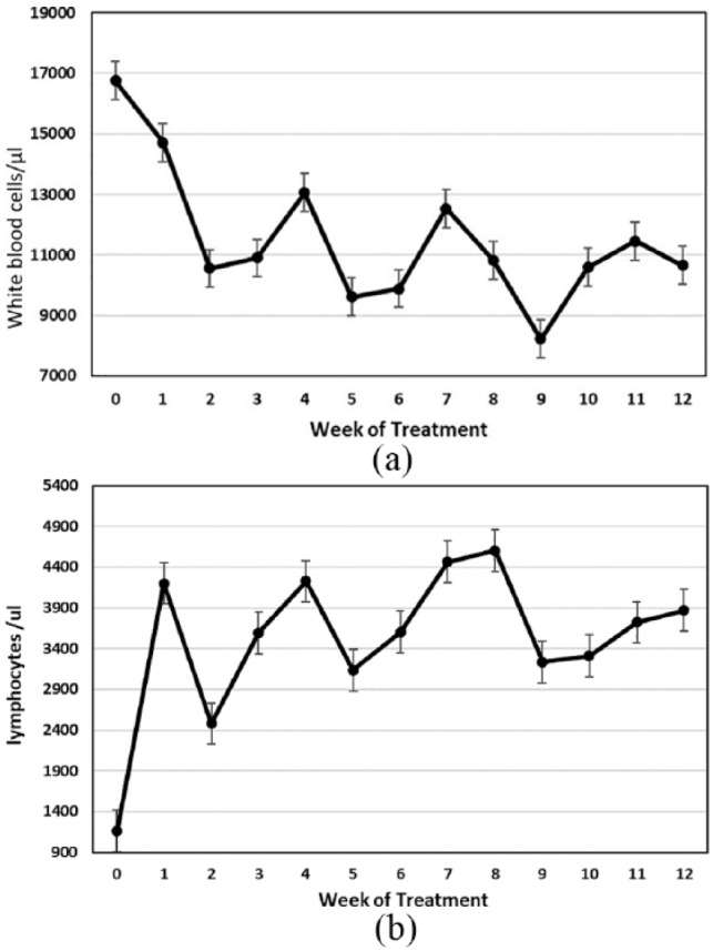


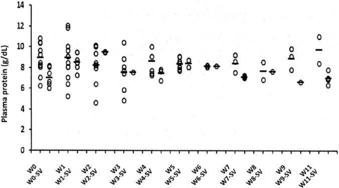


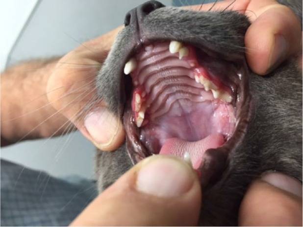


 EN
 SK