Sam Taylor, BVetMed (Hons), CertSAM, MANZCVS, DipECVIM-CA, FRCVS and Emi Barker BSc (Hons), BVSc (Hons), PhD, DipECVIM-CA, MRCVS summarize ideas about the manifestations and diagnosis of this disease and represent a new era of treatment .
Figure 1. “Classic” Cat with “classic” FIP with massive abdominal effusion. Image: Feline Centre, Langford Vets, University of Bristol
FIP is caused by virulent mutations in feline coronavirus (FCoV), which transform it from a mild and enteric infection to a serious systemic disease.
Like other coronaviruses, FCoV is a large enveloped RNA virus - this is important when considering immune system avoidance, environmental survival, detection, treatment and prevention. FIP has a high mortality rate and, until recently, treatments were relatively ineffective.
This article summarizes current views on the manifestations and diagnosis of this disease. It also represents a new era of FIP treatment in the context of the recent availability of legal medicines in the UK.
What causes FIP?
FCoV is an alpha-coronavirus that infects domestic cats and other cats. It is from the same genus as canine enteric coronavirus and swine gastroenteritis virus. FCoV cannot infect humans and is only distantly related to SARS-CoV-2, beta-coronavirus, and pathogen COVID-19.
FCoV, as a feline enteric coronavirus (FECV) biotype, is commonly detected in faeces - especially in cats living in multi-cat households. The infection typically spreads via the fecal-oral route when kittens or young cats are in contact with excreting cats. The chances of survival of this enveloped virus in the environment are generally poor unless the virus is trapped in feces and is sensitive to most disinfectants.
In some cats, and at some point after the initial infection - between viral replication in enterocytes and efficient replication in macrophages and monocytes - the less virulent FECV mutates into a virulent form associated with FIP - that is, the FIP virus biotype (FIPV). Certain mutations associated with this transition have been found in the spike protein gene, although none of them are yet pathognomonic for FIP.
The high frequency of genomic mutations - a hallmark of RNA viruses - can also facilitate the avoidance of the host immune response and drive tissue tropism, leading to various manifestations of the disease. Natural direct transmission of FIPV between cats is considered rare, and it is generally believed that FIPV - and subsequently FIP - results from a new mutation in the FCoV of an individually infected cat.
Figure 2. Mild jaundice and pallor in cats with FIP.
One of the many complexities of FCoV and FIP is that the infection manifests itself in many different ways depending on viral factors such as strain and dose, but also on the cat's immune response and genetic factors.
A strong cell-mediated immune (CMI) response to FCoV appears to provide protection against FIP. In contrast, cats with a predominantly antibody-mediated response with a weak CMI response typically succumb to the effusive "wet" form of the disease due to immune-mediated vasculitis, while cats with moderate CMI develop tissue granulomas typical of the non-fusive "dry" form of FIP.
It is important to keep in mind that the effusive and non-fusive forms of FIP can significantly overlap, leading to a wide range of symptoms; short episodes of the effusive form of FIP may occur rather than the predominantly non-fusive form, and conversely, effusions may form in the terminal stages of the non-fusive form of FIP. In addition, many exuded cats have tissue granulomas.
Clinical signs
Classic presentation of a young cat with protein-rich ascites (Figure 1) may offer easier diagnostics; however, other cats may be more of a diagnostic challenge. Common non-specific symptoms include lethargy, anorexia and weight loss.
Affected cats may be febrile with a moderate fever, typically less than 40 ° C, which often fluctuates and responds poorly to NSAIDs (non-steroidal anti-inflammatory drugs or non-steroidal anti-inflammatory drugs) or antimicrobials, and jaundice, if present, is usually mild (Figure 2).
Figure 3. Hyphema, uveitis and hypopyon in cats with ocular FIP.
High protein viscous effusions are formed in approximately 80% cats with FIP - most (approximately 85%) involve the abdominal cavity, while fewer cases show thoracic effusion (approximately 20%).
Pericardial effusions are occasionally observed, although rarely causing tamponade, and very rarely, scrotal effusions occur in uncastrated cats.
Pyogranulomatous lesions can occur in any tissue, and while they commonly involve the abdominal organs (e.g., mesenteric lymph nodes and kidneys), the disease may be limited to other organs such as the eyes, brain, or spinal cord.
Ocular symptoms include uveitis, ceramic clots, hypopyon, hyphaemia (Figure 3) and retinitis. Neurological symptoms include ataxia, seizures, nystagmus, hyperesthesia, and behavioral / mental changes.
Diagnosis
Although one abnormality does not in itself determine the diagnosis of FIP, nor does their absence rule out the diagnosis, the veterinarian may base the suspicion on FIP with the following findings, bearing in mind signaling, clinical picture, clinical pathology, and imaging results if no alternative diagnosis is present. , more likely explanation:
Figure 4. MRI scan of a cat with FIP showing obstructive hydrocephalus and increased contrast of the meninges.
Blood analysis - lymphopenia, non-regenerative anemia, microcytosis, neutrophilia, hyperglobulinemia, low albumin / globulin ratio (A: G; classically less than 0.4), hyperbilirubinemia, high α-1 acid glycoprotein (often markedly elevated, more than 1.5 mg / ml).
Diagnostic imaging - effusions, abdominal lesions, CNS abnormalities consistent with meningeal thickening and / or obstructive hydrocephalus (Figure 4).
Efusion analysis - non-aseptic pyogranulomatous inflammation with a relatively low cell number (total number of nuclear cells higher than 5 × 10⁹/ l; neutrophils and macrophages) with high protein concentrations (often higher than 35 g / l) and low A: G (Figure 5).
Molecular diagnostics - positive results of FCoV RNA RT-PCR in fluids (eg effusions, aqueous humor, CSF; Note: false positive and negative results are possible in whole blood) or aspirates with a thin needle (FNA) of the affected organ (eg kidneys, liver, mesenteric lymph nodes); the higher the viral load, the more evidence of FIP. Note: RT-PCR cannot confirm the diagnosis of FIP.
High levels of FCoV antibodies indicate only previous FCoV infection and do not indicate a diagnosis of FIP
The definitive diagnosis of FIP is confirmed by positive immunohistochemical staining for coronavirus antigen in macrophages associated with pathological changes in FIP in formalin-fixed tissue samples. However, sampling for histopathology and immunohistochemical staining requires invasive procedures, which may be contraindicated in a sick cat.
Figure 5. Sweat analysis is useful in diagnosing FIP; where possible, take a fluid sample.
Alternatively, the presence of antigen-positive coronavirus cells in cytological specimens (effusion, aqueous humor, cytospin CSF preparations or FNA from any abnormal organs - such as mesenteric lymph node) showing pyogranulomatous changes is very helpful in diagnosis and allows less invasive specimen acquisition.
Some researchers have used cell pellets prepared from centrifuged effusion samples to improve the sensitivity of immunohistochemical staining (Tasker et al, 2021).
However, it is important to diagnose FIP as reliably as possible before treatment, as there are many other differential diagnoses for these clinical symptoms - including neoplasia (especially lymphoma), other infectious diseases (pyothorax, toxoplasmosis, mycobacteriosis, fungal infections)) and primary immune-mediated disease (idiopathic disease). , uveitis, lymphocyte cholangitis). On the Figure 6 is a graph indicating possible diagnostic pathways.
Minimally invasive sampling may include abdominal or thoracic effusions and FNA abnormal organs.
More detailed diagnostic flowcharts for the diagnosis of FIP are available on the website of the European Advisory Committee on Feline Diseases (www.abcdcatsvets.org/feline-infectious-peritonitis).
Figure 6. Access to FIP diagnostics
FIP treatment with antiviral drugs
In recent years, publications have focused on antiviral drugs (GS-441524, a nucleoside analog that inhibits viral RNA polymerase, and GC376, a viral protease inhibitor) with the potential to cure experimentally induced cats (Kim et al, 2016; Murphy et al, 2018) and naturally obtained (Pedersen et al, 2018; 2019; Dickinson et al, 2020) FIP.
Unfortunately, until recently (see below), legal formulations of these drugs were not commercially available, although some owners obtained and administered illegal formulations to their cats of unknown origin and at great expense.
Remdesivir, a prodrug of GS-441524, is an antiviral drug with a broad spectrum of activity against RNA viruses. It was originally developed to treat hepatitis C virus and Ebola virus in humans. Its development was then significantly accelerated due to the worldwide treatment of SARS-CoV-2.
In Australia, remdesivir has been legally available to veterinarians for several months as a "special" formulation that allows veterinarians to gain experience with this drug in the treatment of cats and kittens with FIP, where it has shown promising results. Unlike GS-441524, remdesivir has low oral bioavailability and is administered by intravenous infusion to human patients.
In the UK, remdesivir is legally available through Gilead Sciences, a patent-pending company that manufactures a medicinal product for human use. The currently available formulation is Veklury, a powder for reconstitution with water for solutions for injection to a final remdesivir concentration of 5 mg / ml. After reconstitution, it should be cooled and consumed within 24 hours.
Figure 7. Vials with reformulated remdesivir will be available to treat FIP, legally, in the UK from August 2021.
In cats, SC is usually administered unofficially, although some cats may benefit from initial IV administration. From August 2021, remdesivir will also be available from specialty drug manufacturers as a veterinary ál special ’(Figure 7) in the United Kingdom. The reformulated remdesivir will be supplied in vials containing 100 mg remdesivir, at a concentration of 10 mg / ml, allowing for smaller injection volumes, with a shelf life of at least three months when properly stored.
The experience of our colleagues from Australia (Malik, personal communication) allows us to design benefits according to the above plan. The authors emphasize the need to diagnose FIP before using this drug to ensure its proper use, while acknowledging that the diagnosis of FIP can be anticipated due to clinical or financial diagnostic limitations.
Costs, prolonged nature of the treatment cycle (recommended at least 12 weeks), potential discomfort with SC injections and risk of relapse should be discussed with cat owners before initiating therapy.
Owners may be instructed to give daily injections to their cat, but must be thoroughly trained to avoid unintentional self-administration, incorrect technique that may harm the cat, and to minimize the risk of the cat reacting to injections leading to bite or scratch injuries. This healing process requires committed owners and is an emotional as well as a significant financial commitment.
Depending on the clinical condition of the cat, supportive therapy is still needed (eg IV or SC fluids, antiemetic stimulation and appetite, analgesia, tube nutritional support, sepsis antimicrobials). Cats with uveitis may require topical treatment with corticosteroids and cats with neurological symptoms may need anti-seizure medication.
Figure 8. Bengal kitten with ocular and neurological FIP before (left) and after (right) remdesivir treatment.
Although systemic use of corticosteroids is not generally recommended concomitantly with the use of antivirals, short-term administration of corticosteroids may be considered in cats with a strong suspicion of secondary immune-mediated disease due to FIP (eg immune-mediated haemolytic anemia).
The success rate of treatment is high - 80% to 95% (Malik, personal communication; Figure 8) - and therefore we have reason to be optimistic when discussing treatment with clients, even though we are aware of the commitment and the associated costs and potential for relapse.
Increasing the success of treatment
The treatment is long and remdesivir can be painful when injected. Once opened, the medicine should be stored in the refrigerator and should be warmed to room temperature before injection. Needle size can affect discomfort; for some patients, a faster injection of a larger diameter "green" needle (21G) may be more advantageous, while a smaller diameter orange "orange" (25G) needle may be more advantageous. A new needle should be used for each injection.
Some cats will need to go to the clinic every day for an injection. Gabapentin or trazodone (both 50 mg to 100 mg orally per cat), given two hours before the deadline to reduce anxiety and pain, may benefit some cats. Other cats may need a dose of buprenorphine (0.02 mg / kg to 0.03 mg / kg transmucosally or IM in a clinic before treatment).
To reduce discomfort, the injection site can be trimmed and EMLA topical anesthetic cream applied 45 to 60 minutes before injection. Injection tolerance appears to vary between cats.
Remdesivir in the treatment of FIP
Dosage
FIP s exudates (ie ascites and / or pleural effusion), but without any ocular or neurological impairment: 7 mg / kg until 8mg / kg once a day.
FIP s ocular symptoms (that is, uveitis or other eye disorders, but without neurological impairment): 10 mg / kg once a day.
FIP s neurological symptoms: 12 mg / kg until 15mg / kg once a day.
Method of administration
Most cases: SC injection into the loose skin of the interscapular area.
Very severe cases: 10 mg / kg may initially be given by intravenous infusion (ie diluted in 10 ml saline and given slowly over 10 to 20 minutes) to achieve a rapid antiviral effect; This can be done after three to four days of SC injection as soon as the cat starts eating and its health improves. Note: After intravenous administration, some cats may experience depression for several hours.
Duration of treatment
Treatment of at least 84 days (ie 12 weeks) should be considered. This time is based on a clinical study with GS-441524 and the unofficial use of remdesivir to minimize the likelihood of FIP relapse.
After 84 days, treatment should only be discontinued if the patient is clinically OK and abnormal laboratory parameters have returned to normal.
If the response to treatment is only partial or unsatisfactory, a prolongation of treatment may be necessary.
Monitoring
In the first days, closely monitor for clinical signs: - Improvement should be rapid, within a few days, with weight gain and improvement in clinical symptoms. - Consider verifying the diagnosis if no improvement is seen (with regard to the following symptoms): • Efusion (especially pleural) may worsen for one to two days at the start of treatment and may require therapeutic thoracocentesis or abdominocentesis. This appears to occur most frequently after IV treatment. Consider ultrasound monitoring once or twice daily. • Neurological symptoms may initially appear or worsen in the first days of treatment. This may include the development of seizures that may require medical attention (eg levetiracetam 20 mg / kg to 30 mg / kg every eight hours).
The weight should be checked regularly and the doses adjusted accordingly.
Monitor PCV, total proteins (albumin and globulin), bilirubin and other abnormal parameters until they return to normal. The frequency of monitoring varies between veterinarians; a monthly assessment of biochemistry and hematology is usually performed, but should be adapted to the client's finances and the cat's response and behavior.
Serum globulins may increase initially, but any hyperglobulinemia usually resolves by week 12.
Remdesivir is reported to cause reno / hepatotoxicity in humans, but these have only been observed at higher doses by our Australian colleagues, who resigned when the dose was reduced. * Higher doses may be required depending on the response. Using lower doses to reduce costs may increase the likelihood of treatment failure. Please note that these dosing recommendations may change depending on the growing amount of data and clinical experience of veterinarians using this medicine. It is recommended to consult an expert in cats or internal medicine to discuss the individual case and the appropriate dosage.
Further treatment
The researchers are looking at the beneficial effects of immunostimulants and / or other antiviral medicines, such as interferons or mefloquine, once remdesivir treatment is stopped or if the injections are considered too painful. Most studies published to date have focused on the use of antiviral drugs alone.
Thanks
The authors would like to thank their Australian colleagues David Hughes, Rebecca Brady and Richard Malik for sharing their experiences with remedivirus treatment in cats with FIP. Thanks also to Séverine Tasker and Professor Gunn-Moore for comments on the article.
Do you need advice on FIP treatment?
Stephanie Sorrell and Danièlle Gunn-Moore of the University of Edinburgh will recruit cases to monitor the UK's response to the remdesivir, with more information coming soon.
If advice is needed in the meantime on the diagnosis and treatment of a suspected FIP case, send an e-mail to fipadvice@gmail.com
References
Dickinson PJ, Bannasch M, Thomasy SM, Murthy VD, Vernau KM, Liepnieks M, Montgomery E, Knickelbein KE, Murphy B and Pedersen NC (2020). Antiviral treatment using the adenosine nucleoside analogue GS-441524 in cats with clinically diagnosed neurological feline infectious peritonitis, Journal of Veterinary Internal Medicine34(4): 1,587-1,593.
Kim Y, Liu H, Galasiti Kankanamalage AC, Sahani Weerasekara S, Hua DH, WC Groutas, Chang K and Pedersen NC (2016). Reversal of the progression of fatal coronavirus infection in cats by a broad-spectrum coronavirus protease inhibitor, PLOS Pathogens12(3): e1005531.
Murphy BG, Perron M, Murakami E, Bauer K, Park Y, Eckstrand C, Liepnieks M and Pedersen NC (2018). The nucleoside analog GS-441524 strongly inhibits feline infectious peritonitis (FIP) virus in tissue culture and experimental cat infection studies, Veterinary Microbiology219: 226-233.
Pedersen NC, Kim Y, Liu H, Galasiti Kankanamalage AC, Eckstrand C, Groutas WC, Bannasch M, Meadows JM and Chang KO (2018). Efficacy of a 3C-like protease inhibitor in treating various forms of acquired feline infectious peritonitis, Journal of Feline Medicine and Surgery20(4): 378-392.
Pedersen NC, Perron M, Bannasch M, Montgomery E, Murakami E, Liepnieks M and Liu H (2019). Efficacy, and safety of the nucleoside analog GS-441524 for treatment of cats with naturally occurring feline infectious peritonitis, Journal of Feline Medicine and Surgery21(4): 271-281.
Someone contacted me with 5 cats who got sick after one of their people got sick from COVID-19. The cats had fevers, sneezing, coughing, and making sounds with difficulty. This person wanted to know if cats should be treated for COVID-19 infection. I asked Dr. Pedersen and below is his answer (shared with his consent).
[Dr. Niels Pedersen] - Cats become infected with the SARS-CoV-2 virus, which either does not cause any disease or only a mild and transient upper respiratory tract infection, similar to the very mild case of feline herpesvirus. In rare cases, the disease can be severe in cats. In fact, there is a chance that this group of cats suffers from herpesvirus and not coronavirus Covid-19. Veterinarians can test cats for herpesvirus using PCR, but there is no test available to a veterinarian for Covid-19, and human laboratories refuse to test cats for Covid-19 using human tests.
In any of these infections, cats usually recover in 10-14 days without treatment or with minimal treatment. If the symptoms of the disease they develop are caused by the SARS-CoV-2 virus, they would respond to GS, but why treat them? It would be like treating every person with a positive PCR test for Covid-19 Remdesivir for 5 days for $ 2,000. Of course, cats are not that big and would not cost so much, but they would have to be subjected to daily injections without justification. It is also likely that the veterinarian would still not have access to Remdesivir, as it is currently limited to hospital use for a certain group of patients.
In short, my recommendation would be to test a few worse cases for feline herpesvirus, and they are very likely to be positive. If they test negative, they have a chance of being infected with Covid-19, but in both cases the treatment will be the same. Cats should rest, you should treat the discharge from the eyes and nose locally, and antibiotics should only be used if they are purulent discharge, which indicates a secondary bacterial infection. Rarely, breathing problems, fever and anorexia may occur, suggesting secondary bacterial pneumonia and the need for more aggressive symptomatic treatment. They heal in 10-14 days.
The article uses the term FIP repeatedly for the common non-mutated enteric coronavirus FCoV, probably for simplicity. However, the essence remains the same. (Translator's note)
The scientific community is concerned about a new coronavirus that appeared a year and a half ago in China. Jean-Marc Sabatier * first makes an analogy between coronavirus infectious peritonitis in cats or FIP (non-human transmissible) and SARS-CoV-2, which is caused by Covid-19. A better understanding of the complex physiological phenomena common to both viruses may provide a definitive medical response to a pandemic in the future. An interview.
In collaboration with the heads of the State Virology Laboratory in Wuhan (China), professors Zhijian Cao and Yingliang Wu and veterinarians Pierre Petruzzi (Clinique Vétérinaire de l'Albanne in Barberaz, Savoie) and Philippe Durieux (CHV Les Cordeliers, Meaux), you pointed out the remarkable similarities between cat disease and Covid-19. Can you explain that to us?
Feline infectious peritonitis (FIP) is a feline viral disease caused by alpha-coronavirus, while SARS-CoV-2 responsible for Covid-19 is a beta-coronavirus. These are related viruses. FIP is contagious to cats, but is not transmitted to other species, including humans. The FIP has been well known and documented since the 1980s. FIP viruses are known to be enteric coronaviruses that have become pathogenic due to mutation (s) and affect either young cats (aged 3 months to 3 years) or older cats (aged 10 to 15 years). FIP-related pathologies are similar to Covid-19 diseases.
What does it mean?
The clinical signs of FIP are very diverse, as in Covid-19, and the disease is divided into "dry" and "wet" forms. The dry form can affect the eyes, can cause granulomatous inflammation of the lungs, kidneys, liver, central nervous system, ganglia, intestines with possible neurological disorders… The wet form is manifested by effusions in the abdomen or chest, pleurisy (pleurisy), peritonitis (peritonitis), which are also well defined in the case of Covid-19. Other clinical manifestations are common to both forms of disease: organ inflammation, thrombosis, thrombocytopenia, coagulopathy, etc. Dry and wet forms can coexist or follow each other. FIP virus infection of macrophages is responsible for the fatal granulomatous vasculitis of cats.
What else do we know about this feline disease?
The FIP virus can be latent and harmless for years (up to 14 or 15 years) or it can multiply and become fatal in a short period of time. Progression to severe form takes an average of 2 to 5 weeks. Cats cannot transmit the FIP virus to humans, but cats can be (exceptionally) infected with SARS-CoV-2, which causes Covid-19. The latency observed with FIP raises the question of the possible latency of Sars-CoV-2 given the similarity between the two viruses.
How is the virus transmitted between cats?
The virus is transmitted from mother to kittens. The virus is present in feces and can easily infect other cats, especially in shelters and kennels.
What are the similarities between feline viruses and SARS-CoV-2?
FIP and SARS-CoV-2 viruses are single-stranded, positive RNA-coated coronaviruses. This means that although one is of the alpha genus and the other of the beta genus, both are closely related and have a zoonotic origin. In reality both viruses attack the host renin-angiotensin system (RAS) and over-activation of this system causes comparable disorders and pathologies, even though the two viruses act on different cellular receptors. The FIP virus has a molecule called aminopeptidase N (APN) as a cellular receptor. It is a surface receptor found in the intestines, kidneys, lungs, epithelial and endothelial cells, etc. APN converts angiotensin-3 to angiotensin-4. Angiotensin 3 targets the AT1R receptor, which is responsible for Covid-19.
And what about another virus, SARS-CoV-2?
SARS-CoV-2 binds to the ACE2 receptor and leads to an increase in angiotensin 2, which in turn leads to overactivation of the AT1R receptor, which is very harmful because it is responsible for Covid-19. In other words, the two viruses target the AT1R receptor (feline or human) in different ways. It is important to understand that APN and ACE2 are components of RAS, which is the main physiological system important for the functioning of the body, whether it is renal, cardiovascular or pulmonary functions. It is also involved in the intestinal microbiota and innate immunity, both in cats and humans.
What do you deduce from this?
FIP diseases caused by the FIP virus are comparable to diseases caused by SARS-CoV-2. There is no specific treatment for the cat other than symptomatic medications. RAS inhibitors are needed.We already know, of course, that there is a cure (Translator's note).
Is there no vaccine for FIP?
Vaccination experiments were performed using inactivated FIP virus. Young cats were vaccinated with an avirulent strain of the virus. However, this vaccination did not protect them from the oral route of virulent virus infection. In contrast, vaccinated animals were more easily infected than unvaccinated cats. This lack of protection suggests the existence of a phenomenon called ADE (antibody-dependent amplification). ADE leads to a sequence of events that can be found in several viral diseases (HIV, Dengue, Ebola…). Viruses infect cells by a mechanism that relies on the interaction between so-called "facilitating" antibodies (as opposed to protective "neutralizing" antibodies) and the virus.
This is unexpected. So can the vaccine support the disease?
Indeed, "facilitating" antibodies allow the virus to better penetrate target cells. This mechanism to facilitate virus infection of cells would be problematic if it were also present during SARS-CoV-2 vaccination. Treatment would then be worse than the disease. However, the problem posed by ADE during vaccination can be circumvented.
* Jean-Marc Sabatier, Director of Research at CNRS and Doctor of Cell Biology and Microbiology, working with the Institute of Neurophysiopathology (INP), University of Aix-Marseille. Editor-in-Chief of the international scientific journals "Coronaviruses" and "Infectious Disorders - Drug Targets" (DR)
Feline coronavirus (FCoV) causes a subclinical infection that can develop into FIP's lethal infectious peritonitis. It is caused by the feline infectious peritonitis virus (FIPV), which occurs when a mutation occurs in the spike of the FCoV virus. Despite more than three decades of virus research, there is still no approved treatment for FIP. This is partly due to the fact that FIPV type I causes most clinical infections, but is not very successful in cultures and there are few reagents available for its study, while type II grows well, has more reagents, but generally does not cause clinical infections. disease. The development of new immunotherapies requires significant preclinical study and validation, and due to limitations in the FIPV type I study, no immunotherapies have been developed for clinical FIP. We want to try a new immunotherapy using in vitro FIPV type II research, which could lead to the treatment of FIPV cats caused by FIPV type I. Immunotherapy, which has recently gained attention in the treatment of human cancers, are immune cells treated with a chimeric antigen receptor ( CAR). This approach has not yet been developed for acute viral infections such as FIPV, nor has it been used in cats. CAR consists of two main components: a single chain antibody fragment (ScFv) and a signal domain from the immune costimulatory receptor. We propose to use a FIPV-specific ScFv spike protein expressed in feline immune cells that could be administered to a feline to screen for and destroy FIPV-infected cells. Current human CAR therapies require the use of autologous T cells because allogeneic T cells kill host cells and lead to graft-versus-host disease. NK cells (natural killers) are of increasing interest because NK cells do not induce graft-versus-host disease and can therefore be used in "conventional" therapies. However, little is known about feline NK cells. We hypothesize that genetically modified feline NK cells expressing CAR targeting the feline infectious peritonitis virus type II spike protein will recognize and kill FIPV-infected target cells in vitro.
To achieve our goals:
determine the cytotoxic activity of a subset of NK cells in comparison with NKT and T cells for the validation of feline NK cells as potential CAR carriers,
for cloning and sequencing, we will use currently available hybridoma spike anti-FIPV type II antibodies to construct a CAR directed against spike type II protein, and
in vitro we validate the NK cell assay and data demonstrating the effect of CAR-NK cells killing FIPV type II infected cells.
The completion of this study will provide data important for the development of CAR-NK therapy for FIPV type I, a completely new treatment for this devastating disease.
Remdesivir suppresses host cell defense. This knowledge may be important for the development of drugs to fight various viruses. Photo: Bernard Chantal / shutterstock
When a cell is infected, SARS-CoV-2 not only causes the host cell to produce new viral particles. The virus also suppresses host cell defense mechanisms. The nsP3 viral protein plays a central role in this. Researchers at Goethe University, in collaboration with the Paul Paul Scherer Institute in Switzerland, found through structural analyzes that the degradation product of the virostatic agent remdesivir binds to nsP3. This points to another previously unknown potent mechanism of remdesivirus, which may be important for the development of new drugs to combat SARS-CoV-2 and other RNA viruses.
The virostatic agent remdesivir was developed to disrupt an important step in the propagation of RNA viruses, including SARS-CoV-2: the reproduction of the virus's own genetic material. This is represented by the RNA matrix by which the host cell directly produces viral proteins. However, to accelerate the production of their own proteins, RNA viruses also copy the RNA matrices themselves. For this purpose, they use their own specific protein (RNA polymerase), which is blocked by remdesvirus. More specifically, it is not the remdesivir itself that does, but rather the substance that is synthesized from the remdesivir in five steps as the remdesivir penetrates the cell.
In the second of these five steps, a product is formed from remdesivir, a substance with a somewhat impractical name, GS-441524 (in scientific terminology: a metabolite of remdesivir). GS-441524 is also a virostatic agent. As researchers in a group led by Professor Stefan Knapp of the Institute for Pharmaceutical Chemistry at Goethe University in Frankfurt found out, GS-441524 targets a SARS-CoV-2 protein called nsP3. nsP3 is a multifunctional protein whose tasks include suppressing the host cell defense response. The host cell is not helpless in the face of the virus because, among other things, it activates the inflammatory mechanisms by which it calls the endogenous immune system of the cell. However, the nsP3 protein helps the virus to suppress this call for help.
Professor Stefan Knapp explains: “GS-441525 inhibits the activities of the nsP3 domain, which is important for virus reproduction and which communicates with human cellular defense systems. Our structural analysis shows how this inhibition works, which allows us to lay an important foundation for the development of new and more effective antiviral drugs - effective not only against SARS-CoV-2. The target structure of GS-441524 is very similar in other coronaviruses, such as SARS-CoV and MERS-CoV, as well as in a series of alphaviruses, such as chikungunya virus. Therefore, the development of such drugs could help prepare for future viral pandemics. "
Translator's note: The nsP3 protein also contains the feline coronavirus FCoV / FIPV.
Publication: Xiaomin Ni, Martin Schröder, Vincent Olieric, May E. Sharpe, Victor Hernandez-Olmos, Ewgenij Proschak, Daniel Merk, Stefan Knapp, Apirat Chaikuad: Structural Insights into Plasticity and Discovery of Remdesivir Metabolite GS-441524 Binding in SARS-CoV ‑ 2 Macrodomain. ACS Med. Chem. Lett. 2021, 12, 603-609 https://pubs.acs.org/doi/10.1021/acsmedchemlett.0c00684
Some time ago, a Latin teacher at Mount Holyoke College who loved cats decided to change her profession. She chose the prestigious American Veterinary School of the time, Cornell, and found a place in America’s best veterinary hospital, Angell. She became the first specialist in cat medicine. In 1962, she reported her observations of a strange and always fatal cat disease, which she called "chronic fibrinous peritonitis." Two years later, a veterinarian in PennVet informed about a cat with a similar disease. Humans have lived with domestic cats for thousands of years and have been writing about them for centuries. It is unlikely that a serious disease such as FIP will go unnoticed and unrecorded for so long. I believe that an important coronavirus mutation occurred in the late 1950s. Some experts believe that there is a combination of the intestinal form of canine and feline coronavirus. (read here)
Today, veterinarians call the widespread and rarely dangerous intestinal (enteric) feline coronavirus FCoV (= feline enteric coronavirus alias FECV) and its dangerous mutated form, FIPV (feline infectious peritonitis virus). When a coronavirus mutation occurs, they are often viral spike proteinswhich changes the selection of the target (cells) they attack. (read here) A group of Dr. Niels Pedersen of Davis came up with the hypothesis that something like this happened to cats. (read here) He confirmed this in 2009. (read here)
When the virus reaches the mutation of interest, this new mutated form prevails. This is not the case with FIP coronavirus. A mild form of feline coronavirus FCoV still predominates. It is never in the virus's interest to kill the host it inhabits. Most veterinarians and scientists now believe that the mutated FIPV form of feline coronavirus must come from the mild intestinal form of FCoV in every new cat that becomes ill with FIP (or manages to overcome it). In most cats, this mutation is unlikely to ever occur. We currently believe that this will happen in about 8-10% cats. Of these cats, some will overcome the virus on their own and survive, some will not. Until recent discoveries at the Veterinary School in Davis, California, all cats that began to show signs of the disease died. Genetic factors unique to your cat are undoubtedly another factor that depends on whether or not FIP breaks out in a cat. It is also known that experimental transmission of mutated FIPV coronavirus from a sick cat to a healthy cat causes FIP to break out in most cats. (read here)
How common is the common mild intestinal form of feline coronavirus?
Common, mild, feline coronavirus (FCoV) is quite common in cats around the world. Some studies show that almost half of the world's domestic cats have been exposed to it. In some urban areas, up to nine out of ten cats have encountered the virus. However, it is quite important which cats are actually sampled. (read here, here, here and here) In these studies, the number of FCoV-positive cats living in groups is much higher than in those living in individual households. Cats in remote isolated areas appear to live their lives without coronavirus. (read here and here)
Detection of previous FCoV exposure by routine antibody-based tests does not necessarily mean that the virus is still present in your cat. And a lack of antibodies can also occur at the onset of a true FCoV infection. Whatever the actual numbers, the virus is much more common than veterinarians would like. When cats are not more concentrated than nature would like, the mild form of feline coronavirus and the mutated form of FIP are not significant problems. Only your cat's 4% wild European ancestors show signs that they have ever encountered a coronavirus - and even that has probably happened to an accidental encounter with a domestic cat. (read here)
Does the common mild intestinal form of feline coronavirus (FCoV) cause the disease?
This mild form of coronavirus is most common in cats less than two years old. Many cats (probably most) do not show any symptoms related to the health condition. For those who show symptoms, these are usually mild diarrhea lasting only a few days. Most cats eventually eliminate the active virus from their bodies - although it is not certain whether the virus in some cats does not persist in an inactive form, incapable of replication, the so-called provirus. We all carry viruses in our bodies.
Is my cat's age important for the common coronavirus mutation to FIPV?
Yes.
Most cats that develop FIP are young - usually between the ages of three and sixteen months. More than half are less than one year old. In one California study, laboratory-bred cats developed increased resistance to FIP infections at 6 to 12 months of age. This means that FIP occasionally develops in older cats. Veterinarians do not know whether older cats' immune systems are more likely to kill coronavirus mutants (destroy them before they can multiply) or whether the virus is less likely to mutate older cats or whether other unknown factors are involved. (read here)
How does a common coronavirus infection become a case of FIP?
Nature created viruses to randomly change their structure in an endless battle to defeat the immune system and continue to invade a new susceptible host. It is a random but very effective approach. Most of these random genetic changes in the composition of the virus (mutation) are unsuccessful. The viruses that carry these failed mutations are destroyed and no one has heard of them. But sometimes, by complete coincidence, there is a mutation that brings some benefits to the virus. When this happens, this mutated virus becomes the predominant form. Coronavirus is known for frequent mutations that allow them to jump between species or attack new parts of the body that they could not before. An attack on a new species or area of the body often involves changes in the surface spike viral proteins that I have already mentioned. In the case of an FCoV to FIPV mutation, this allows the virus to infect a new cell type, macrophages and monocytes in your cat. This transformation or change in the virus's preferences (tropism) causes the cat's immune system to start up, causing severe inflammation because the cells of the immune system attack the cat's cells that carry the virus (the release of pro-inflammatory cytokines). (read here, here and here)
I do not believe that a mild mutation in feline enteric coronavirus (FCoV) to a highly pathogenic (dangerous) feline infectious peritonitis virus (FIPV) offers any benefits to coronavirus. Killing a host is never an advantage for the virus. I therefore believe that FIP only occurs in a small number of cats that carry FCoV. FIPV mutations are likely to occur in many more cats than those who have developed FIP. But in these cats, their body recognizes and destroys these mutants shortly after they appear. Some sources report that FIP develops in 5-10% cats infected with FCoV. However, the actual numbers are probably much lower than in the general cat population. The reason is that a large proportion of healthy cats never visit a veterinarian, or if they do, it is only for regular vaccinations. If you find out which patients come to the clinic, most of them are sick.
Are there more forms of FIP?
Yes.
Veterinarians usually distinguish between two forms of FIP, wet and dry. In the "wet" form, inflammatory fluids are produced on the surface of the cat's abdominal and / or thoracic organs. In the "dry" form, inflammation occurs in the body's organs without the production of excess fluids. The same cat occasionally has symptoms of both forms.
Wet or effusive form of FIP
This was the first form of FIP that veterinarians such as Jean Holzworth recognized. With the effusive or moist form of FIP, fluid accumulates in your cat's chest or abdomen, or both. This leaked fluid is mainly the result of the cat's immune system attacking what it perceives as a threat from a foreign attacker (presence of proteins that do not belong to the cat's body = antigens). Most veterinarians believe that this accumulated fluid (exudate) is not caused by any direct damage that the virus causes to the cells in the body of the cat it inhabits. It is the cat's own cytokine-induced inflammatory response that releases a mixture of fluid (blood serum) and proteins (globulin and fibrin) as well as white blood cells, which would normally be limited to your cat's bloodstream. This fluid has a characteristic dense ("fibrinous") consistency with a yellowish appearance, which always hurts my heart when I see it slowly being sucked into the syringe.
Your veterinarian will probably tap (pull) to check the fluid to confirm suspicions, and then perform tests to confirm that the fluid is indeed exudate. In X-rays, this liquid has a characteristic grainy appearance (the appearance of "cut glass"). When fluid is found in your cat's abdomen, other possible causes will fly through your veterinarian's mind. Lymphoma tumors, heart or liver disease, or even a ruptured bladder are occasionally the cause of abdominal fluid. Other laboratory tests rule out these causes. When there is fluid in the cat's chest (pleural effusion), other tests distinguish inflammatory fluid (exudate) from lymph nodes. When cats have fluids in their chest, your veterinarian may note that your cat's heart sounds are muffled and difficult to hear through a stethoscope (difficult to aculture). When a cat's liver is affected, its gums, in addition to being pale from anemia, may have a slight yellowish tinge (jaundice). When an effusive FIP develops in a kitten, the pup may appear to be wilting or has stopped growing satisfactorily.
Dry, or non-fusible, pyogranulomatous form of FIP
Granulomas are made up of cells from the immune system that gather around objects and diseased organisms that the body is trying to separate or eliminate. When FIP coronavirus-infected cells are primarily located in organs, this is the way the cat attacks them. These events can take place in your cat's liver, kidneys, lymph nodes, eyes or nervous system, or any combination of these. Needless to say, the symptoms seen by cat owners and veterinarians are very variable. Veterinarians call this form the "dry" form of FIP because, unlike the effusive form, excess fluid does not accumulate - or at least does not predominate. The dry form is sometimes called "parenchymatous". Functional cells of various organs are called parenchyma.
These changes caused by the dry form of FIP tend to start gradually. They are so non-specific that they could have many explanations that are not related to the FIP. Inflammatory changes in the eye (uveitis) are a textbook symptom of a non-fusive form of FIP. This symptom is likely to trigger a FIP alarm sooner than most others. This is especially true if it is accompanied by other changes in the nervous system or eyes. These neurological problems may include bleeding, seizures, or just strenuous and unstable gait in the back (ataxia). It is not uncommon for both forms of FIP for the owner to tell me that the cat has experienced a major stressful event in recent months. Events such as a diet, a new domestic cat, a move, a rival cat in the neighborhood, or another problem that has been thought to be resolved. Another typical anamnesis is that the cat was recently acquired from a shelter, home with numerous cats or a private kennel. (read here and here)
How is the common milder coronavirus (FECV) transmitted between cats?
The more common, milder, non-mutated form of feline coronavirus FCoV lives in the cells that line your cat's gut. Upon release, these viruses are excreted in large amounts in cat feces. (read here) Cats that modify or share a toilet are the most common ways of transmitting this virus to another cat. Although no one has documented the survival of feline coronavirus in the environment, coronavirus generally survives in the environment for a maximum of several days. Therefore, most veterinarians do not believe that contaminated objects in your cat's environment are the source of many (if any) infections. But regular litter changes in households with multiple cats is always a good idea. When you bring a new cat into your home, a PCR test (FCoV RT-PCR) of her feces can tell your veterinarian if there is FCoV in the stool at that time. However, FCoV shedding is often interrupted. And at the onset of infections, test results may be negative, even if the virus is already present in the cat's stool - but in a number that is too low for the PCR test to detect.
How is dangerous FIP-mutated coronavirus transmitted between cats?
Veterinarians do not believe that the natural transmission of FIP virus from cat to cat is common. FIV-infected cats are known to excrete very little or no mutated virus in the faeces. (read here) Most veterinarians currently believe that each case of FIP requires a new mutation in the common FCoV virus after it infects a new cat. One article questions this. However, no one was able to duplicate these results. (read here)
When a new mutation produces a dangerous feline infectious peritonitis virus (FIPV), a certain percentage of cats develop FIP. But this will not happen with all cats. Experimentally, scientists can extract the FIP virus from a sick cat and inject it directly into another cat. This often leads to an outbreak of FIP in the second cat. However, we do not think that this is happening in the real world. In contrast, the complex interactions between the virus and the cat's immune system, the myriad ways in which the virus can mutate, and the unique genetics of each cat make the utter statements unreasonable. (read here) Few things in medicine are absolute.
How long after this mutation can my cat get sick?
I have already mentioned that the vast majority of cats that are exposed to the common feline coronavirus have never become seriously ill. When the virus stays in the intestines of your cat, where it belongs, you can most often notice only a short period of soft stools or diarrhea. Infection of the cat's intestinal lining (its enterocytes) most likely occurs immediately after the cat's coronavirus ingestion. However, it may take several days for a sufficient number of viruses to be detected in laboratory tests. (read here) After this initial infection, most cats seem to eliminate the virus from their bodies relatively quickly. However, some cats become intermittent or persistent carriers of the non-mutated form of the virus, and several develop FIP. (read here)
The time that elapses from infection with a mild coronavirus form to a possible mutation and possible outbreak of FIP is highly variable. It would be very difficult to design valid studies to address this. In the laboratory, FIP-related disease can begin within a few weeks. But it was then that the already mutated FIP virus was experimentally transmitted from cat to cat. Apart from the laboratory environment and "purposefully" cats, it tells us little about what is happening in the real world. Veterinarians really do not know the dynamics of FIP in a domestic or breeding situation when "wild" strains of the virus and genetically diverse cats are present. Between the time the cat was probably exposed to the "wild" coronavirus and the time it begins to show signs of disease, there are often long breaks - several months or even years. As I mentioned, many veterinarians associate FIP with stress or trauma. Factors such as overcrowding, frequent pregnancies, genetic predisposition, inbreeding, and surgery have been discussed as triggers that could upset the balance in favor of FIP. This is especially sad for me, because when a cat gets sick, my clients are already emotionally attached to it.
What are the symptoms of FIP?
I mentioned that the initial symptoms of FIP are often insignificant and can be easily overlooked. Lethargy, rough hair, little interest in grooming or playing, increased sleepiness, picky eating and weight loss are often the first reasons to visit a vet. However, these symptoms can have many other causes.
During a veterinary inspection, it is common for FIP cats to have low temperatures. Keep in mind, however, that normal cats often have mild fevers due to travel, stress, fear, or being manipulated by strangers. You may have mentioned to the receptionist that your cat has more problems in the toilet. Sick cats care less about their hygiene. Therefore, your veterinarian may suspect a bacterial infection and treat the cat with antibiotics. But antibiotics do not reduce these fevers. When these cats fight the FIP virus for a longer time, it is common for them to become anemic (non-regenerative anemia = there are too few reticulocytes in their blood).
Symptoms of the effusive (wet) form of FIP
Once the cat has entered the effusive wet form of FIP, the mediator compounds (inflammatory cytokines) released by the feline immune system result in inflammation of the blood vessels on the external surfaces of the body's organs (polyserositis). (read here) When this happens in the abdomen, the cats develop a fluid-filled, enlarged pear-shaped abdomen. When this happens in the chest (chest), breathing becomes strenuous. When fluid accumulates in your cat's abdomen, its weight may not change, even if it is a proper eater. This is due to the weight of the excess fluid in the abdomen. However, when you run your finger over the cat's spine, you will notice that it is bony and many cats have also lost the muscle mass of their legs. This condition is accompanied by malaise and cachexia.
Symptoms of non-fusible dry form of FIP
With this less common non-fusible (= granulomatous FIP) fluid, they do not tend to accumulate in your cat's chest or abdomen. This form of FIP is also developing more slowly. The non-fusible, dry form of FIP is also more likely than the wet form to be initially confused with diseases other than FIP that exhibit similar symptoms. Neurological health problems, such as epilepsy, seizures, tremors, balance problems, personality changes, or increased sensitivity to touch resulting from FIP, can be easily confused with other causes. So are vision problems. It is common for the dry form of FIP to affect the nerves and brain. But other organs may also be affected: the eye (s) (read here), liver, kidneys, respiratory tractread here) intestinal tract, skin. (read here)
What other diseases can be confused with FIP?
Diseases that can be mistaken for FIP include toxoplasmosis, cryptococcosis, hemobartonella, middle ear infections, trauma, drug intoxication, lymphoma, FeLV, FIV and tumors. Occasionally, cats show signs of both effusive and non-fusive forms. (read here)
How does my vet confirm that my cat has FIP?
The wet form of FIP is relatively simple for your veterinarian. Young cats, which come with mild fevers and fluid build-up in the chest or abdomen or both, are likely to suffer from the disease. Blood samples received by your veterinarian usually show mild to moderate anemia. Blood albumin levels are often low, A: G ratios are low, blood globulin levels are high, giving an albumin to globulin ratio <0.6. However, this test serves more as an exclusion test than as a test to confirm the diagnosis of FIP. (read here) Total lymphocyte counts are often low and neutrophil counts increased. Examination of the specific gravity of the fluid confirms that it is an exudate (> 1030).
When a cat develops a dry form of FIP, diagnosis is much more difficult. High levels of globulin in the blood (hyperglobulinemia) - often in combination with low levels of albumin in the blood suggest FIP. Although tissue biopsies can confirm the disease with certainty, their implementation is often difficult or impossible. Advanced diagnostic centers use useful tools for magnetic resonance imaging (MRI) and CSF analysis. (read here and here)
Most veterinarians diagnose FIP in dry form based on the predominance of evidence, not 100% certainty. Prevalence is a very relative term and is probably responsible for the FIP miracle drugs that are sometimes mentioned. Just because a cat is coronavirus positive and has some of the common symptoms of the dry form of FIP, doesn't mean it has FIP. None of the more common tests alone can confirm that the cat has a dry form of FIP. But a few tests, along with the symptoms your cat shows, are usually enough for your veterinarian to make a reasonable diagnosis. Even the presence or absence of antibodies against the coronavirus or the virus itself is insufficient, as many healthy cats appear in this way, so that the levels of antibodies against the virus and the coronavirus do not correspond well with FIP.
Some, including Idexx Laboratories, believe that high FCoV antibody titers increase the suspicion of FIP. In 2014, the company introduced its FIP Virus RealPCR ™ test, which claimed to be able to recognize (distinguish) common FCoV coronavirus from mutated FIP coronavirus. The test was designed based on some German studies. (read here, here and here) Despite some recent studies not sponsored by the company, they raised considerable doubts about the value of their patented test. (read here)
Protease inhibitors and similar compounds are the only drugs that currently offer hope to cats that fight FIP. Their existence dates back to studies conducted in the early 1990s to screen drugs for their ability to treat human hepatitis C and to suppress human AIDS. The research was later prompted by the 2003 SARS coronavirus epidemic. The general interest in antiviral drugs motivated a veterinary school in Manhattan, Kansas, to investigate some of these compounds. (read here) Some were already known to be effective against coronavirus. As it was a group of biochemists at a veterinary school, their interest naturally turned to the hitherto incurable FIP feline coronavirus and feline calicivirus. In 2015, they confirmed that of the 13 compounds studied, several were effective against feline coronavirus. Among them was GC-376. (read here) This information reached a veterinary school in Davis, California, where a veterinary virologist, Dr. Niels Pedersen, began attempts to administer GC376 to cats owned by wet or dry FIP clients. (read here) The results were very promising; although the drug appeared to be more effective in treating wet than dry forms of the disease. In another study from 2018, this group in Davis, in collaboration with Gilead Sciences, identified another closely related compound, GS-441524, which was also effective against FIP. (read here and here) Patent for GS-441524 owned by Gilead. This particular compound is a metabolite of its precursor remdesivir (aka GS-5734), which the company currently markets for the treatment of human Covid-19. Most information about the use of Remdesivir in cats is spread over the Internet. There are probably correct but also wrong advice on the Internet. Contact Bova Australia for more information.
GS-441524
Dr. Pedersen has documented the effectiveness of the drug in many cases. The problem was getting it. Gilead is a publicly traded company. Shareholders expect maximum profit. The company argues that selling products such as Remdesivir / Veklury® for human use is much more profitable than drugs for veterinary use. As I mentioned, remdesivir is a prodrug or prodrug that the body then metabolises to GS-441524. The precursor form of the drug will help him enter the right cells of the immune system, where the immunosuppressive coronaviruses reside. In the studies of dr. Pedersen on cats, GS-441524 was administered unchanged FIP, with a unprecedented success rate. Gilead's refusal to release GS-441524 for the treatment of FIP in cats quickly led desperate cat owners to turn to Chinese biochemical companies on the black market, which operate in violation of international patent law.
GC376
In other studies at veterinary schools in Kansas and California, another similar compound, a protease inhibitor, GC376, appears to help cats infected with FIP. A compound with similar potential is GC373 (read here and here) GC376 is licensed by the company Anivive Lifesciences. I don't know what their marketing plans are with this drug, but usually profit maximization comes into play.
One of the Chinese drug suppliers, MUTIAN Biotechnology Co. Ltd., based in Nantong, sells a drug called Mutian® xraphconn. The last time I checked their website, it was no longer available in capsular form. Previously, however, the FDA's list of non-approved drugs included active ingredients such as: fruiting body inonotus obliquus, nicotinamide ribotide, crocin, S-adenosylmethionine, and hake / silymarin inotodiol. The FIP is unlikely to cure anything. I was told that this product was sold as a "dietary supplement intended exclusively for cats with FIP," to strengthen their immune system and overall well-being. " Mutian is also available as an injectable form. As for the likely composition of these products, you will probably need to go to a website Sock FIP and FIP Warriors. But Gilead has his suspicions.
I was told that the Mutian solution for injection was selling for about $ 4,000 at a time for a 6.6 / 3 kg cat. Friends in Russia told me that the price there in 2021 had dropped to $ 1,000. This is probably due to competing suppliers in China, but I have no further information. Sock FIP and FIP Warriors would be much better sources of this kind of information. Mutian has stated that the composition of their product is a trade secret. But I also read that Gilead, the manufacturer of the remdesivir, sued them for patent infringement. Cat owners must be aware of the type of people they are dealing with, as well as the possibility that these drugs could be obtained by the US Customs Service if discovered.
It is almost certain that more antiviral compounds effective in the treatment of FIP-positive cats will be discovered in the near future. (read here and here) I believe there will be some among the 3CLpro inhibitors. Pfizer is already testing two of them on humans. So treatment options for FIP + cat owners should soon be much greater. I do not believe that some of these future drugs will be cheap. However, the costs will hopefully fall sharply as one drug competes with the other in the human coronavirus treatment market.
Davis also considered Merck's drug MK-4482 / EIDD-2801 (Molnupiravir®), which is being developed for oral use against coronavirus. However, I do not believe that the owner of the Merck patent would make this drug available to them. I may be wrong. But in April 2021, one in two clinical trials of the compound in people with Covid-19 was stopped because of its ineffectiveness in severely ill hospitalized patients. I see on the Internet that experiments with people in less severe cases of Covid-19 can continue. Pharmaceutical companies tend to be very cautious about disclosure - especially if they are negative and can affect their stock prices.
None of these drugs is completely risk-free. Skin reactions, tenderness and ulcers occasionally occur at the injection site. Rarely were they serious. Most of this can be caused by errors in injection technology. Minor kidney damage was once attributed to one of the Chinese drugs. The severity of your cat's condition at the time these medications are started also affects the success of the treatment. Some of these cats require infusion therapy. When secondary infections are present, they need to be treated with antibiotics and nutritional support. These measures, which may even include a blood transfusion, can give your cat the best chance of survival. It is always wise to perform a complete blood test (CBC and biochemistry, including A: G ratio) shortly before and regularly during treatment. They allow your veterinarian to scientifically monitor your cat's overall health and evaluate the course of treatment. However, a good appetite, a return to your cat's normal weight and a slow return to her old cheerful nature are always the best signs. If the cat does not want to eat, several days of corticosteroids (eg prednisolone) are allowed. Non-steroidal appetite stimulants such as Entyce® are available, but we have no idea if they interact negatively with any of these drugs. If you have been taking this medicine or mirtazapine, let me know the result. Elimination of effusion fluid is only desirable if it affects your cat's ability to breathe. Some cat owners use tonics such as SAMe, silymarin /, S-adenosylmethionine, probiotics, vitamin B3, etc. to support the liver or kidneys. However, none of these products affect the results of the treatment.
According to Dr. Pedersen et al., Cats less than seven years old are more likely to be cured than cats that develop FIP at an older age. And as I mentioned, cats with a dry form of FIP will be more likely to succeed than cats with a dry form - especially those that already have neurological and / or eye problems. FIP relapses have also been reported after a 12-week GS-441524 cycle. Sometimes another 8 weeks of medication was enough, and sometimes not. Sometimes cats that have developed a neurological form (dry form) of FIP and have been successfully treated with authentic or Chinese GS-441524 to remove the FIPV virus have not fully recovered from the disability. This is especially true if it involves paralysis and weakness of the legs due to damage to the central nervous system. Some believe that a higher dose of the drug can prevent this. However, in general medicine, peripheral nerves (hands, feet, etc.) often regenerate slowly: but once central nervous system neurons are destroyed, the changes may no longer be irreversible. (read here) However, most cats recover - if not completely, enough to lead a happy future.
Drugs with no proven effect
I mentioned that the cytidine nucleoside analogue Molnupiravir (EIDD-2801), which is or was developed by Merck for severe coronavirus disease in humans, may be effective in cats with FIP. But we still don't know for sure, or at least I don't.
Japanese veterinarians generally publish high-quality veterinary studies. One veterinarian in Japan thought that the combination of an anti-rheumatoid arthritis drug, Humira®, in combination with an antifungal drug, itraconazole, could be of some benefit to FIP cats. (read here) It may prove useful, maybe it won't. I don't know of any controlled studies. Itraconazole is primarily used in cats to treat severe skin conditions.
Curcumin
Curcumin is obtained from spices, curcuma. It is sold in health food stores with various health claims. One study from 2020 reported that curcumin had laboratory suppressive effects on the FIP virus. However, they never got to try it in FIP + cats. (read here) Curcumin is gaining great fame. However, as far as I know, it has never been found to be beneficial or curative for any human disease. (read here) We really have no idea what he could offer to FIP-positive cats.
Mefloquine
In 2020, four veterinarians at the University of Sydney looking for treatment options for FIP were investigating how cats metabolize the compound mefloquine used to treat human malaria. (read here) Sold as Lariam® in the USA. They were interested in the medicine because a previous study had shown that it was effective against feline coronavirus. (read here) I do not know whether these studies are ongoing. Clinical trials have also been launched in Russia to determine whether the drug could be beneficial for patients with Covid-19. No results were reported.
A treatment I don't trust
For many years, I believe that occasional "drugs" against FIP are likely to be cases where cats have only had a non-fatal intestinal coronavirus, but have FIP-like signs due to some other unrecognized health problems. No report I know of has ever verified that the coronavirus left the gut and attacked the cat's body. (monocyte / macrophage tropism). This must happen until the cat develops FIP.
Doxycycline
Some claim that a doxycycline antibiotic with or without prednisolone is beneficial to FIP + cats. Doxycycline is a suitable antibiotic for secondary bacterial infections that your FIP cat may encounter. It is also a great drug for the treatment of hemobartonella, which some cats may have positive for FIP. But no one has ever confirmed that it has any effect on the FIP virus. If you are giving your cat doxycycline in capsular or tablet form for any reason, always give it enough water or a piece of soft food, as it may settle in your cat's esophagus and cause damage. This is a good approach when giving your cat any kind of pill.
Prednisolone and other corticosteroids
It is true that prednisolone and similar compounds (corticosteroids) reduce inflammation. However, they have never been shown to affect the downward spiral of the FIP. However, corticosteroids are an excellent stimulator of appetite and positive mood, and in some cases provide FIP-positive cats with a short dose of energy and motivation. Similar effects occur in humans. (read here) It is therefore not harmful to administer them for several days under the supervision of a veterinarian.
Paradoxically, while some veterinarians try to stimulate your cat's immune system with drugs such as interferon and polyprenyl immunostimulant, others administer corticosteroids such as prednisolone or dexamethasone to weaken the cat's immune system.
In 1996, the founders of the Russian start-up company Micro-plus published an article about the amazing abilities of their compound, polyprenylphosphate (phosprenyl, aka fosprenil), to treat a wide range of diseases. They claimed to be "highly effective" in the treatment of rabies, cancer, canine distemper, hepatitis, enteritis (parvo?) And potentially useful in the treatment of human HIV and human herpes. They obtained a Russian patent for the compound. Polyprenyl immunostimulant (PI) is still commonly sold in Russian pet stores, where it is used to treat anything.
Russian veterinarian about Phosprenyl {2013)
I remain skeptical that polyprenyl immunostimulant has some value to your cat. Mithridates the Great last mentioned a potion with such wide effects in 100 BC If you want, read more about Big Theriak. Nevertheless, in 2009, veterinarians at the University of Tennessee said PI was likely to benefit several cats that were thought to have a dry form of FIP. (read here) Naturally, everyone was excited and full of hope. Even the Winn Feline Foundation contributed financially. But since this initial announcement in 2009, I have not known of any other publications on this drug in human or animal medicine. The effective drugs that veterinarians and cat owners so urgently need are absorbed much faster.
Omega feline interferon
Interferons play a role in your cat's natural resistance to corona and other viruses. (read here) Thus, it could be assumed that administering interferon to cats with FIP could help them. A Japanese study once suggested this. (read here) However, it was a poorly constructed study. Only a few animals were treated and observed, and no evidence was ever provided that any of the cats had FIP. But since the vets at the time had so little to offer cats with FIP, it caused a lot of excitement at the time.
A recent German study found that feline interferon administration had no effect on the life expectancy of FIP-infected cats studied, nor on their quality of life. (read here) You can, of course, say that the Germans did not administer interferon in sufficient doses, or administered it incorrectly or at the wrong stage of the disease, etc. So there are still veterinarians who give or recommend it. Virbac Corporation's European division still sells a recombinant feline interferon product (Virbagen Omega®). It is not currently sold in the US, but some North American cat owners have managed to get it. A British veterinarian published an article in 2020 suggesting that its use was beneficial in treating a cat infected with the dry form of FIP. However, in addition to the omega interferon, the cat received the Chinese drug Mutian. (read here) My belief, the belief of the NIH and others, is that these commercially available interferon products do not have the ability to treat coronavirus and probably not the course of FIP, FeLV or FIV. (read here and here and here) Another with similar claims, Feliferon®, is sold in Russia. I have no idea what's in it.
Alternative and complementary medicine
One person's despair is another person's opportunity. There have always been, and always will be, herbalists, naturopaths, obscure art practitioners, and even several veterinarians who claim to be able to cure incurable diseases. They will raise your hopes by telling you what you want to hear; and then when the money changes hands, they will disappoint you. Tragedy and loss are painful for each of us. But false expectations do not delay the necessary. These options only provide a false sense of comfort, support and consolation to some cat owners facing FIP. These cat owners should have the right to use them if they wish
What to believe? What information can I rely on?
Whether you can trust my information is something you will have to decide for yourself. But in general, I take my information from a group in Davis, California under Dr. Niels Pedersen. Over the years, I have learned to trust him and the publications and judgment of UC Davis. Equally credible is the information coming from Cornell. When evaluating the quality of a research paper and its findings, I always check who paid for it and the reputation of the journal in which it was published. I am also considering who will benefit financially from these findings. You can find a scientific journal that will publish almost anything today. These two veterinary colleges in the USA and the British Royal Veterinary Academy (RVC) are placed in the top three from year to year. Another veterinary institution with very high standards is the veterinary school in Shut up.
There is no consensus in the RVC on what can currently be done for cats with FIP. Neither what constitutes a sufficiently valid diagnosis of the dry form of FIP nor any treatment of any form after acceptance of the diagnosis. Some doctors still recommend a combination of interferon and prednisolone. Others do not believe in either. Until the results of the UC Davis studies became known, most veterinarians at the institution sucked out pleural effusions as needed and did whatever they thought could do the cat better. They offered consolation to cat owners as well as cats themselves and possible euthanasia.
I have been told that most veterinarians in the RVC are now likely to alert cat owners to the FIP Warriors website - warning that they may spend a lot of money and that the medicine they receive may not be what they thought they paid for. Regulations and thinking in Britain are such that most veterinarians there weigh in direct involvement for fear that they could be "removed from the register" (loss of license). So some veterinarians don't even mention China. Those I spoke to, as well as UC Davis, are opposed to the use of these Chinese drugs in an effort to achieve coronavirus-negative breeding due to fears of drug-resistant FIP strains that will no longer respond to GS-441524 and similar drugs. I agree. Some have fewer reservations about it. (read here)
There are also veterinarians in the RVC who question the likelihood that any common feline coronavirus has the ability to mutate to a variant of FIPV. This is the current theory accepted here in America and the one I have presented to you. These veterinarians from the UK are interested in what are the chances of the same mutation being repeated over and over again in different cats? And they ask, "Isn't it much more likely that cats have several different forms of coronavirus and only one virus (or a group of related viruses) has the ability to cause FIP?" I don't know the answer.
Can my cat be vaccinated against FIP?
Yes
Zoetis produces a FIP vaccine, Felocell FIP (IN). It is a vaccine that is given to the cats' noses. The company suggests that it should only be given to cats aged 16 weeks or older as 'help' in preventing the disease. Contains attenuated FIP virus. This particular strain of the virus is believed not to survive at the cat's core (internal) body temperature and to proliferate only in the cat's upper respiratory tract and nose, where it is likely to be harmless.
There is not enough data to tell if this vaccine has any practical use to protect your cats. First, many believe that most cats are exposed to the feline coronavirus before the age of 16 weeks - when they are still too young to receive a vaccine that produces antibodies to the coronavirus. However, we do not know how long these antibodies last or whether they are more effective in preventing FIP than the antibodies that most coronavirus-exposed cats already carry. There are also veterinarians who are concerned that antibodies destroy tissue in FIP-infected cats and that stimulating antibody production may be a negative event. Some early studies concluded that the vaccine was beneficial (read here), while others do not. (read here) If you decide to have your cat vaccinated with this vaccine, you should know if it already has anti-coronavirus antibodies in your blood as a result of previous exposure to feline coronavirus. In these situations, the vaccine is unlikely to be beneficial. (read here)
I have more cats. What should I do to protect them if FIP breaks out at one?
As a rule, I do not recommend isolating a sick cat from a stable household with multiple cats. Watch her, make sure she is not bullied at the time of feeding, meet her special needs if they do occur, but separation from roommates and changes in the household can be another stress for many cats. Assuming mutation theory is correct, if your cat family has been together for some time, all cats have been exposed to feline coronavirus, and are immune or possibly healthy carriers of the non-mutated form of FCoV. Try to reduce conflicts between cats. Kennels should consider a break in breeding and keep in mind that siblings of FIP + cats are at greater risk of developing the disease. In these situations, consideration should also be given to avoiding the stress associated with castration, overcrowding, sales or unnecessary vaccination.
The University of Colorado Veterinary School has the staff of the philosopher Bernard Rollin. He warns against keeping suffering animals alive for too long. He wrote an article about end-of-life problems in old cats, but many also apply to the situation of a cat with FIP. His and my beliefs are that when deciding to end your cat's life, you owe it to your friend to choose what will cause him the least pain and suffering, even if it means more immediate pain and sadness for you. You can read the article read here. If you want, read my thoughts on losing pets here. Over the years, many people have written to me that they blamed themselves for something they did or did not cause to lose their cats. But this is rare, and never so when it comes to the FIP.
Sarah Jones 1, Wendy Novicoff 2, Julie Nadeau 3 and Samantha Evans 1,*
1 Department of Veterinary Life Sciences, College of Veterinary Medicine, Ohio State University, Columbus, OH 43210, USA 2 Department of Orthopedic Surgery and Public Health, Faculty of Medicine, University of Virginia, Charlottesville, VA 22903, USA 3 Hamilton Region Veterinary Emergency Clinic, Hamilton, ON L8P 4W3, Canada * The author to whom the correspondence should be addressed.
Academic Editor: Clive JC Phillips Animals 2021, 11 (8), 2257; https://doi.org/10.3390/ani11082257 Delivered: April 22, 2021 / Revised: May 29, 2021 / Accepted: July 27, 2021 / Published: July 30, 2021
Feline infectious peritonitis (FIP) is a fatal feline disease caused by feline coronavirus. The aim of this study was to formally evaluate the administration of unlicensed antiviral therapy GS-441524 from group sources for cats with suspected infectious peritonitis in cats (FIP). Members of social networking support groups and GS-441524-based drug distribution groups were interviewed via the Internet. Of the 393 surveys that met the inclusion criteria, 73.7% owners were receiving this therapy from the United States. Only 8.7% owners reported that they received significant help from their veterinarian in treating their cat. The average cost of treatment was $ 4,920. The majority of owners (88.2%) reported a marked improvement in clinical symptoms within one week of starting therapy. At the time of the survey, 96.7%s (380 cats) lived, of which 54.0%s were considered cured and another 43.3%s were monitored over a 12-week observation period. A total of 12.7% cats had a relapse of clinical signs of FIP and 3.3% cats died despite GS-441524 therapy. The reported complications were mostly related to the administration of subcutaneous injections of GS-441524 acidic solution to the owner, such as vocalization, pain, wrestling and wounds at the injection site. Limitations of this study include retrospective design, bias in case selection, reliance on owner-reported data, and inability to validate unlicensed drugs; however, important lessons can be learned from the experience of these owners. Although unconventional and certainly not without health and legal risks, unlicensed, home therapy similar to GS-441524, according to information from the owners, can obviously bring benefits in the treatment of cats with a suspected diagnosis of FIP.
Abstract
The aim of this study was to formally evaluate the administration of unlicensed antiviral therapy similar to GS-441524 from group sources for cats with suspected infectious peritonitis in cats (FIP), a recently fatal disease. Members of FIP support groups on social networks and drug distributor groups with GS-441524 were interviewed via the Internet. The survey focused on owners who treated their cats for at least 12 weeks with unlicensed drugs with GS-441524. Of the 393 polls analyzed that met the inclusion criteria, 73.7% owners were receiving this therapy from the United States. Only 8.7% owners stated that a veterinarian helped them provide treatment to their cat. The average cost of treatment was $ 4,920. The majority of owners (88.2%) reported a marked improvement in clinical symptoms within one week of starting therapy. At the time of the survey, 96.7%s (380 cats) lived, of which 54.0%s were considered cured and another 43.3%s were monitored over a 12-week observation period. A total of 12.7% cats had a relapse of clinical signs of FIP and 3.3% cats died despite GS-441524 therapy. The reported complications were mostly related to the administration of subcutaneous injections of GS-441525 acidic solution to the owner, such as vocalization, pain, discomfort and wounds at the injection site. Limitations of this study include retrospective design, bias in case selection, reliance on owner-reported data, and inability to validate unlicensed drugs; however, important lessons can be learned from the experience of these owners. Although unconventional and certainly not without health and legal risks, unlicensed, home therapy with GS-441524, according to owners, can obviously bring benefits in the treatment of cats with suspected FIP.
Keywords: FIP; GS-441524; COVID-19; black market; coronavirus; cat; remdesivir
Introduction
Since March 2020, the spread of the coronavirus known as SARS-CoV2 has evolved into the global COVID-19 pandemic. A similar but cat-specific virus known as feline coronavirus (FCoV) is the cause of a fatal feline disease known as feline infectious peritonitis (FIP). Antiviral drugs that inhibit viral RNA replication offer potential therapeutic and preventive benefits to COVID-19, including the adenosine nucleoside monophosphate prodrug GS-5734, more commonly known as remdesivir (marketed by Gilead Sciences, Inc. (Foster City, CA, USA)). [1]. A 2018 study published by Murphy et al. GS-441524, a less chemically complex parent nucleoside (also patented by Gilead), was highly effective against experimentally induced FIP at a dose of 4.0 mg / kg subcutaneously every 24 hours for 12 weeks (84 days) in 10 laboratory cats [ 2]. Later in 2019, the same group conducted a clinical study using GS-441524 to treat 31 cats owned by FIP clients, and the results indicated that this once fatal disease is clinically reversible using this nucleoside analog [3]. The name of GS-441524 is (2R, 3R, 4S, 5R) -2- (4-aminopyrrolo [2,1-f] [1,2,4] triazin-7-yl) -3,4-dihydroxy-5 - (hydroxymethyl) tetrahydrofuran-2-carbonitrile, which is known by many other synonyms [4]. For the sake of brevity, we list several unlicensed drug formulations that claim to contain this compound as "similar to GS-441524", although not manufactured by Gilead Sciences.
FCoV (feline coronavirus) is an extremely common virus that is considered ubiquitous worldwide [5], with a seroprevalence between 26 and 87% (depending on the study and region of the world) in cats from a high-concentration environment (kennels or breeders). and animal shelters) [6]. Active FCoV infections cause mild to subclinical transient GI infections in most patients [7,8]. While 70% of these cats is transiently infected with FCoV, 13% remains permanently infected and chronically excretes the virus in its feces [9]. FIP arises from a mutated form of FCoV in approximately 5% cats [8] and is estimated to kill 0.3 to 1.4% cats worldwide each year [3]. The two main clinical forms of FIP include the effusive ("wet") form, in which the patient develops effusions in the body cavities, and the non-fusive ("dry") form. Until recently, the development of FIP in cats was uniformly fatal and it was often difficult to make an accurate diagnosis of anemorrhea (before death) [10].
Probably due to the ambition to continue research into human RNA viruses that could be inhibited by GS-441524, Gilead stopped further development of GS-441524 therapy in cats. This has sparked demand for GS-441524 in cat owners diagnosed with FIP cats and facing the same death. As a result, several unlicensed drug manufacturers (sometimes referred to as the "black market") have begun selling the compound to cat owners over the Internet, which is not currently authorized by the U.S. patent by Gilead Sciences [4]. The evolution continued into an extensive online network for the treatment and support of FIP, based on crowd-sourcing, in veterinary medicine to an unprecedented extent. There are currently several large groups on social networks that help cat owners obtain medication with GS-441524, plan a protocol, and treat their cats with suspected FIP. Because GS-441524 remains an unlicensed therapy in the United States and elsewhere in the world, most of this drug is manufactured, purchased, administered, and monitored without much, if any, veterinary supervision.
The aim of this study was to formally evaluate the administration of this unlicensed therapy for FIP from various sources and to determine which factors may lead to the success of the treatment. To this end, we conducted a survey of members of the largest of the above groups on social networks and drug distribution groups with GS-441524. What we discovered was truly amazing; Not only do laymen routinely attempt this therapy on their cats, but according to the owners, it is effective and leads to a cure. This report analyzes additional patient characteristics, treatments, and outcomes.
Materials and methods
The questionnaire was created using Google Forms (docs.google.com/forms). The questionnaire was distributed on the group's prominent social networking site ("FIP Warriors") dedicated to the treatment and distribution of GS-441524. It has been advertised and targeted at owners of cats suspected of having FIP (based on the variable criteria used by their individual veterinarians) and have undergone at least some GS-441524 treatment. Some of these patients underwent a full 84 days of treatment, while others started the treatment protocol and subsequently died of the disease or were euthanized due to complications. Inclusion criteria were responses for any cat who underwent treatment of GS-441524 suspected of having FIP and who had completed at least 84 days of treatment (regardless of outcome) or who started treatment and subsequently died. The exclusion criteria were responses for patients who completed <84 days of therapy (due to the existence of a half-duration treatment protocol) or who did not answer most of the survey questions.
It took the cat owners about thirty minutes to complete the questionnaire, and the questions consisted of a multiple-choice format and a short-answer format. Some questions arose from previous answers in a hierarchical arrangement. Permission to share and distribute the survey was obtained from social group administrators, and was often shared and published on all related sites. Survey data were collected from July 31, 2020 to October 25, 2020. The study was approved by the Ohio State University Institutional Review Board (Protocol No. 2021E0162).
All data were analyzed in Minitab version 19 (State College, PA, USA). Descriptives and frequencies were calculated for all categorical variables. Basic descriptive statistics were calculated for all continuous variables. Two-sample t-tests or one-way ANOVA with alpha set at 0.05 and 80% power were used to compare differences between groups.
The results
Owner / patient characteristics
During the study period, 411 surveys were collected, of which 393 cases met the inclusion criteria and were known (alive vs. dead). The remaining 18 surveys were incomplete to obtain sufficient information. Of these 393 cases, 64.9% males (52.3% castrated males and 12.6% intact males) and 35.1% females (25.9% sterilized females and 9.2% intact females).
Most owners were from the US (73.7%), the next highest number came from Canada (6.6%) (table 1). The mean age at diagnosis was 1.65 years (approximately 1 year and 8 months) with a standard deviation of 2.28 years and a range of 2 months to 18 years. More than two thirds of cats (67,2%) were crossed breeds, long-haired domestic or short-haired domestic. The remaining 32,8% consisted of a wide range of purebred breeds, with the most common breed being reported as Siamese (5,6% of all cats) (table 2). This supports a 16-year retrospective study conducted in 2006 by Pesteanu-Somogyi et al., Which found that purebred cats were significantly more likely to be diagnosed with FIP [11]. Nearly a quarter of owners reported that a veterinarian told them directly (14.5%) or indirectly (12.0%) about the unlicensed GS-441524, while 30,36% owners said they found the information in their own online survey, and 23,21% reported that they learned about GS-441524 therapy on social networking group websites for owners of cats with FIP (table 3).
Table 1: Country of origin of survey respondents.
Country
Number of answers
%
USA
289
73.72
Canada
26
6.63
Great Britain
15
3.83
Italy
13
3.32
Bulgaria
8
2.04
Croatia
4
1.02
Netherlands
4
1.02
Romania
4
1.02
Australia
3
0.77
Ireland
3
0.77
China
2
0.51
Germany
2
0.51
Hong Kong
2
0.51
Mexico
2
0.51
New Zealand
2
0.51
Poland
2
0.51
Portugal
2
0.51
Czech Republic
1
0.26
Hungary
1
0.26
Malaysia
1
0.26
Peru
1
0.26
Serbia
1
0.26
Singapore
1
0.26
Sweden
1
0.26
Switzerland
1
0.26
Turkey
1
0.26
In sum
392
100%
Not specified
1
Table 2. Breeds of cats treated with GS-441524.
Tribe
Number of answers
%
Mixed
264
67.18
Siamese cat
22
5.6
Maine Coons
12
3.05
Bengal cat
11
2.8
British Shorthair
10
2.54
Siberian cat
9
2.29
Ragdoll
8
2.04
Devon Rex
7
1.78
Persian
7
1.78
Russian blue
7
1.78
Scottish Fold
5
1.27
Sphynx
5
1.27
Burma
4
1.02
Exotic shorthair
3
0.76
Himalayan cat
2
0.51
Norwegian Forest Cat
2
0.51
Snowshoe
2
0.51
Abesín cat
1
0.25
Balinese cat
1
0.25
Bombay cat
1
0.25
Burmese cat
1
0.25
Lykoi
1
0.25
Mandalay (Bombay)
1
0.25
Munchkin
1
0.25
Ocicat
1
0.25
Oriental shorthair
1
0.25
Ragamuffin
1
0.25
Selkirk Rex
1
0.25
Singapore
1
0.25
School
1
0.25
In sum
393
100.0
Table3. How cat owners first learned of the existence of GS-441524 therapy for FIP. Owners could cite more than one source, so the total number of responses is higher than the total number of respondents.
The way
Number of answers
%
Internet, not Facebook
119
30.36
Facebook
91
23.21
Veterinarian (directly)
57
14.54
A friend
56
14.29
Veterinarian (indirectly)
47
11.99
Rescue station
12
3.06
Breeder
8
2.04
Conference
2
0.51
While most owners (291 responses, 74.3%) reported that they had at least some help from a veterinarian (usually diagnostic monitoring), only 34 (8.7%) reported that they received significant treatment assistance from their veterinarians such as assistance. with drug injections or oral administration of the compound. It should be noted that 101 (25.7%) owners stated that they did not receive any veterinary assistance other than the initial diagnosis. These owners have treated the cat all by themselves, which raises animal welfare concerns if the owners are untrained or inexperienced in the field of injection or post-injection wound management. The average cost reported for the various GS-441524 treatment brands was $ 4,920, with a standard deviation of $ 3,115 and a median of $ 4,000. These costs ranged from the lowest $ 500 to the highest $ 21,000.
Disease characteristics
Most owners (224 responses, 57.0%) reported that their cat had signs of effusive ("wet") FIP, often with apparently visible effusions observed by the owners. A large contingent (169 responses, 43.2%) of the owners reported symptoms of neurological and / or ocular disease. The most common clinical signs were lethargy / apathy (347 responses, 88.3%), decreased appetite (308 responses, 78.4%) and fever (258 responses, 65.7%) (table 4).
Table 4. Clinical signs reported in cats treated with GS-441524. Owners could provide more than one answer, so the total number of responses is higher than the total number of respondents.
Clinical signs
Number of answers
%
Lethargy
347
88.3%
Anorexia
308
78.4%
Weight loss
282
71.8%
Fever
258
65.7%
Refusal to eat (anorexia)
188
47.8%
Enlarged abdomen
167
42.5%
Concealment / Avoidance
147
37.4%
Outflow
116
29.5%
Difficult walking
103
26.3%
Neurological symptoms
103
26.3%
Breathing difficulties / cough
95
24.2%
Jaundice
53
13.5%
The most common signs of response to therapy were increased appetite (287 responses, 73.8%), improved movement / walking / jumping (205 responses, 52.7%), improved energy (153 responses, 39.3%), and fever resolution (104 responses). 26.7%). Among the 50 cats that relapsed (12.7%), more than half (69.0%) developed worsening symptoms related to appetite, lethargy, weakness, and fever. Another 14,3% owners observed neurological or ocular symptoms.
Treatment characteristics
Many different unlicensed brands of GS-441524 drugs have been reported. The country of origin of these compounds is not easily traceable. Mutian (19,4%) was most commonly reported, followed by owners who used several different brands (18,1%) during treatment, mainly because the original brand was no longer available when they needed to re-order (table 5). Perhaps due to confusion among survey respondents regarding the difference in "dose" (mg / kg) versus "dose" (mg total), only 203 owners specified the initial and final dosing for their cats. The mean initial dose was 6.7 mg / kg (range 2 mg / kg to 16 mg / kg) with a standard deviation of 2.24 and a median of 5 mg / kg, and the mean final dose was 8.5 mg / kg ( range 3 mg / kg to 25 mg / kg) with a standard deviation of 3.34 and a median of 8 mg / kg. Initial dosing was loosely based on a previous study by UC Davis [3], and owners sought help from group social group webmasters for individual changes based on patient response and recent laboratory findings. It should be noted that no dosage was allegedly reduced during the treatment period. Using a paired t-test, the difference between initial and final dosing was very significant (p <0.001). This dose differs from the 2018 study, which found that the optimal dose was 4.0 mg / kg subcutaneously every 24 hours for at least 12 weeks [3]. The initial and final doses were significantly different for cats with neurological or ocular symptoms reported by owners compared to cats that did not have neurological or ocular impairment (mean dose 8.01 mg / kg vs. 5.44 mg / kg at baseline; mean dose 10 , 47 mg / kg versus 6.62 mg / kg at the end of treatment).
Table 5. Reported brand GS-441524. Many respondents used more than one brand during treatment.
GS Brand (Concentration)
Number of answers
%
Mutian (various dosages and formulations)
76
19.13
More brands *
71
18.11
Shire (15 mg / mL)
62
15.82
Capella (15 mg / mL)
38
9.69
Hero-White (17 mg / mL)
38
9.69
Other *
36
9.18
Oscar (15 mg / mL)
32
8.16
Hero-Blue (15 mg / mL)
20
5.1
Pine (15 mg / mL)
19
4.85
In sum
392
100.00
Not specified
1
—
Of the 393 patients, 348 (88.5%) were treated for 12 weeks (84 days), with 45 patients (11.4%) receiving extended treatment (an average of 4 additional weeks). Prolongation of treatment was based on whether symptoms completely resolved at the end of standard 12 weeks of therapy and did not include cats that relapsed during or after the observation phase. Most patients (71.7%) were treated with GS-441524 injection, 8.2% received oral formulations, and 20.1% received a combination of oral and injectable formulations. Treatment with other drugs and supportive therapies were also included in the survey (table 6). The most common adjunctive therapies included vitamin B1 or B12 (41.8%) and steroid use (37.9%). Only 8.2% owners reported using gabapentin.
Table 6. Supportive therapies reported for cats treated with GS-441524. Owners could provide more than one answer, so the total number of responses is higher than the total number of respondents.
Supportive therapy
Number of answers
%
Vitamin B1 or B12
138
41.8%
Steroids
125
37.9%
Other vitamins and supplements
103
31.3%
Fluids (SQ or IV, unspecified)
90
27.4%
Antibiotics
74
22.4%
Light therapy
39
11.9%
Vomiting agents
36
11.1%
Appetite stimulators
34
10.3%
Gabapentin
27
8.2%
The owners of most patients who received injections reported vocalization at the time of injection (309 responses, 82.0%) and / or pain at the injection site (287 responses, 76.1%). More than half (203 responses, 51.7%) reported scarring and scabs at the injection site (s) (table 7).
Table 7. Complications and adverse events reported in cats treated with GS-441524. Owners could provide more than one answer, so the total number of responses is higher than the total number of respondents.
Complications
Number of answers
%
Vocalization during injection
309
82.0%
Injection site pain
287
76.1%
The cat fought during the application
236
62.6%
Rattles / scars
203
51.7%
Lesions / wounds at the application site
157
42.2%
Bleeding at the application site
147
39.4%
Increased activity
133
35.5%
Swelling at the application site
96
25.7%
Increased appetite
54
14.3%
The results
In the case of cats that met the inclusion criteria at the time of the survey, 380 (96.7%) lived and 13 (3.3%) died. Regarding the current treatment phase, 54.0% were reported as "cured", 43.3% was at the observation period (surviving at least 12 weeks of treatment) and 2.7% was again treated for relapse of FIP symptoms. Among the 224 cats with signs of effusive ("wet") FIP, eight cats (3.6% cats in this group) died by the time the owner completed the questionnaire. Of the 169 cats with non-fusion (dry) FIP, five (2,96%) died by the time the questionnaire was completed. Of the 169 cats in which neurological or ocular symptoms were reported, eight died (4.7% cats in this group). Four of these cats that died had neurological / ocular as well as symptoms of effusive (wet) FIP, and four cats that died had neurological / ocular as well as symptoms of non-fusive (dry) FIP. The difference in mortality between these groups was not statistically significant.
The majority of owners (88.2%) reported a marked improvement within one week of starting treatment with GS-441524, and the mean time to "return to normal behavior" was 3.85 weeks (range 1 to 16 weeks). Owners of cats with neurological or ocular symptoms reported that 66% responded to treatment within one week and 16.3% of these cats showed significant improvement within one day. Recurrence of suspected FIP (defined as recurrence of symptoms after the 12-week treatment period) was reported in 50/393 cases (12.7%), with 48 of these 50 cat owners (96%) reporting only one relapse. Five cats (10%) with reported relapse died or were euthanized, all after one relapse. Of the 45 cats (90%) with FIP relapse that were alive at the time of the survey, 9 were currently treated, 17 were in the 12-week observation period, and 19 were considered cured. Four (8%) out of fifty cats with reported relapse showed neurological or ocular symptoms at the time of diagnosis; this was not a statistically significant difference from the number of cats without neurological or ocular signs at the time of diagnosis.
Discussion
We present the first scientific report on unlicensed antiviral therapy used for the successful home treatment of feline infectious peritonitis (FIP) by cat owners. To the best of the author's knowledge, this is the first widespread use of an unlicensed group drug for the treatment of a veterinary disease. We do not advocate unauthorized use of this drug; Our aim was to present this information in order to better inform cat owners and veterinarians who are facing decisions on the treatment of suspected FIP cases.
Males were above average (64.9%) in our study, similar to the previous findings of Riemer et al. which showed that male sex correlated significantly with FIP [12]. Nearly three-quarters (72.7%) of surveyed owners were based in the United States. This may be due in part to the fact that the survey is written in English, and although the social network targeted here is an international platform, members appear to be based mostly in the United States. The average age of the cats in our study was one year and eight months, which corresponds to Riemer et al. (2016) who found that FIP is significantly more common in cats less than two years old [12]. More than two-thirds (67.2%) of the reported cats were mixed breeds or domestic cats, which is slightly less than reported by Riemer et al. in cats with FIP in 2016 (78.8%) [12]. The survey found that 32.8% cats treated with GS-441524 were purebreds, with Siamese (5,6%), Maine Coon (3,05%), Bengal (2,8%) and British Shorthair (2,54%) being the most frequently reported breeds. The over-representation of purebred cats reported in our study may be due to the increased prevalence of FIP, as previously reported by Pesteanu-Somogyi et al. (2006) [11]; due to its location in an environment with a higher number of cats; or perhaps because purebred cat owners are more affluent on average and invest more money in their cats.
Interestingly, about a quarter (26%) of owners reported that after being diagnosed with FIP, they treated their cats all by themselves, without the help or knowledge of their veterinarians. It is important to note that although most respondents to our survey did not receive treatment assistance from their veterinarians, we suspect that the vast majority received a presumed FIP diagnosis through a veterinarian in the form of clinical and laboratory data (eg complete blood count, biochemistry, analysis). fluids, etc.), as they are required by social group administrators before starting therapy. These owners were able to find the support needed to administer GS-441524 therapy at home through the online help of groups on social networks, especially from group administrators. Only 8.7% reported that their veterinarian significantly helped them in treating their cat GS-441524 by helping to administer the compound instead of offering only routine examinations and re-diagnostic tests. Most owners (65.6%) nevertheless continued their veterinarian's care, which usually consisted of repeat blood tests and further diagnosis. It is not clear from our results what proportion of those who visited their veterinarians informed the veterinary team about home treatment GS-441524. It is known that group administrators monitor the progress of individual cats through a treatment protocol in a table. They monitor various parameters such as weight, hematocrit, white blood cell count, lymphocyte count, neutrophil count, monocyte count, albumin, globulin, serum A: G ratio (albumin / globulin ratio), total bilirubin, BUN (blood urea nitrogen), creatinine and ALT (alanine transaminase). Based on progress in monthly blood count values, owners may consult their assigned administrator whenever the attending veterinarian is unable to assist, either due to limited knowledge of treatment or reluctance to assist for fear of losing a medical license. This finding raises important questions as to whether administrators could be accused of performing unlicensed veterinary medicine in an effort to assist patients with FIP and could unknowingly cause harm, or what role veterinarians could and should play in this effort.
Initial unofficial reports from owners obtaining the drug from unlicensed sources in desperation to help their sick cats came with rumors of $ 10,000 - $ 20,000, and some of our survey respondents said they had paid those prices. Although it is certainly still an expensive treatment, the owners in our survey reported an average cost of just under $ 5,000 ($ 4,920). This difference may be partly due to the increase in companies producing this unlicensed drug, which reduces costs through competition. We suspect that the cost of treatment has decreased over time, but we cannot definitively determine this because our survey did not ask about the calendar date of therapy.
Mutian, an international brand and probably the best-known formulation of GS-441524, does not list GS-441524 on its label and is instead listed as a "dietary supplement" for the treatment of FIP [13]. However, this was the GS-441524 brand most often mentioned by the owners, suggesting that both the owners and the webmasters believe that the brand contains GS-441524. The owners of the survey reported higher starting doses than in the original study (4.0 mg / kg) [3], and even higher final doses, probably based on their cat's response to treatment. The mean starting dose was 6.7 mg / kg and the mean final dose was 8.5 mg / kg; this difference was statistically significant (p <0.001). Owners of cats that showed neurological or ocular symptoms of FIP were instructed by group administrators to start with higher doses (8 mg / kg). It is unclear whether this practice is justified, given the lack of published evidence of the use of GS-441524 therapy in neurological or ocular FIP, except for a single case of ocular disease treated with 8 mg / kg Mutian and feline interferon omega [14]. Cats initially diagnosed with non-neurological / neocular FIP and who later developed neurological or ocular symptoms were also advised to increase the dose. Many owners also increased the dose if their cat did not show a significant improvement at the 4 or 8 week time point, i.e., had an A: G of 0.5 or less. According to our findings, some owners decided to start at the lower end of the dosing range first to see if this would be enough to treat the clinical symptoms of their cats, especially due to cost concerns. Original studies at GS-441524 for FIP reported less success in treating FIP in cats starting at 2.0 mg / kg and determined an optimal dose of 4.0 mg / kg given subcutaneously every 24 hours [3]. The difference in effective dosing between a controlled study by UC Davis [3] and those reported by owners in our study who received unlicensed therapy similar to GS-441524 may be due to insufficient quality control from various black manufacturers. It is possible that unlicensed formulations contain a less pure compound or are otherwise less effective than a pharmaceutical grade compound. Therefore, it is difficult to compare these dosages directly.
Based on the original treatment protocol used in the UC Davis study, the recommended duration of treatment is 84 days (12 weeks) [3]. From our group site observations, the decision to discontinue treatment and begin an 84-day (12-week) observation period is based not only on complete remission of clinical symptoms reported by the owner (except for minor residual ocular or neurological symptoms), but also at week 12 of blood tests to normalize serum protein levels. A volunteer veterinarian on the social networking site will often review these owners' blood tests and approve the termination of GS-441524 therapy. Although some cats appear to achieve clinical remission much earlier than 12 weeks, website owners and administrators are reluctant to discontinue treatment in less than 12 weeks for fear that the cat will relapse. Treatments shorter than 12 weeks have not yet been formally evaluated. No survey respondents in our study reported premature (<12 weeks) termination of therapy, except when the cat died or was euthanized.
The protocol created by the social group administrators recommends all owners to start treatment with the injectable drug containing GS-441524. This is considered better due to concerns about enteral absorption of drugs in cats with gastrointestinal symptoms (vomiting, diarrhea) or anorexia. After two to four weeks of injection treatment, the cats are re-evaluated on a case-by-case basis to see if they can switch to the oral formulation. There are patient compatibility issues with both forms of the drug. The concentration of the compound in the oral preparation is low, which requires the administration of several capsules at once, which can be difficult for the owners. Although it is easier to ensure that the patient receives the full dose in the injectable form, the pH 1.5 required to keep the drug in a stable state [3] is very acidic, which is likely to lead to pain and vocalization after the injection. Most owners in our survey (71.7%) stated that they used only the GS-441524 injection form, while only 8.2% stated that they used only the oral formulation. Probably due to the acidic nature of the injection form, most patients (89% of those who received the injections) vocalized after the injection, and for most (83% of those who received the injections) the application was painful at the injection site (s). Injection site pain and morbidity accounted for the majority of reported adverse drug reactions. Many cats (46% from those who received injections) developed ulcers or wounds at previous injection sites, and 43% cats (those who received injections) reported bleeding at the injection site.
Injectable and oral forms of vitamin B1 or vitamin B12 were the most commonly reported supportive therapy used by owners. This was probably to help cats that were anemic, had gastrointestinal symptoms and / or had anorexia [15]. In addition to its haematological effects, vitamin B12 affects other body systems, such as neurological and cardiovascular systems, and may help prevent human disease [16], which could be used therapeutically or prophylactically in cats with suspected FIP. Not surprisingly, the second most common adjunctive therapy was the administration of steroids (probably glucocorticoid; not reported), which are traditionally part of standard FIP care [17]. Various nutraceuticals have been reported and either subcutaneous or intravenous fluid therapy has been commonly used as supportive care (27,4%, our research does not distinguish). Frequent use of antibiotics (presumed due to injection site wounds / abscesses, prophylactic use due to neutropenia or other empirical uses) raises serious concerns about how these drugs were obtained and whether adequate antimicrobial handling was followed. Interestingly, "light therapy" (also known as photobiomodulation) was used by 11.9% owners. This therapy is occasionally used in veterinary medicine as an alternative method to treat inflammatory conditions, provide analgesia and to accelerate wound healing [18,19]. It is suspected that light therapy was mostly used to treat wounds that developed as a result of the injectable treatment. Pain control was not reported as often as expected. NSAIDs (non-steroidal anti-inflammatory drugs) have been mentioned rarely, probably due to concomitant use of steroids. Gabapentin was reported by only 8.2% owners and this drug could be used either for sedation / chemical restraint of the patient or for analgesia.
From the data presented, we considered the most remarkable number of cats, which allegedly responded well to treatment, and at the time of the survey their owners stated that they were in complete clinical remission (without the clinical symptoms previously associated with the diagnosis of FIP). Only a small percentage continued treatment due to relapse (2.7%), which is defined as recurrences of clinical symptoms leading to a diagnosis of FIP or the onset of new clinical symptoms of FIP. This has been the case for various clinical manifestations of FIP, including effusive, non-fusive, ocular and neurological FIP. Almost all (96.7%) cats were alive at the time their owners submitted the questionnaire. It has previously been believed that while GS-441524 has been shown to be an effective treatment for effusive and non-fusive forms of FIP, ocular and neurological forms are much more difficult to treat due to GS441524's inability to cross the blood-blood or blood-brain barrier [2]. In addition, previous measurements of GS-441524 in ventricular and cerebrospinal fluid (CSF) samples showed significantly lower concentrations than serum samples after subcutaneous injections of GS-441524 [2]. In our study, 42,3% owners reported that their cat had neurological or ocular symptoms and only 4.7% allegedly did not survive. Although further research is needed in this area, our study provides evidence that GS-441524 therapy is an option for these patients as well. This may be because the initial and final dosages of cats in this group treated at home by their owners were significantly higher than the originally published dosages [3].
Due to the number of different unlicensed brands of GS-441524 antiviral drugs and the number of owners who used multiple brands during treatment, our study was unable to determine whether one brand is more effective than the other. This was exacerbated by the fact that most of the cats in the study were alive at the time of writing, suggesting the very high effectiveness of most, if not all, of the unlicensed GS-441524 brands. While acknowledging the inherent bias in this study, despite the lack of oversight in the manufacture of drugs with GS-441524, it appears to be very successful in treating FIP according to the data provided by the owners. Nevertheless, it is reasonable to assume that these different formulations may differ in concentration, purity, pH, efficacy, absorption pattern, side effects, and other characteristics, making them a valuable basis for further study. Please note that we cannot directly confirm that any of the marks listed here contain any compound similar to GS-441524; this is an important area of active investigation of our group and others. It is still possible that some of these labels contain other antiviral drugs based on nucleoside analogues, the protease inhibitor GC376 [20] or something else entirely.
One obvious limitation of our study is that there is no definitive anti-mortem diagnostic test for feline infectious peritonitis (other than invasive surgery and biopsy, which is performed very rarely), and respondents to our survey chose to join FIP social groups and respond. for our research. Therefore, although 380 of the 393 cats whose owners participated in the survey were alive, there is no way to confirm the diagnosis of FIP in these cats or in 13 dead cats because no autopsies were reported. Undoubtedly, some of these patients may have been misdiagnosed and their owners were misdirected to GS-441524. Thus, a positive response to therapy in some patients could only be a coincidence. Similarly, cases that were reported not responding well to GS-441524-like therapy (ie, 13 cats that died in this study) may have been because the patient was indeed suffering from another disease. However, given the cost and difficulty of obtaining this treatment, it is reasonable to assume that for many patients, most other diagnoses and therapies have already been performed and exhausted, and that FIP was indeed the most likely differential diagnosis. It should be noted that while there is currently no single, minimally invasive, definitive diagnostic test for FIP, the response to GS-441524 treatment could be considered a supportive or even diagnostic method for FIP. The majority of owners (88.2%) reported a dramatic improvement in clinical symptoms within one week of starting treatment with a mean "return to normal" time of 3.85 weeks. Therefore, in the future, cats with suspected FIP could begin treatment with GS-441524, and a rapid response to drugs could be useful both diagnostically and therapeutically. However, it is important to ensure continuous assessment of renal and hepatic function, as nucleoside analogues can be nephrotoxic and hepatotoxic [21].
In connection with the above limitation, the selection deviation probably played a large role in the apparent success of the therapy in our study. The survey was distributed on the main page of the FIP support group, as well as on pages for cats that were considered either "cured", with relapse and prolonged treatment, suffering from neurological and ocular FIP, or suffering from effusive (wet) FIP. Responses to the survey were requested from owners whose cats had completed at least 12 weeks of treatment. Although this met our goal of obtaining information from Internet users about unlicensed GS-441524, the results undoubtedly show some bias against owners whose cats started treatment with GS-441524 but subsequently died of FIP because they may have left the groups or decided not to fill out. research due to negative emotional states, including lack of enthusiasm for failed treatment. It is reasonable to assume that these owners are more likely to leave the group after losing their cat due to illness, or that the group's website algorithm may not show them these posts as often. This effect is exacerbated by the fact that the FIP Group's original website was removed by the social network operator in August 2020. The remaining members of the group are likely to be affected by "enthusiasm", with only the most enthusiastic and positive members of the group remaining. These factors most likely increased the success reported here; However, a recent study in China found similar, very high survival in cats treated with GS-441524 and GC376 (29/30 clinically cured cats) [22]. These data on the results reported by the owners should therefore be considered preliminary and further research on the true efficacy of GS-441524 therapy in the field is urgently needed.
Other limitations of the study include that it took about 30 minutes to complete the survey, at which point owners may be tired of answering questions. Bias could also play an important role in questions that provided long lists of possible answers [23]. In addition, relying on owners (with unknown medical education) to remember the events of a stressful period when caring for a sick cat can lead to inaccuracies in remembrance. Partly due to these issues, our group is continuing an ongoing prospective analysis of unlicensed therapy similar to GS-441524 for FIP. Nevertheless, the data provided here point to an unprecedented event in veterinary medicine and lessons can be learned from the experience of these owners.
Importantly, although GS-441524 is structurally very similar to remdesivirus and there is good reason to believe that it may be effective against other coronaviruses, no survey respondents reported the use of unlicensed GS-441524 for cats in the treatment of people with SARS-CoV-2. . This was slightly surprising given that the same despair of FIP treatment in cats, which led to the development of the black therapeutic market with GS-441524, can be found in millions of families around the world with a loved one suffering from COVID-19 during the survey period. . However, we have no information on such use, either from our survey data or from unofficial monitoring of these FIP groups. From a regulatory point of view, the unauthorized use of GS-441524 drugs in humans is likely to imply a higher level of legal interest.
Conclusions
Although unconventional and certainly not without health and legal risks, unlicensed, home therapy with GS-441524, according to the owners, effectively addresses clinical signs in cats suspected of having FIP. According to the owners, this unlicensed treatment is clearly effective in various clinical manifestations of the disease (effusive, non-fusive, neurological and ocular FIP). The future of this particular drug for the treatment of FIP is unclear, as the state of manufacture, distribution and patent rights of GS-441524 is constantly evolving. However, this treatment is a test case for social media and crowd-rearing entities to fill the void left by the corporate rejection of the "miracle cure" for the previously 100% deadly disease. This is the first such large-scale case in veterinary medicine, but in today's highly interconnected internet it will almost certainly not be the last.
References
Frediansyah, A .; Nainu, F .; Dhama, K .; Mudatsir, M .; Harapan, H. Remdesivir and its antiviral activity against COVID-19: A systematic review. Clin. Epidemiol. Glob. Health2021, 9, 123–127. [Google Scholar] [CrossRef] [PubMed]
Murphy, BG; Perron, M .; Murakami, E .; Bauer, K .; Park, Y .; Eckstrand, C .; Liepnieks, M .; Pedersen, NC The nucleoside analog GS-441524 strongly inhibits feline infectious peritonitis (FIP) virus in tissue culture and experimental cat infection studies. Vet. Microbiol.2018, 219, 226–233. [Google Scholar] [CrossRef] [PubMed]
Pedersen, NC; Perron, M .; Bannasch, M .; Montgomery, E .; Murakami, E .; Liepnieks, M .; Liu, H. Efficacy and safety of the nucleoside analog GS-441524 for the treatment of cats with naturally occurring feline infectious peritonitis. J. Feline Med. Surg.2019, 21, 271–281. [Google Scholar] [CrossRef] [PubMed]
Pubchem. PubChem Compound Summary for CID 44468216 [Internet]. National Center for Biotechnology Information. 2021. Available online: https://pubchem.ncbi.nlm.nih.gov/compound/gs-441524 (accessed on 28 May 2021).
Pedersen, NC An update on feline infectious peritonitis: Virology and immunopathogenesis. Vet. J.2014, 201, 123–132. [Google Scholar] [CrossRef] [PubMed]
Drechsler, Y .; Alcaraz, A .; Bossong, FJ; Collisson, EW; Diniz, PPV Feline coronavirus in multicat environments. Vet. Clin. U.S. Small Anim. Pract.2011, 41, 1133–1169. [Google Scholar] [CrossRef] [PubMed]
Ettinger, SJ; Feldman, EC; Cote, E. Textbook of Veterinary Internal Medicine — eBook; Elsevier Health Sciences: Amsterdam, The Netherlands, 2017. [Google Scholar]
Lewis, CS; Porter, E .; Matthews, D .; Kipar, A .; Tasker, S .; Helps, CR; Siddell, SG Genotyping coronaviruses associated with feline infectious peritonitis. J. Gen. Virol.2015, 96, 1358–1368. [Google Scholar] [CrossRef] [PubMed]
Addie, DD; Jarrett, O. Use of a reverse-transcriptase polymerase chain reaction for monitoring the shedding of feline coronavirus by healthy cats. Vet. Rec.2001, 148, 649–653. [Google Scholar] [CrossRef] [PubMed]
Pedersen, NC An update on feline infectious peritonitis: Diagnostics and therapeutics. Vet. J.2014, 201, 133–141. [Google Scholar] [CrossRef] [PubMed]
Pesteanu-Somogyi, LD; Radzai, C .; Pressler, BM Prevalence of feline infectious peritonitis in specific cat breeds. J. Feline Med. Surg.2006, 8, 1–5. [Google Scholar] [CrossRef] [PubMed]
Riemer, F .; Kuehner, KA; Ritz, S .; Sauter-Louis, C .; Hartmann, K. Clinical and laboratory features of cats with feline infectious peritonitis — A retrospective study of 231 confirmed cases (2000–2010). J. Feline Med. Surg.2016, 18, 348–356. [Google Scholar] [CrossRef] [PubMed]
MUTIAN. Cat FIP Symptoms & Treatment: Free Online Support. Available online: https: /www.mutian.us/ (accessed on 1 March 2021).
Addie, DD; Covell-Ritchie, J .; Jarrett, O .; Fosbery, M. Rapid Resolution of Non-Effusive Feline Infectious Peritonitis Uveitis with an Oral Adenosine Nucleoside Analogue and Feline Interferon Omega. Viruses2020, 12, 1216. [Google Scholar] [CrossRef] [PubMed]
Maunder, CL; Day, MJ; Hibbert, A .; Steiner, JM; Suchodolski, JS; Hall, EJ Serum cobalamin concentrations in cats with gastrointestinal signs: Correlation with histopathological findings and duration of clinical signs. J. Feline Med. Surg.2012, 14, 686–693. [Google Scholar] [CrossRef] [PubMed]
O'Leary, F .; Samman, S. Vitamin B12 in health and disease. Nutrients2010, 2, 299–316. [Google Scholar] [CrossRef] [PubMed]
Hartmann, K .; Ritz, S. Treatment of cats with feline infectious peritonitis. Vet. Immunol. Immunopathol.2008, 123, 172–175. [Google Scholar] [CrossRef] [PubMed]
Venter, M. Light therapy in veterinary practice. Vet. Nurs. J.2013, 28, 320–323. [Google Scholar]
Pedersen, NC; Kim, Y .; Liu, H .; Galasiti Kankanamalage, AC; Eckstrand, C .; Groutas, WC; Bannasch, M .; Meadows, JM; Chang, K.-O. Efficacy of a 3C-like protease inhibitor in treating various forms of acquired feline infectious peritonitis. J. Feline Med. Surg.2018, 20, 378–392. [Google Scholar] [CrossRef] [PubMed]
Fan, Q .; Zhang, B .; Ma, J .; Zhang, S. Safety profile of the antiviral drug remdesivir: An update. Biomed. Pharmacother.2020, 130, 110532. [Google Scholar] [CrossRef] [PubMed]
Yin, Y .; Li, T .; Wang, C .; Liu, X .; Ouyang, H .; Ji, W .; Liu, J .; Liao, X .; Li, J .; Hu, C. A retrospective study of clinical and laboratory features and treatment on cats highly suspected of feline infectious peritonitis in Wuhan, China. Sci. Rep.2021, 11, 5208. [Google Scholar] [CrossRef] [PubMed]
Koppell, JG; Steen, JA The Effects of Ballot Position on Election Outcomes. J. Politics2004, 66, 267–281. [Google Scholar] [CrossRef]
EOs function as antibacterial, antiviral, antifungal and insecticidal agents.
TEO has been shown to be effective against several RNA viruses, including CoV.
The anti-infective activity of TEO against FCoV was assessed in CRFK cells.
TEO has been shown to inhibit FCoV replication in vitro.
TEO showed virucidal activity against FCoV up to 92,86%.
Abstract
Feline peritonitis (FIP) is a deadly systemic disease of cats caused by coronavirus (CoV) (FIPV). Despite their clinical significance and impact on cat health, therapeutic options for the treatment of FIP in cats are currently limited. The emergence of pandemic coronavirus severe respiratory syndrome type 2 (SARS-CoV-2), the etiological agent of coronavirus disease of 2019 (COVID-19), capable of infecting a wide range of animal species, including cats, has sparked interest in developing new molecules with antiviral activity to treat infections. CoV in humans and animals.
Essential oils (EOs) have attracted significant attention with their antiviral properties, which integrate and in some cases replace conventional drugs. Thymus vulgaris EO (TEO) is known to be effective against several RNA viruses, including CoV. In the present study, the antiviral activity of TEO against FIPV was evaluated in vitro.
TEO at a concentration of 27 μg / ml was able to inhibit virus replication with a significant reduction of 2 logs10 TCID50/ 50 μl. In addition, virucidal activity was tested by TEO at concentrations of 27 and 270 μg / ml, above the cytotoxic threshold, to determine a reduction in viral titer up to 3.25 log10 TCID50/ 50 μl within 1 hour of time contact. These results open up several perspectives in terms of future applications and therapeutic options for coronaviruses, as FIPV infection in cats could be a potential model for the study of antivirals against CoV.
The 2019 coronavirus disease pandemic (COVID-19) caused by severe respiratory syndrome coronavirus (SARS-CoV-2) (WHO, 2020) stimulated research into treatment and immune prophylaxis using previous knowledge gathered on SARS-CoV-1 and animal CoV (Decaro et al., 2020). To date, no specific drug has been approved for the treatment of patients with COVID-19. However, remdesivir, an inhibitor of RNA-dependent RNA polymerase (RdRp), has shown promising results (Kabir et al., 2020). In addition, studies are currently underway to evaluate the efficacy of teicoplanin and monoclonal and polyclonal antibodies against SARS-CoV-2 (Kabir et al., 2020).
Drugs studied in cats for the treatment of feline infectious peritonitis (FIP) have also been tested against COVID-19 in human patients (Pedersen et al., 2018; Pedersen et al., 2019).
Coronaviruses have long been known for the existence of FIP, a deadly systemic disease in cats. FIP virus (FIPV) is a virulent pathotype of feline enteric coronavirus (FCoV) (Kummrow et al., 2005). Despite its impact on cat health, therapeutic options for the treatment of FIP in cats are limited and effective vaccines are not available. In addition, adverse effects of vaccines have been reported (Tizard, 2020). The development of new molecules with antiviral activity for the treatment of CoV infections is currently perceived as a priority in both human and animal medicine. FIPV infection in cats is considered a potential model for studying antiviral drugs against CoV (Amirian and Levy, 2020).
Herbal medicines have aroused the interest of both consumers and scientists (Hosseinzadeh et al., 2015) and essential oils (EO) extracted from aromatic and medicinal plants have increased special attention for their beneficial properties (de Sousa Barros et al., 2015). EOs have been reported to exhibit significant antiseptic, antibacterial, antiviral, antioxidant, antiparasitic, antifungal, and insecticidal effects (Chouhan et al., 2017; Ma and Yao, 2020). Recently, in catfish experimentally intoxicated with Thiamethoxam (TMX), administration of EO Thymus vulgaris (TEO) partially reduced the toxic effects of TMX (El Euony et al., 2020). EOs are also a potential reservoir of innovative therapeutic solutions that integrate and in some cases replace conventional drugs (Reichling et al., 2009). For example, EO Laurus nobilis (SARS-CoV-1) inhibited SARS CoV type 1 (SARS-CoV-1) (Loizzo et al., 2008) and the EO mixture was effective against avian coronavirus infectious bronchitis virus (IBV) (Jackwood et al. , 2010). ). EOs have also recently been shown to have antiviral activity against SARS-CoV-2 (Asif et al., 2020). TEO has been shown to be effective against several RNA viruses, including CoV (Lelešius et al., 2019; Nadi et al., 2020).
The composition of TEO (Specchiasol Bussolengo, Verona - Italy) was determined in three independent experiments by gas chromatography-mass spectrometry (GC-MS) (Rosato et al., 2020). Details on sample preparation, apparatus, and GC-MS analysis methods have already been published (Salvagno et al., 2020; Rosato et al., 2018). Data from GC / MS analyzes were expressed as % areas ± Structural Equation Modeling (SEM). In all cases, the SEM was less than 10%. Statistical analysis for SEM was performed using Microsoft Excel Office 2010 (Windows 7 Home Premium, Microsoft Corporation, USA). A total of 26 components were identified in the TEO sample, corresponding to 98.7% of the whole mixture. The detailed chemical composition of TEO is given in Additional Table 1.
TEO at a concentration of 928 mg / ml was first diluted in dimethyl sulfoxide (DMSO; Sigma-Aldrich, St. Louis, Missouri, USA) and then in Dulbecco-MEM (D-MEM).
Crandell Reese cat kidney cells (CRFK) were cultured in DMEM and strain FCoV-II 25/92 (Buonavoglia et al., 1995) with a titer of 105.25 TCID50/ 50 μl.
TEO cytotoxicity was assessed by XTT assay (Denizot and Lang, 1986) using the In Vitro Toxicology Assay Kit (Sigma - Aldrich Srl, Milan, Italy) after exposure of cells to various concentrations of compounds (7.25, 14.5, 29, 58, 116, 232 , 464, 928, 1856 μg / ml) for 72 hours. Cytotoxicity was evaluated by spectrophotometric measurement of the absorption signal (optical density, OD). In all experiments, untreated cells were used as a negative control for 0% cytotoxicity. Cells treated with equivalent dilutions of DMSO were used as a vehicle control. After logarithmic conversion of TEO concentrations, the data obtained in the cytotoxicity assays were analyzed using a non-linear curve fitting procedure. The accuracy of the match was tested by non-linear regression analysis of the dose-response curve. The maximum non-cytotoxic concentration was considered to be the concentration of compound at which the viability of treated CRFK cells decreased by no more than 20% (CC20) in view of the negative control.
CC value20 TEO was evaluated at 27 μg / ml and calculated based on the mean ± standard deviation (SD) from three experiments. In all experiments, DMSO showed no effect on cells.
Based on the results of the cytotoxicity test, the antiviral activity of TEO against the FCoV-II 25/92 strain was evaluated at 27 μg / ml and also below the cytotoxic threshold (13.5 μg / ml). Using a substance below the cytotoxic threshold allows us to reduce toxicity and achieve effective results at lower cost. Confluent CRFK cell monolayers 24 h in 24-well plates were infected with 100 μl FCoV-II containing 10,000 TCID50 with a multiplicity of infection (MOI) of 0.14. After virus adsorption for 1 hour at 37 ° C, the inocula were removed, the cell monolayers were washed once, and TEO was added. In untreated infected cells, D-MEM was used to replace the inoculum and used as a viral control. After 72 hours, aliquots of supernatants were taken for viral titration (Lanave et al., 2019) and RNA quantification (Gut et al., 1999).
The virucidal activity of TEO against FCoV-II was assessed by pre-treatment with the virus (10,000 TCID50) with TEO at a concentration of 27 μg / ml and above the cytotoxic threshold (270 μg / ml), because when used as a virucide, the molecule is not in direct contact with cells. Specifically, 100 μl of FCoV-II was treated with TEO (1 mL) at room temperature. Virus control was used for the experiments. After 10 minutes, 30 minutes, and 1 hour, aliquots of each virus-TEO and virus control mixture were subjected to virus titration (Lanave et al., 2019).
Data from antiviral and virucidal activity assays were expressed as mean ± SD and analyzed by analysis of variance (ANOVA) using Tukey's test as post hoc test (statistical significance set at 0.05).
Statistical analyzes were performed using GraphPad Prism v.8.0.0 software (GraphPad Software, San Diego, CA, USA).
Virus titers of TEO-treated CRFK cells and untreated infected cells (viral control) were expressed as log10 TCID50 / 50 μl and plotted against drug concentrations. By comparing the viral titer of untreated infected cells (4.25 log10 TCID50/ 50 μl) with infected TEO-treated cells at 13.5 and 27 μg / ml there was a decrease of 0.25 (p> 0.05) and 2.25 log10 TCID50/ 50 μl (p <0.0001), respectively (Fig. 1A). This suggests that TEO at a concentration of 27 μg / ml is able to significantly inhibit virus replication. The antiviral activity of TEO against FIPV corresponds to the results obtained with Thymus vulgaris hydrosols in vitro against swine reproductive and respiratory syndrome virus (PRRSV) (Kaewprom et al., 2017).
Figure 1. Viral titers of supernatants collected 72 hours after infection from untreated FCoV-infected CRFK cells (10 TCID50) and treated with Thymus vulgaris essential oil (TEO) at various concentrations (13.5 and 27 μg / ml). 0 = Untreated CRFK cells infected with FCoV, NA = Nucleic acid
Viral titers were evaluated by endpoint dilution, expressed as log10 TCID50/ 50 μl and plotted against TEO at various concentrations (A). Viral nucleic acids were quantified by qPCR, expressed as log10 viral NA / 10ul and plotted against TEO at various concentrations (B). Bars in the figures indicate diameters. Error bars indicate standard deviation.
Viral nucleic acids (NA) were expressed as log10 viral NA / 10μl of infected cells treated with TEO and viral control and plotted against non-cytotoxic concentrations treated. By comparing the viral load of untreated infected cells (6.53 log10 viral NA / 10μl) with infected TEO-treated cells at 13.5 and 27 μg / ml, there was a decrease of 0.61 (p = 0.0005) and 1.34 (p <0.0001) log10 NAs / 10 μl, in that order (Figure 1B).
The virucidal activity of TEO was evaluated at different concentrations and for different contact times with FCoV-II (Fig. 2). After 10 minutes of TEO at 27 and 270 μg / ml, a reduction of 1.5 (p = 0.0008) and 2.5 (p <0.0001) log10 TCID50/ 50μl compared to virus control (4.25 log10 TCID50/ 50 μl) (Fig. 2A). After 30 minutes, TEO at 27 and 270 μg / ml induced a decrease of 1.25 (p = 0.0007) and 3.375 (p <0.0001) log10 TCID50/ 50 μl compared to virus control (3.75 log10 TCID50/ 50 μl) (Fig. 2B). After 1 hour of TEO at 27 and 270 μg / ml, there was a decrease of 1.25 (p = 0.0007) and 3.25 (p <0.0001) log10 TCID50/ 50μl compared to virus control (3.50 log10 TCID50/ 50μl). Viral inactivation occurred in a dose-time manner, starting at 33,33% and reaching 92,86%, when TEO was used at the highest concentration (270 μg / ml), after 1 hour (Fig. 2C). The virucidal activity of TEO can be explained by its ability to damage the viral envelope, thereby preventing adsorption and penetration into host cells (Reichling et al., 2009), as shown by electron microscopy of the herpesvirus envelope after pretreatment with EO (Shogan et al., 2006). TEO could therefore be a valuable means of disinfecting surfaces and could be an additive in the preparation of food products.
Figure 2. Virucidal effect of TEO at various concentrations (27 and 270 μg / ml) against FCoV (10000 TCID50). The virus was incubated with TEO for 10 minutes (A), 30 minutes (B) and 60 minutes (C) at room temperature and subsequently titrated in CRFK cells. FCoV viral titers were expressed as log10 TCID50/ 50 μl and plotted against TEO at various concentrations. Bars in the figures indicate diameters. Error bars indicate standard deviation.
Thymus vulgaris is an aromatic plant of Mediterranean origin that contains EO and lipophilic substances (Nabavi et al., 2015) and its extracts are rich in thymol, carvacrol, p-cymene and γ-terpinene (Kowalczyk et al., 2020).
Thymus vulgaris has been shown to have antiviral activity against herpes simplex virus (HSV) (Nolkemper et al., 2006), influenza virus (Vimalanathan and Hudson, 2014), Newcastle disease virus (Rezatofighi et al., 2014), PRRSV (Kaewprom et al., 2017) and IBV (Lelešius et al., 2019), although the antiviral mechanism has yet to be elucidated. In contrast, inhibition of human immunodeficiency virus replication in vitro by TEO has been elucidated (Feriotto et al., 2018).
The chemical composition of TEO revealed the presence of 26 molecules, of which the main fractions were thymol, p-cymene, γ-terpinene, β-linalool, caryophyllene. To reduce the cytotoxicity of TEO, it would be interesting to identify active molecules and test them individually. As expected, the main component of TEO used in this study was thymol. The thymol fraction has been shown to be effective against HSV (Sharifi-Rad et al., 2017) and influenza virus (Alburn et al., 1972). Additional less abundant TEO fractions should be tested to evaluate their antiviral activity.
We demonstrated the in vitro antiviral and virucidal effect of TEO against FCoV in CRFK cells. These studies open up several perspectives in terms of future applications and therapeutic options for human and animal coronaviruses.
Declaration of conflict of interest
The authors declare that there is no conflict of interest.
Appendix A. Additional information
Table 1: Chemical composition of Thymus vulgaris essential oil
The legend SEM = Structural Equation Modeling SI / MS = Similarity Index / Mass Spectrum LRI = Linear retention index AI = Arithmetic Index NA = not assessed
OI El Euony, SS Elblehi, HM Abdel-Latif, MM Abdel-Daim, YS El-Sayed
Modulatory role of dietary Thymus vulgaris essential oil and Bacillus subtilis against thiamethoxam-induced hepatorenal damage, oxidative stress, and immunotoxicity in African catfish (Clarias garipenus)
G. Feriotto, N. Marchetti, V. Costa, S. Beninati, F. Tagliati, C. Mischiati
Chemical composition of essential oils from Thymus vulgaris, Cymbopogon citratus, and Rosmarinus officinalis, and their effects on the HIV-1 Tat protein function
J. Sharifi-Rad, B. Salehi, P. Schnitzler, SA Ayatollahi, F. Kobarfard, M. Fathi, M. Eisazadeh, M. Sharifi-Rad
Susceptibility of herpes simplex virus type 1 to monoterpenes thymol, carvacrol, p-cymene and essential oils of Sinapis arvensis L., Lallemantia royleana Benth. and Pulicaria vulgaris Gaertn
Feline peritonitis (FIP), caused by a genetic mutant of feline enteric coronavirus known as FIPV, is a deadly disease in cats for which no FDA-approved vaccine or treatment is currently available. The spread of FIPV in affected cats leads to a number of clinical symptoms, including cavitation effusions, anorexia, fever and lesions of pyogranulomatous vasculitis and perivasculitis with or without central nervous system and / or eye involvement. There has been a critical need for effective and approved antiviral therapies against coronaviruses, including FIPV and zoonotic coronaviruses such as SARS-CoV-2, caused by COVID-19. For SARS-CoV-2, preliminary evidence suggests that there may be potential clinical and pathological features common to feline coronavirus disease, including enteric and neurological impairment. We examined 89 selected antiviral compounds and identified 25 compounds with antiviral activity against FIPV, which represent different classes of drugs and mechanisms of antiviral action. Based on successful combination therapy strategies in human patients with HIV infection or hepatitis C virus, we have identified drug combinations targeting different phases of the FIPV life cycle that lead to a synergistic antiviral effect. Similarly, we suggest that combination anti-cancer therapy (cACT) with multiple mechanisms of action and penetration into all potential anatomical sites of viral infection should be applied to the treatment of other coronaviruses, such as SARS-CoV-2.
Author Summary
We tested in vitro antiviral activity against FIPV in 89 compounds. The antiviral activity of these compounds consisted either of a direct effect on viral proteins involved in viral replication or an indirect inhibitory effect on normal cellular processes usurped by FIPV to promote viral replication. Twenty-five of these compounds showed significant antiviral activity. We have also found that certain combinations of these compounds are more effective than monotherapy alone.
Dictionary
In short
English expression
Slovak translation
PI
protease inhibitor
protease inhibitor
NPI
nucleoside polymerase inhibitor
nucleoside polymerase inhibitor
NNPI
non-nucleoside polymerase inhibitor
a non-nucleoside polymerase inhibitor
CPE
cytopathic effect
cytopathic effect
cACT
combined anti-coronaviral therapy
combined anticoronavirus therapy
cART
combined anti-retroviral therapy
combination antiretroviral therapy
CRFK cells
Crandell-Rees Feline Kidney cells
Crandell-Rees feline kidney cells
Introduction
Feline infectious peritonitis (FIP) is a highly fatal disease without an effective FDA-approved vaccine or treatment. Although the pathogenesis is not fully understood, FIP is generally thought to be the result of specific mutations in the viral genome of the minimally pathogenic and ubiquitous feline enteric coronavirus (FECV) that result in the virulent FIP virus (FIPV) [1–3]. These FECV mutations lead to a change in the tropism of the virus-infected host cell from intestinal enterocytes to peritoneal-type macrophages. FIPV productive macrophage infection, targeted extensive anatomical dissemination, and immune-mediated perivasculitis lead to the highly fatal systemic inflammatory disease FIP [4]. As a result of viral dissemination, FIP may present with clinical signs reflecting inflammation at various anatomical sites, which may potentially include the abdomen and intestines, the thoracic cavity, the central nervous system, and / or the eyes [5-8]. Due to its high mortality, FIP remains a devastating viral disease in cats and a challenge in making an accurate etiological diagnosis with a current lack of available and effective treatment options [7, 9]. The development of an effective FIP vaccine has been complicated by the role of antibody-dependent amplification (ADE) in the pathogenesis of FIP, where the presence of non-neutralizing anti-coronavirus antibodies has been shown to exacerbate FIP [10–12].
In mammals, coronaviruses infect and generally cause disease of the intestinal tract or respiratory system of infected hosts [13]. However, FIP often manifests itself as a multisystem inflammatory disease syndrome due to the widespread spread of FIPV-infected macrophages. The recent pandemic occurrence of SARS-CoV-2 in infected human patients results in various disease syndromes, collectively referred to as COVID-19. Although SARS-CoV-2 has overt tropism for respiratory epithelium leading to interstitial pneumonia, recent evidence suggests that COVID-19 may also present as a digestive disease and clinically manifest as diarrhea [14, 15]. The tropism for these tissues reflects the membrane expression of the ACE2 protein, the cellular target of SARS-CoV-2 [16]. Furthermore, SARS-CoV-2 has been shown to be able to infect and cause inflammatory disease in tissues outside the intestinal tract and respiratory tract, including the brain, eyes, reproductive organs, and cardiac myocardium [17-21]. Brain stem neuroinvasion and subsequent encephalitis caused by SARS CoV-2 may contribute to respiratory failure in patients with COVID-19 [20,22]. Experimentally, SARS CoV-2 is able to create a productive infection in cats [23]. Therefore, feline FIPV infection and SARS CoV-2 infection in human patients are more similar than originally thought.
There is an immediate and critical need for available and effective antiviral therapies for the treatment of these coronavirus diseases. FIPV-infected cats could serve as a translational model and provide useful insights useful for SARS-CoV-2-infected patients with COVID-19. Recent antiviral clinical trials in both experimental and naturally infected FIPV cats have provided hope for the treatment and cure of FIP with GS-441524, the nucleoside analog and metabolite of the prodrug Remdesivir (Gilead Sciences) or GC-376, a 3C-like FIPV protease inhibitor (Anivive) [24– 26]. Remdesivir, a prodrug of GS-441524, has recently been shown to be promising in the treatment of human patients infected with SARS-CoV-2 [27,28]. Despite these recent clinical successes, these antiviral compounds have yet to be approved and are not currently available for clinical veterinary use in cats with FIP.
Identifying and developing effective antiviral therapies can be costly and time consuming. Targeted screening and reuse of drugs already approved by the FDA or approved for research use can play an effective role in drug discovery. Using putative antiviral compounds selected on the basis of their proven efficacy in the treatment of other RNA viruses, we identified a subset of compounds with potent anti-FIPV activity and characterized their in vitro safety and efficacy profiles. Based on the great success of combination antiretroviral therapy (cART) against HIV-1 and combination treatment of hepatitis C virus [29], we have developed methods to identify effective combination therapies against FIPV. Initial monotherapies against HIV-1, such as azidothymidine (AZT), often led to viral escape mutations. The concomitant use of multiple antiviral compounds appears to block this adaptive viral evolutionary mechanism, as the development of HIV-1 is effectively arrested by modern cARTs [30]. The success of cART is the result of a pharmacological focus on multiple stages of the virus life cycle simultaneously, while achieving a synergistic antiviral effect [31].
Given the impressive success of cART, it could appear that the current targeting of FIPV at different stages of the viral life cycle by combined anti-coronavirus therapy (cACT) may offer a higher level of lasting and more complete success, compared to the monotherapies themselves. The inclusion of an antiviral agent in cACT capable of penetrating the blood-brain (BBB) and blood-eye barriers, and reaching pharmacologically relevant tissue concentrations, may facilitate the eradication of FIPV throughout the system. We describe a set of in vitro assays that facilitate rapid screening and identification of effective anti-coronavirus compounds. Active antiviral agents with different mechanisms of action and presumed distribution in the body were combined into cACT and tested for compound synergy. We hypothesized that the combined use of two or more effective antiviral monotherapies with different mechanisms of action would facilitate the identification of synergistic combinations providing excellent anticoronavirus efficacy compared to their use alone. Identification of successful cACT may also provide guidelines for the treatment of other emerging viral diseases, such as SARS-CoV-2.
The results
Compound testing
To identify compounds with anti-FIPV activity, a group of 89 compounds were tested in vitro (Additional table 1) from different classes of drugs and with different presumed mechanisms of action. Test compounds included nucleoside polymerase (NPI) inhibitors, non-nucleoside polymerase inhibitors (NNPIs), protease inhibitors (PIs), NS5A inhibitors, a set of novel anti-helicase chemical "fragments", and a set of compounds with unspecified mechanisms of action. Of this group of 89 compounds, a total of 25 different compounds were shown to have antiviral activity against FIPV, including NPI, PI, NS5A inhibitors, and two compounds with unspecified mechanisms of action (referred to as "others", Figure 1). These successful antivirals included toremifene citrate, daclatasvir, elbasvir, lopinavir, ritonavir, nelfinavir mesilate, K777 / K11777, grazoprevir, amodiaquin, EIDD 1931, EIDD 2801 and GS-441524 from three different Chinese manufacturers (Table 1). We tested several nucleoside analog compounds provided by Gilead Sciences structurally related to nucleoside analogs GS-441524 and Remdesivir for their antiviral properties and found several with potential (contained in the 25 identified compounds above), but did not follow these substances further. Thirteen antiviral agents were selected for further analysis. This total includes the previously identified 3-C protease inhibitor, GC-376 (Anivive).
Name of the compound
Drug category
EC50 (µM)
GC376
PI
0.04
EIDD 1931
NPI
0.09
Elbasvir
NS5A Inhibitor
0.16
EIDD 2801
NPI
0.4
GS-441524+
NPI
0.66
K777 / K11777
PI
0.67
Toremifene citrate
Other*
5
Amodiaquine
Other**
6.5
Lopinavir
PI
8.08
Ritonavir
PI
8.7
Grazoprevir
PI
12.13
Nelfinavir mesylate
PI
13.47
Table 1. EC50 of compounds with anti-FIPV activity. PI = Protease inhibitor; NPI = Nucleoside polymerase inhibitor + MedChem Express, HY-103586 * Selective estrogen receptor modulator ** 4-Aminoquinoline
Figure 1 Test compounds by mechanism of action (A) All test compounds (B) Compounds found to have anti-FIPV activity in in vitro assays
Figure 2 An example of a test matrix using crystal violet staining to identify anti-FIPV activity at 10μM. The upper left row are control wells with CRFK cells only and without drug or FIPV. The upper right row is a positive control using GS-441524 with the known complete protection of CRFK cells from FIPV-induced cell death. The entire bottom row of wells represents CRFK cells infected with FIPV and without treatment. The remaining rows are test wells, with the left half evaluating cytotoxicity at 10μM (no FIPV infection) and the right half evaluating anti-FIPV activity at 10μM for any given compound. Loss of staining indicates loss of cells. Daklatasvir and velpatasvir demonstrated anti-FIPV activity, as evidenced by increased crystal violet staining (relatively intact cell monolayers) compared to control wells containing only FIPV (bottom row of the plate). Drugs 32, 33 and 34 showed absent to minimal antiviral activity, while drug 34 (Ravidasvir) also showed cytotoxicity at 10 μM based on the dramatic well cleaning observed in the left half of the FIPV-free matrix.
Determination of antiviral activity
Antiviral activity (EC50) was determined for 10 antiviral compounds. For these compounds, the EC50 ranged from 0.04μM to 13.47μM (Table 1, Figure 3). One of the antiviral agents, Daclatasvir, showed unacceptable cytotoxicity at 20 μM and was excluded from further testing. GS-441524 originating in China (MedChemExpress, HY-103586) was shown to have a comparable EC50 compared to previously published values for GS-441524 originating from Gilead Sciences [25].
Figure 3 Representative examples of non-linear EC50 regression assays for compounds with anti-FIPV activity. Serial dilutions of each compound with anti-FIPV activity were performed to identify half-maximal effective concentration (EC50). The GS-441524 results reported herein represent a compound derived from MedChemExpress.
Cytotoxicity safety profiles
Cytotoxicity safety profiles (CSPs) were determined for ten different antiviral compounds in CRFK cells. At 5 μM, the seven test compounds showed essentially no cytotoxicity, while two of the antivirals, amodiaquine and toremifene, had 11 and 12% cytotoxicity, respectively (Fig. 4; Table 2). The 50% cytotoxic concentration (CC50) for GC376 is reported as> 150μM [32]. Interestingly, based on the Promega CellTox-Green Cytotoxicity assay, the cytotoxicity of both EIDD compounds was essentially undetectable up to 100μM. However, visual inspection of the EIDD wells just prior to fluorescent dye application and matrix reading revealed differences in cell morphology (cytopathic effect) between untreated CRFK cells and treated cells. Untreated CRFK cells showed adherent spindle morphology in a single monolayer, while EIDD wells showed a marked decrease in confluence compared to variable cell morphology, including cell rounding (cytopathic effect). The discrepancy between the subjective visual evaluation of the EIDD wells and the fluorescence assay is a mystery. It is possible that an overall reduction in the number of cells in the EIDD wells led to the loss and degradation of the nucleic acid necessary for fluorescent binding and detection in the CellTox assay.
Compound
Drug category
5μM
10μM
25μM
50μM
100μM
Elbasvir
NS5A Inhibitor
0.67
0.46
1.4
2
4.9
K777 / K11777
PI
0.61
0.29
2.3
9
16
Toremifene citrate
Other *
12
22
23
35
39
Amodiaquine
Other **
11
12
19
23
26
Lopinavir
PI
0.67
15
13
16
18
Ritonavir
PI
0.49
0.84
2
33
26
Grazoprevir
PI
0.55
0
0.81
3.9
19
EIDD1931
NPI
1.2
0.94
0.6
0.78
2.8
EIDD2801
NPI
0
0.95
0.63
1.7
0.8
GS-441524+
NPI
0.21
0
0
0
0
Table 2 Percent cytotoxicity by compound and concentration. PI = Protease inhibitor; NPI = Nucleoside polymerase inhibitor * Selective estrogen receptor modulator ** 4-Aminoquinoline + NMPharmTech
Figure 4 Representative cytotoxicity profiles. Bar graphs of percent cytotoxicity +/- standard deviation (SD) for four compounds with anti-FIPV activity. Percent cytotoxicity values were determined by normalizing cytotoxicity for control wells with positive toxicity (set to 100% cytotoxicity) and untreated CRFK cells (set to 0% baseline cytotoxicity).
Quantification of inhibition of viral RNA production in monotherapy
A real-time RT PCR assay was used to measure the ability of each antiviral agent to inhibit coronavirus replication in monotherapy (Viral RNA knock-down assay). The compounds demonstrating the greatest inhibition of FIPV RNA production were GC376, a 3C-like coronavirus protease inhibitor, GS-441524, EIDD-1931 and EIDD-2801, the last three being nucleoside analogs (Fig. 5, Table 3). Substances with the least inhibitory effect on viral RNA production include elbasvir, nelfinavir and ritonavir. Ritonavir, a protease inhibitor, is used in combination with lopinavir to treat HIV-1 infection (Kaletra, AbbVie). Lopinavir monotherapy has unsatisfactory oral bioavailability in humans, but when used in combination, ritonavir has been shown to significantly improve lopinavir plasma concentrations [33]. Therefore, despite the relatively minimal inhibition of FIPV identified with ritonavir as monotherapy, this compound has been further tested, including combined anti-cancer evaluation.
Compound
Virus titer reduction fold
GC376 (20μM)
25000
GC376
7300
GS-441524 (NMPharmTech)
5280
EIDD-1931
3700
GS-441524 (MedChemExpress)
3500
EIDD-2801
2110
Lopinavir
309
Toremifene
10
K777
7
Grazoprevir
5
Amodiaquine
4
Elbasvir
2
Nelfinavir mesylate
1
Ritonavir
1
Table 3 Multiple reduction in viral RNA copy number for anti-FIPV compounds on monotherapy * Unless otherwise indicated, all compounds were used at 10 μM.
Figure 5 Multiple reduction in FIPV RNA copy number using antiviral compounds as monotherapy. FIPV-infected CRFK cells were incubated for 24 hours with compounds with detected anti-FIPV activity. The viral copy number was then determined by RT-qPCR and normalized to feline GAPDH copy number to determine the fold reduction effect for each compound. All compounds were tested at 10μM unless otherwise noted. All experimental treatments were performed in triplicate wells, and the fold decrease was calculated by dividing the average experimental normalized FIPV copy number by the average normalized FIPV copy number determined for untreated FIPV-infected wells. 1GS-441524 - NMPharmTech (China). 2GS-441524 - MedChemExpress (China).
Quantification of inhibition of viral RNA production in cACT
To identify drug combinations with synergistic antiviral activity versus monotherapy, combinations of two or more compounds were selected based on (i) established combinations used in other viral infections such as HIV-1 and HCV, (ii) drugs with different mechanisms of action, (iii) potential changes in the systemic distribution of the compound (eg ability to cross the blood-brain or blood-eye barrier according to chemical classification) and (iv) minimal cytotoxicity (based on CSP). For each cACT, any resulting reduction in FIPV copy number in excess of the calculated additive effect for each drug used in the monotherapy regimen was considered synergistic (Table 4). The combination of GC376 and amodiaquin achieved the greatest synergistic effect with the highest overall fold reduction in viral RNA with a 76-fold reduction in viral RNA compared to the additive effect (Fig. 6). This particular synergistic combination was one of the surprising results, given that amodiaquin alone showed only limited inhibition of FIPV viral RNA copies as determined by qRT-PCR.
Compound 1
Compound 2
Compound 3
Virus titer reduction
Add
cACT / add
GC376 (20 μM)
Amodiaquine
—
1897000
25004
76
GC376 (20 μM)
Amodiaquine
Toremifene
256000
25014
10
GC376 (20 μM)
K777
—
248000
25007
10
GC376 (20 μM)
Toremifene
—
128000
25010
5.1
GC376 (20 μM)
Nelfinavir mesylate
—
91100
25001
3.6
Elbasvir (5 μM)
Lopinavir
—
14000
311
45
Elbasvir (5 μM)
GC376 (20 μM)
—
12600
25002
0.50
GC376 (10 μM)
Amodiaquine
—
11700
7304
1.6
GC376 (10 μM)
Grazoprevir
—
8290
7305
1.1
GC376 (20 μM)
GS-441524 (NMPharmTech)
8260
30280
0.27
GC376 (10 μM)
Amodiaquine
Elbasvir (5 μM)
7570
7306
1.0
GC376 (10 μM)
GS-441524 (MedChem)
—
6910
10800
0.64
GC376 (20 μM)
Ritonavir
—
6560
25001
0.26
K777
Lopinavir
—
5530
316
18
GC376 (10 μM)
GS-441524 (NMPharmTech)
—
4340
12580
0.34
GC376 (20 μM)
Lopinavir
—
3400
25309
0.13
Lopinavir
Ritonavir
—
3130
310
10
Lopinavir
Ritonavir
Toremifene
1740
320
5.4
GC376 (10 μM)
EIDD-2801
—
255
9410
0.03
K777
Toremifene
—
25
17
1.5
Table 4 Multiple reduction in FIPV viral RNA copy number in combination therapy (cACT). The expected additive effect reflects the sum of the fold reductions in viral RNA of each compound used in monotherapy (Table 3). * Unless otherwise indicated, all compounds were used at 10μM. cACT / add - ratio of FIPV titer reduction in combination therapy versus the sum of fold reduction titers in monotherapy Add - the sum of fold reduction titers in monotherapy
Figure 6 Selected examples of fold FIPV RNA copy reduction using combination therapy (cACT). The bars represent the mean fold decrease in three wells of treated CRFK cells compared to the mean fold decrease in three untreated wells infected with FIPV. All compounds were tested at 10μM unless otherwise noted.
Given the strong anti-FIPV activity of GC-376, as well as its potential availability for advancement in in vivo pharmacokinetic studies, clinical trials, and promising use in cACT, this compound was selected for a series of “viral RNA knock-down” assays in monotherapy and combination therapy (Fig. 7). Overall, GC376 demonstrated excellent anti-FIPV activity at 20μM both in monotherapy and combination therapy in vitro. The most significant reduction in FIPV RNA occurred when GC376 was combined at 20μM with amodiaquine at 10μM. The experiment combining GC376 with amodiaquine was repeated, and both results are shown for comparison in Fig. 7C.
Figure 7 GC376 antiviral activity in monotherapy and in combination therapy at 10 and 20 μM (A) FIPV RNA reduction quantified by RT-qPCR using GC376 as monotherapy at 10 and 20 μM. There is a significant difference between the two concentrations, with 20μM being better than 10μM. (unpaired t-test; p <0.0001). (B) Combination in vitro therapy using GC376 at 10 μM. (C) Combination in vitro anti-FIPV therapy using GC376 at 20 μM.
Discussion
Because there is currently no effective vaccine against FIP, there is a strong clinical and worldwide need for effective antiviral treatment options for FIPV-infected cats. We tested 89 compounds, which resulted in the identification of 25 antiviral agents with antiviral activity and strong safety profiles against feline coronavirus, FIPV. We also identified combinations of antiviral agents (cACT) that resulted in greater efficacy or synergism over monotherapy alone. Of particular interest was the finding regarding the use of elbasvirus, which repeatedly demonstrated excellent protection of CRFK against CPE-induced FIPV at concentrations below 1 μM based on multiple assays (EC50 0.16 μM). In principle, however, no difference in viral RNA copy number was found between infected cells treated with or without elbasvirus. Further visual analysis of FIPV-infected CRFK cells treated with elbasvirus revealed an atypical cell morphology relative to uninfected cells, which was characterized by variable enlargement, cell rounding, and partial cell detachment (cytopathic effect). These "atypical cells" were rarely detached from the culture plate, and as a result, absorbance values were comparable to uninfected control wells. This dichotomous result between platelet analysis and viral RNA knock-down assay suggests that the antiviral effect of elbasvir occurs after viral replication and, as a result, elbasvir may not protect cells from viral RNA accumulation. Elbasvir is used to treat patients infected with hepatitis C virus (HCV) and is thought to target the HCV NS5A protein, which prevents replication and also to complete virions [34]. Although no NS5A homolog has been identified for FIPV, it is possible that elbasvir exhibits a similar antiviral effect by preventing the assembly of FIPV virions without blocking viral RNA synthesis in CRFK cells. Additional evaluation of treated FIPV-infected CRFK cells by transmission electron microscopy may shed light on the effect of elbasvirus on protecting CRFK from FIPV-related damage and death.
Co-administration of ritonavir with lopinavir has been shown to significantly increase lopinavir plasma concentrations in rats, dogs and humans [33]. Ritonavir is a potent inhibitor of CYP3A, which is the primary enzyme responsible for protease inhibitor metabolism, and therefore its co-administration with other protease inhibitors results in increased systemic concentrations of co-administered protease inhibitors such as lopinavir [38, 39]. The increase in the antiviral effect of lopinavir associated with ritonavir was relatively minimal in the viral RNA elimination assays with only a 10-fold inhibition of FIPV over the additive effect. This may be the result of an in vitro testing artifact on a feline kidney cell line (ie CRFK cells) that lacks the enzyme CYP3, an enzyme that typically occurs at sites of high protease inhibitor metabolism at the first pass effect (ie enterocytes). and hepatocytes) [39]. These results suggest that in vitro tests alone may not fully predict the effect of antiviral agents in FIPV-infected cats in vivo.
Grazoprevir, a serine protease inhibitor NS3 / 4, has been used in combination with elbasvirus, an NS5A inhibitor, to treat HCV-infected patients (Zepatier, Merck) [40]. Here, we demonstrated that grazoprevir has anti-FIPV activity when used as monotherapy. The cysteine protease inhibitor K777 / K11777 has been investigated for its ability to block coronavirus (MERS-CoV and SARS-CoV-1) and ebolavirus entry and has been found to completely inhibit coronavirus infection, but only in target cell lines without virus-activating serine proteases. [41]. For other cell lines, K777 inhibited coronavirus cell entry in combination with a serine protease inhibitor [41]. It is possible that limited inhibition of FIPV RNA K777 production could be increased if combined with a serine protease inhibitor.
Amodiaquine is an antimalarial drug and belongs to the class of 4-aminoquinoline drugs. Amodiaquine, along with related 4-aminoquinolines such as chloroquine and hydroxychloroquine, was originally developed to treat malaria [42], and like chloroquine and hydroxychloroquine, it has a wide range of anatomical distributions, including the eyes and brain [43-49]. Penetration of antiviral agents into the CNS and / or ocular compartments is particularly important in FIPV-infected cats with neurological and / or ocular disorders. While several studies have defined the antiviral properties of chloroquine and hydroxychloroquine [28, 50, 51], the antiviral activity of amodiaquin has also been investigated with the identification of antiviral activity against dengue virus, Ebola virus and severe fever with viral thrombocytopenia syndrome (SFTS) [52–55]. The mechanism of action of amodiaquine may involve an increase in cytoplasmic lysosomal and / or endosomal pH, which prevents the release of viable virions into the cytoplasm [56]. Due to its unique drug class status and presumed ability to cross the blood-brain barrier [57], amodiaquine is a promising candidate for the combined treatment of neurological and / or ocular forms of FIP.
Toremifene citrate, a selective estrogen receptor modulator (SERM), is used to treat metastatic breast cancer in human patients. Recently, toremifene has been evaluated for its antiviral properties and has demonstrated anticorrosive activity against zoonotic coronaviruses, Middle East Respiratory Syndrome (MERS-CoV) and SARS-CoV-1 coronaviruses [58]. Toremifene has also been shown to be active against Ebola virus (EBOV) [59, 60]. Although the exact mechanism of antiviral action is not defined, the antiviral effect of toremifene against EBOV appears to be to destabilize the EBOV glycoprotein [59].
Interestingly, GC376 demonstrated confusing differences between 10 μM and 20 μM in combination therapy. When used at 10 μM with other compounds, synergism and in some cases a decrease in antiviral effect compared to additive values ranging from 0.03 to 1.6 were absent (Table 4). When used at 20 μM, there were still cases where combination with another compound resulted in a reduced antiviral effect compared to GC376 used as monotherapy at 20 μM. However, many more variations in the 20 μM combinations showed a fold effect compared to additive values ranging from 0.13 to 76 (Table 4). A specific example is the contrast between GC376 at 20 μM compared to GC376 at 10 μM combined with amodiaquine at 10 μM. The first caused the greatest inhibition of viral RNA as well as the greatest multiple of the additive (synergistic) effect, while the second caused almost a loss of synergism with a value of the additive multiple of 1.6.
The identification of effective antiviral strategies for the treatment of FIPV-infected cats has translational implications for the ongoing SARS-CoV-2 pandemic. FIPV infection in cats resembles coronavirus infection in ferrets [61, 62] and is compared with the pathogenesis of other chronic macrophage-dependent diseases such as tuberculosis [63]. Because the clinical and pathogenic details of SARS-CoV-2 infection in humans are still emerging, there appears to be some overlap with FIPV in anatomical distribution, clinical manifestations, and likely response to certain antiviral therapies. In cats, the feline enteric coronavirus biotype (FECV) is restricted to the gastrointestinal tract due to enterocyte tropism. Clinical symptoms in FECV-infected cats range from mild gastrointestinal disease (diarrhea) to the absence of symptoms. The mutated FIPV coronavirus feline biotype acquires macrophage tropism and preferentially targets serous abdominal and thoracic surfaces with a subset of cats that demonstrate CNS or eye involvement [7]. Similarly, in patients with COVID-19, there are reports of diarrhea and a subset of patients with CNS involvement [14]. Although the cellular receptor for SARS-CoV-2 has been identified as ACE2 [64], the cellular receptor for FIPV serotype I has yet to be determined. The cellular receptor for less clinically relevant FIPV serotype II has been identified as feline aminopeptidase peptidase (fAPN) [4]. A study using RNAseq to evaluate gene expression profiles of ascites cells obtained from cats with FIP did not identify ACE2 expression, suggesting that ACE2 is unlikely to be a FIPV serotype I receptor [63]. A more detailed examination of the identity of the FIPV serotype I receptor is needed.
Clinical successes with GS-441524 or GC-376 in cats with experimental and naturally occurring FIPs indicate that FIP can be effectively treated, but treatment of dry (granulomatous), neurological and ocular FIPs remains a challenge. The protease inhibitor 3C-like protease, GC-376, appears to be relatively effective in the treatment of FIPV effusion infection limited to body cavities, but may be less effective in the treatment of neurological or ocular forms of the disease [24]. These different results may be the result of ineffective penetration of the blood-brain and blood-eye barriers, making GC-376 a promising candidate for combination therapy with a CNS-penetrating antiviral drug.
Materials and methods
FIPV inoculation for in vitro experiments
Crandell-Reese cat kidney cells (CRFK, ATCC) were cultured in T150 flasks (Corning), seeded with FIPV serotype II (WSU-79-1146, GenBank DQ010921) and propagated in 50 ml of Dulbecco's modified Eagle's medium (DMEM) with 4 , 5 g / l glucose (Corning) and 10% fetal bovine serum (Gemini Biotec). After 72 hours of incubation at 37 ° C, extensive cytopathic effect (CPE) and large cell clearing / separation areas were noted. The flasks were then flash frozen at -70 ° C for 8 minutes, thawed briefly at room temperature, and the cells and supernatant were then centrifuged at 1500 g for 5 minutes, followed by a second centrifugation step at 4000 g for 5 minutes to isolate cell - free viral volumes. The supernatant containing the virus base was divided into 0.5 and 1.0 ml aliquots in 1.5 ml cryotubes (Nalgene) and archived at -70 ° C. After freezing, one tube was allowed to thaw and the virus titer was determined using biological assays (TCID50) and real-time RT PCR methods (below).
The tissue culture dose-50 infectious dose (TCID50) was determined using a viral plaque assay. CRFK cells were grown in a 96-well tissue culture plate (Genesee Scientific) until CRFK cells reached approximately 75-85% confluence. Serial 10-fold dilutions were prepared from FIPV stock solution and 200 μl samples from each dilution were added to 10-well replicates. 72 hours after infection, cells were fixed with methanol and stained with crystal violet (Sigma-Aldrich). Individual wells were visually evaluated for virus-induced CPE, evaluated as CPE positive or negative, and TCID50 was determined based on the log equation.10TCID50 = [total number of # wells CPE positive / # replicates] + 0.5 to reflect infectious virions per milliliter of supernatant [68].
Quantification of FIPV by qRT-PCR
Cell-free viral RNA was isolated from the starting virus using the QIAamp Viral RNA Mini Kit (Qiagen) according to the manufacturer's instructions. The isolated RNA was treated with DNase (Turbo DNase, Invitrogen) and then reverse transcribed using the High-Capacity RNA-to-CDNA Kit (Applied Biosystems) according to the manufacturers' protocols. Copy numbers of FIPV and feline GAPDH cDNA were determined using the Applied Biosystems' QuantStudio 3 Real-Time PCR System and PowerUp SYBR Green Master Mix according to the manufacturer's protocol for a 10 μL reaction. Each PCR reaction was performed in triplicate with aqueous template as a negative control and plasmid DNA as a positive control. A control reaction excluding reverse transcriptase was included in each set of real-time PCR assays. cDNA templates were amplified using the FIPV forward primer, 5'-GGAAGTTTAGATTTGATTTGGCAATGCTAG, and the FIP reverse primer, 5'-AACAATCACTAGATCCAGACGTTAGCT (terminal part of the FIPV 7b gene) [25]. Real-time PCR for the feline housekeeping gene GAPDH was performed simultaneously using primers, 5'-GAPDH, 5'-AAATTCCACGGCACAGTCAAG, and 3'-GAPDH, 5'-TGATGGGCTTTCCATTGATGA. Cycling conditions for both FIPV and GAPDH amplicons were as follows: 50°C for 2 min, 95°C for 2 min, followed by 40 cycles of 95°C for 15 s, 58°C for 30 s, 72°C for 1 min. The final step included a dissociation curve to evaluate the specificity of primer binding. FIPV and GAPDH copy numbers were calculated based on standard curves generated in our laboratory. FIPV cDNA copies determined by real-time RT PCR were normalized to 106 copies of feline GAPDH cDNA.
Development of anti-helicase chemical fragments
The drugs studied and described in this study were already known antiviral agents. In contrast, the helicase enzyme FIPV was cloned, expressed and used as a target for coronavirus and enzyme-specific viral discovery. The AviTag-FIP Helicase-HisTag target DNA sequence was optimized and synthesized. The synthesized sequence was cloned (Adeyemi Adedeji) into the Avi-His tagged pET30a vector to express the protein in E. coli. E. coli strain BL21 (DE3) was transformed with a recombinant plasmid. One colony was inoculated into 1 liter of auto-induced medium containing the antibiotic and the culture was incubated at 37 ° C at 200 rpm.
When the OD600 reached about 3, the cell culture temperature was changed to 15 ° C for 16 hours. Cells were harvested by centrifugation. The cell pellets were resuspended in lysis buffer followed by sonication. The centrifuge precipitate was dissolved with a denaturing agent. The target protein was obtained by one-step purification on a Ni column. The target protein was sterilized with a 0.22 μm filter. The yield was 7.2 mg at 0.90 mg / ml and was stored in PBS, 10% glycerol, 0.5 mM L-arginine, pH 7.4. The concentration was determined by the Bradford protein assay with BSA as a standard. Protein purity and molecular weight were determined by SDS-PAGE with Western blot confirmation.
Surface plasmon resonance (SPR) fragments were screened on a ForteBio Pioneer FE SPR platform. A HisCap sensor chip containing an NTA surface matrix was used. Channels 1 and 3 were filled with 100 μM NiCl 2, followed by injection of 50 μg / ml FIP protein. Channel 2 was left protein-free as well as NiCl 2 as a reference. Channel 1 was immobilized to a density of 88000 RU, while channel 3 contained approximately 12000 RU. Channel 1 was used. The buffer used for immobilization was 10 mM HEPES, pH 7.4, 150 mM NaCl and 0.1% Tween-20. DMSO was added to a final concentration of 4% for this assay. The proprietary compound library was diluted in the same DMSO-free buffer to a final DMSO concentration of 4% DMSO. Compounds from the library were tested at a concentration of 100 μM using the OneStep gradient injection method. The findings were selected based on RU and kinetics and used for cell screening.
Viral plaque test
To screen for antiviral activity of compounds, infected CRFK cells were treated with compounds in six-well replicates and compared to positive control wells (infected cells), negative controls (uninfected cells) and treatment controls (infected cells treated with a known active antiviral compound) simultaneously on each tissue plate. culture. CRFK cells were grown in 96-well tissue culture plates (Genesee Scientific) containing 200 μl of culture medium. At 7575-85% cell confluence, the medium in uninfected control wells was aspirated and replaced with 200 μl of fresh medium. The medium in the infected wells was aspirated and replaced with FIPV inoculated medium at a multiplicity of infection (MOI) of 0.004 infectious virion per cell. The tissue culture plate was incubated for 1 hour with periodic gentle agitation ("number eight" manipulations) every 15 minutes to facilitate virus-cell interaction. One hour after infection, each putative antiviral compound was added to six wells infected with FIPV (to determine the antiviral activity of the compound) and six uninfected control wells (to screen for cytotoxicity of the compound in CRFK cells). All compounds were initially screened at 10 μM, except for the "chemical fragment" compounds supplied by M. Olsen (Midwestern University), which were evaluated at 50 μM. Tissue culture plates were incubated at 37 ° C for 72 hours and then fixed with methanol and stained with crystal violet. Plates were scanned for absorbance at 620 nm using an ELISA plate reader (FilterMax F3, Molecular Devices; Softmax Pro, Molecular Devices). For each treatment condition, individual well absorbance values were recorded along with the mean absorbance value and mean error of the mean for 6-well experimental replicates.
For substances that demonstrated antiviral efficacy at initial screening at 10 or 50μM (protected from CPE-associated virus), the EC50 was determined by performing a series of progressive 2-fold dilutions of the compounds in a viral plaque assay. To determine the EC50, CRFK cells were grown in 96-well tissue culture plates similar to the antiviral screening assay. Except for uninfected control wells, all remaining wells were infected with FIPV as described above. A two-fold dilution series ranged from 20μM to 0μM and each concentration was performed in six well replicates. The number of dilution steps ranged from 6 to 14 and was compound dependent. Six well replicates of uninfected CRFK cells served as a control for normal CRFK cells; six FIPV-infected CRFK cell replicates served as untreated FIPV-infected control wells; and six well replicates of FIPV-infected CRFK cells treated with GS-441524 served as control wells for protection against virus-induced cell death based on published data on the efficacy of using GS-441524 in vitro in CRFK cells [26].
Tissue culture plates were incubated for 72 hours and then fixed with methanol, stained with crystal violet, and the absorbance at 620 nm was scanned using an ELISA plate reader. Individual absorbance values along with the mean absorbance value and standard deviation for 6-well experimental replicates were recorded for each treatment condition. The EC50 was calculated by plotting a non-linear regression equation (dose-response curve) using Prism 8 software (GraphPad).
Viral RNA knock-down test
Real-time RT-PCR assays were used to quantify inhibition of viral RNA production by the compound. CRFK cells were cultured in a 6-well tissue culture plate (Genesee Biotek). At approximately 75-85% cell fusion, the culture medium was replaced with fresh medium and the cells were infected with FIPV serotype II at an MOI of 0.2 (MOI based on TCID50 bioassay / pfu). The plates were incubated for one hour with periodic gentle shaking every 15 minutes. Wells infected with FIPV were treated with one (monotherapy), two or three (combined anti-cancer therapy) antiviral compounds; each experimental treatment was performed three times. The compound dose was based on the EC50 of the compounds and ranged from 0.001 to 20 μM. For each experimental set, three culture wells with FIPV-infected and untreated CRFK cells served as virus-infected controls. Infected cell cultures were then incubated for 24 hours and total RNA associated with the cells was isolated using a PureLink-RNA mini kit (Invitrogen). RNA was treated with DNAse (TurboDNAse, Ambion), reverse transcribed into cDNA using the High-Capacity RNA-to-cDNA Kit (Applied Biosystems), and FIPV cIPNA and feline GAPDH cDNA were measured by real-time qRT-PCR as described above. . The fold reduction in viral titer was determined by dividing the normalized mean FIPV RNA copy number for untreated FIPV-infected CRFK cells into the normalized mean FIPV RNA copy number for CRFK treated cells with the desired compound (s). The expected additive effect was determined by adding a fold reduction for each monotherapy used in combination. The composite additive effect was determined by dividing the predicted additive effect by the combined multiple reduction value for a particular combination therapy.
Determination of cytotoxicity safety profiles (CSP).
The cytotoxicity of the compound in feline cells was assessed using a commercially available kit (CellTox Green Cytotoxicity Assay, Promega) according to the manufacturer's instructions. Untreated CRFK cells were used as negative controls and the cells were treated with a cytotoxic solution provided by the manufacturer as positive toxicity controls. Briefly, in addition to control wells, CRFK cells were plated in 96-well tissue culture plates (Genesee Scientific) in four well replicates with 5, 10, 25, 50 or 100 μM concentrations of the desired compound and incubated for 72 hours. After 72 hours, all wells were stained with the DNA kit, incubated at 37 ° C protected from light for 15 minutes, and the fluorescence intensity at 485-500 nm Ex / 520-530 nm EM was subsequently determined using a plate reader (FilterMax F3, Molecular Devices; Softmax Pro, Molecular Devices). The cytotoxicity of a compound at a particular concentration was thought to be proportional to the fluorescence intensity based on the selective penetration and binding of the dye to the DNA of degenerated, apoptotic or necrotic cells. The extent of cytotoxicity was determined by adjusting the fluorescence value for cells treated with the positive control reagent to 100% and untreated feline cells as 0% cytotoxicity. The average fluorescence value for the four wells containing each compound concentration was then interpolated as a percentage (percent cytotoxicity) ranging from 0 to 100%.
Conflict of interests
The authors declare that there has been no conflict of interest.
We appreciate funding provided by the Winn Feline Foundation (MTW 17-020; MTW 19-026) and the University of California, Davis, Center for Companion Animal Health (CCAH; 2018-92-F; 2018-94-FE) through multi-FIP research donations individual donors and organizations (SOCK FIP, Davis, CA) and foundations (Philip Raskin Fund, Kansas City, KS).
Additional information
Complete list of compounds tested in vitro for FIPV activity
Nucleoside polymerase inhibitors
12x GS Nuc Analogs
Nucleoside analog
NPI
GS-441524 (China-sourced)
Adenosine analog nucleoside
NPI
3-Deazaneplanocin A Hydrochloride
Adenosine analog nucleoside
NPI
Adefovir
Adenosine analog nucleoside
NPI
Galidesivir
Adenosine analog nucleoside
NPI
GS-441524 (Manufactured in China)
Adenosine analog nucleoside
NPI
MK-0608
Adenosine analog nucleoside
NPI
NITD008
Adenosine analog nucleoside
NPI
Didanosine
Adenosine analog nucleoside
NPI
Tenofovir alafenamide
Adenosine analog nucleoside
NPI
Tenofovir disoproxil fumarate
Adenosine analog nucleoside
NPI
EIDD 1931
Nucleoside analog Cytidine
EIDD 2801
Nucleoside analog Cytidine
2′-C-methylcytidine
Nucleoside analog Cytidine
NPI
Gemcitabine Hydrochloride
Nucleoside analog Cytidine
NPI
2-C-methylguanosine
Nucleoside analogue Guanosine
NPI
7-methylguanosine
Nucleoside analogue Guanosine
NPI
Entecavir
Nucleoside analogue Guanosine
NPI
Mizoribine
Nucleoside analogue Guanosine
NPI
Ribavirin
Nucleoside analogue Guanosine
NPI
PSI-6206
Nucleoside analog Uridine
NPI
6-Azauridine
Nucleoside analog Uridine
NPI
Balapiravir
Nucleoside analog Cytidine
NPI
Sofosbuvir
Nucleoside analog Uridine
NPI
Favipiravir
Nucleoside analog Purine
NPI
Total 36
Protease inhibitors
Grazoprevir
NS3 / 4A protease inhibitor
PI
Rupin trivir
Rhinoviral 3CP inhib
PI
Lopinavir
Antiretroviral PI
PI
Ritonavir
Antiretroviral PI
PI
Nelfinavir
Antiretroviral PI
PI
Disulfiram (tetraethyliuram disulfide)
Papain-like protease inhib
PI
K777 / K11777
Cysteine protease inhibitor
PI
Telaprevir
NS3 / 4A protease inhibitor
PI
Camostat mesylate
Serine protease inhibitor
PI
Paritaprevir
Serine protease inhibitor
PI
GC376
Coronavirus protease inhibitor
PI
Total 11
NS5A inhibitors
Velpatasvir
NS5A Inhibitor
NS5A Inhibitor
Ravidasvir / PPI-668
NS5A Inhibitor
NS5A Inhibitor
Ledipasvir
NS5A Inhibitor
NS5A Inhibitor
Ombitasvir
NS5A Inhibitor
NS5A Inhibitor
Pibrentasvir
NS5A Inhibitor
NS5A Inhibitor
Daclatasvir
NS5A Inhibitor
NS5A Inhibitor
Elbasvir
NS5A Inhibitor
NS5A Inhibitor
Total 7
NNPI
Dasabuvir
Non-nucleoside polymerase inhibitor
NNPI
Total 1
other
Monensin
Ionophore
other
Phenazopyridine hydrochloride
Crystalline solid
other
Pyrvinium pamoate hydrate
Androgen receptor inhibitor
other
Toremifene citrate
Selective estrogen receptor modulator
other
AM580
Retinobenzoic derivative
other
Homoharringtonine
Translation elongation inhib
other
Amodiaquine
4-aminoquinolone
other
Total 7
Midwestern Chemical Fragments
F0472-0017
Midwestern
F6190-0257
Midwestern
F6279-0675
Midwestern
F2167-1080
Midwestern
F6190-0740
Midwestern
F6438-2155
Midwestern
F3411-5663
Midwestern
F6233-0011
Midwestern
F9995-2543
Midwestern
F2124-0890
Midwestern
F2711-2577
Midwestern
F2130-0055
Midwestern
F2493-3358
Midwestern
F2459-0974
Midwestern
F2124-0465
Midwestern
F1899-2269
Midwestern
F2185-1982
Midwestern
F2189-0717
Midwestern
F2147-0158
Midwestern
F1371-0192
Midwestern
F2156-0057
Midwestern
F9995-2431
Midwestern
F3349-0218
Midwestern
F2156-0059
Midwestern
F2156-0070
Midwestern
F5856-0194
Midwestern
F2147-0975
Midwestern
Total 27
References
Chang HW, de Groot RJ, Egberink HF, et al. Feline infectious peritonitis: Insights into feline coronavirus pathobiogenesis and epidemiology based on genetic analysis of the viral 3c gene. J Gen Virol 2010; 91: 415–420.CrossRefPubMedWeb of ScienceGoogle Scholar
Pedersen NC, Liu H, Dodd KA, et al. Significance of coronavirus mutants in feces and diseased tissues of cats suffering from feline infectious peritonitis. Viruses 2009; 1: 166–184.CrossRefPubMedWeb of ScienceGoogle Scholar
Vennema H, Poland A, Foley J, et al. Feline infectious peritonitis viruses arise by mutation from endemic feline enteric coronaviruses. Virology 1998; 243: 150–157.CrossRefPubMedWeb of ScienceGoogle Scholar
Rottier PJM, Nakamura K, Schellen P, et al. Acquisition of Macrophage Tropism during the Pathogenesis of Feline Infectious Peritonitis Is Determined by Mutations in the Feline Coronavirus Spike Protein. J Virol 2005; 79: 14122–14130.Abstract / FREE Full TextGoogle Scholar
Diaz JV., Poma R. Diagnosis and clinical signs of feline infectious peritonitis in the central nervous system. Can Vet J 2009; 50: 1091–1093.PubMedGoogle Scholar
Foley JE, Lapointe JM, Koblik P, et al. Diagnostic features of clinical neurologic feline infectious peritonitis. J Vet Intern Med 1998; 12: 415–423.CrossRefPubMedGoogle Scholar
Pedersen NC. An update on feline infectious peritonitis: Diagnostics and therapeutics. Vet J 2014; 201: 133–141.CrossRefPubMedGoogle Scholar
Pedersen NC. A review of feline infectious peritonitis virus infection: 1963-2008. J Feline Med Surg 2009; 11: 225–258.CrossRefPubMedGoogle Scholar
Hohdatsu T, Yamada M, Tominaga R, et al. Antibody-Dependent Enhancement of Feline Infectious Peritonitis Virus Infection in Feline Alveolar Macrophages and Human Monocyte Cell Line U937 by Serum of Cats Experimentally or Naturally Infected with Feline Coronavirus. J Vet Med Sci 1998; 60: 49–55.CrossRefPubMedGoogle Scholar
Takano T, Yamada S, Doki T, et al. Pathogenesis of oral type I feline infectious peritonitis virus (FIPV) infection: Antibodydependent enhancement infection of cats with type I FIPV via the oral route. J Vet Med Sci 2019; 81: 911–915.CrossRefPubMedGoogle Scholar
Vennema H, de Groot RJ, Harbor DA, et al. Early death after feline infectious peritonitis virus challenge due to recombinant vaccinia virus immunization. J Virol 1990; 64: 1407–1409.Abstract / FREE Full TextGoogle Scholar
Wei X, Wang X, Peng W, et al. The Lancet Respiratory Medicine Clinical characteristics of SARS-CoV-2 infected pneumonia with diarrhea. 4.Google Scholar
Hosoda T, Sakamoto M, Shimizu H, et al. SARS-CoV-2 enterocolitis with persisting to excrete the virus for about two weeks after recovering from diarrhea: A case report. Infect Control Hosp Epidemiol 2020; 1-4.Google Scholar
Hamming I, Timens W, Bulthuis MLC, et al. Tissue distribution of ACE2 protein, the functional receptor for SARS coronavirus. A first step in understanding SARS pathogenesis. 2004; 631–637.Google Scholar
Zhang J, Wu Y, Wang R, et al. Bioinformatic analysis of reproductive system Bioinformatic analysis reveals that the reproductive system is potentially at risk from SARS-CoV-2. 2020; 1-15.Google Scholar
Zheng YY, Ma YT, Zhang JY, et al. COVID-19 and the cardiovascular system. Nat Rev Cardiol. Epub ahead of print 2020. DOI: 10.1038 / s41569-020-0360-5.CrossRefGoogle Scholar
Chen L, Deng C, Chen X, et al. Ocular manifestations and clinical characteristics of 534 cases of COVID-19 in China: A cross-sectional study. medRxiv 2020; 2020.03.12.20034678.Google Scholar
Mao L, Wang M, Chen S, et al. Neurological Manifestations of Hospitalized Patients with COVID-19 in Wuhan, China: a retrospective case series study. medRxiv 2020; 2020.02.22.20026500.Google Scholar
Chen C, Zhou Y, Wang DW. SARS-CoV-2: a potential novel etiology of fulminant myocarditis. Heart 2020; 10-12.Google Scholar
Li YC, Bai WZ, Hashikawa T. The neuroinvasive potential of SARS-CoV2 may be at least partially responsible for the respiratory failure of COVID-19 patients. J Med Virol 2020; 24-27Google Scholar
Shi J, Wen Z, Zhong G, et al. Susceptibility of ferrets, cats, dogs, and other domesticated animals to SARS-coronavirus 2. Science. Epub ahead of print 2020. DOI: 10.1126 / science.abb7015.Abstract / FREE Full TextGoogle Scholar
Pedersen NC, Kim Y, Liu H, et al. Efficacy of a 3C-like protease inhibitor in treating various forms of acquired feline infectious peritonitis. J Feline Med Surg 2018; 20: 378–392.CrossRefGoogle Scholar
Murphy BG, Perron M, Murakami E, et al. The nucleoside analog GS-441524 strongly inhibits feline infectious peritonitis (FIP) virus in tissue culture and experimental cat infection studies. Vet Microbiol 2018; 219: 226–233.Google Scholar
Pedersen NC, Perron M, Bannasch M, et al. Efficacy and safety of the nucleoside analog GS-441524 for the treatment of cats with naturally occurring feline infectious peritonitis. J Feline Med Surg 2019; 21: 271–281.Google Scholar
Holshue ML, DeBolt C, Lindquist S, et al. First case of 2019 novel coronavirus in the United States. N Engl J Med 2020; 382: 929–936.CrossRefPubMedGoogle Scholar
Wang M, Cao R, Zhang L, et al. Remdesivir and chloroquine effectively inhibit the recently emerged novel coronavirus (2019-nCoV) in vitro. Cell Res 2020; 30: 269–271.CrossRefPubMedGoogle Scholar
Nakagawa F, May M, Phillips A. Life expectancy living with HIV: Recent estimates and future implications. Curr Opin Infect Dis 2013; 26: 17–25.CrossRefPubMedGoogle Scholar
Simonetti FR, Kearney MF. Review: Influence of ART on HIV genetics. Current Opinion in HIV and AIDS. Epub ahead of print 2015. DOI: 10.1097 / COH.0000000000000120.CrossRefGoogle Scholar
Arts EJ, Hazuda DJ. HIV-1 antiretroviral drug therapy. Cold Spring Harb Perspect Med; 2. Epub ahead of print 2012. DOI: 10.1101 / cshperspect.a007161.Abstract / FREE Full TextGoogle Scholar
Kim Y, Liu H, Kankanamalage ACG. Reversal of the Progression of Fatal Coronavirus Infection in Cats by a Broad-Spectrum Coronavirus Protease Inhibitor. 2016; 1-18.Google Scholar
Kumar GN, Jayanti VK, Johnson MK, et al. Metabolism and disposition of the HIV-1 protease inhibitor lopinavir (ABT-378) given in combination with ritonavir in rats, dogs, and humans. Pharm Res 2004; 21: 1622–1630.CrossRefPubMedWeb of ScienceGoogle Scholar
Sulejmani N, Jafri SM, Gordon SC. Pharmacodynamics and pharmacokinetics of elbasvir and grazoprevir in the treatment of hepatitis C. Expert Opin Drug Metab Toxicol 2016; 12: 353–361.Google Scholar
Julg B. Atripla ™ - HIV therapy in one pill. 2008; 4: 573–577.Google Scholar
Clay PG, Taylor TAH, Glaros AG, et al. “One pill, once daily”: what clinicians need to know about Atripla ™. 2008; 4: 291–302.Google Scholar
Hill A, Van Der Lugt J, Sawyer W, et al. How much ritonavir is needed to boost protease inhibitors? Systematic review of 17 dose-ranging pharmacokinetic trials. Aids 2009; 23: 2237–2245.CrossRefPubMedWeb of ScienceGoogle Scholar
A. H, GR G, RJ B. Ritonavir: Clinical pharmacokinetics and interactions with other anti-HIV agents. Clin Pharmacokinet 1998; 35: 275–291.CrossRefPubMedWeb of ScienceGoogle Scholar
Kwo P, Gane EJ, Peng C, et al. Effectiveness of Elbasvir and Grazoprevir Combination, With or Without Ribavirin, for Treatment-Experienced Patients With Chronic Hepatitis C Infection. Gastroenterology 2017; 152: 164–175.e4.Google Scholar
Zhou Y, Vedantham P, Lu K, et al. Protease inhibitors targeting coronavirus and filovirus entry. Antiviral Res 2015; 116: 76–84.CrossRefPubMedGoogle Scholar
Alessandro SD, Scaccabarozzi D, Signorini L, et al. The Use of Antimalarial Drugs against Viral Infection. 2020; 1-26.Google Scholar
Briceño E, Reyes S, Sotelo J. Therapy of glioblastoma multiforme improved by the antimutagenic chloroquine. Neurosurg Focus. Epub ahead of print 2003. DOI: 10.3171 / foc.2003.14.2.4.CrossRefPubMedGoogle Scholar
Ekong M, Igiri A, Ekanem T, et al. The effect of amodiaquine on some brain macromolecules of Wistar rats. Int J Biol Chem Sci. Epub ahead of print 2009. DOI: 10.4314 / ijbcs.v2i4.39762.CrossRefGoogle Scholar
Mackenzie AH. Pharmacologic actions of 4-aminoquinoline compounds. Am J Med. Epub ahead of print 1983. DOI: 10.1016 / 0002-9343 (83) 91264-0.CrossRefPubMedWeb of ScienceGoogle Scholar
Kuroda K. Detection and distribution of chloroquine metabolites in human tissues. J Pharmacol Exp Ther.Google Scholar
Maguire A, Kolb H. THE EFFECT OF A SYNTHETIC ANTIMALARIAL (AMODIAQUINE) ON THE RETINA. Br J Dermatol. Epub ahead of print 1964. DOI: 10.1111 / j.1365-2133.1964.tb15487.x.CrossRefPubMedWeb of ScienceGoogle Scholar
McAnally D, Siddiquee K, Goma A, et al. Repurposing antimalarial aminoquinolines and related compounds for treatment of retinal neovascularization. PLoS One. Epub ahead of print 2018. DOI: 10.1371 / journal.pone.0202436.CrossRefGoogle Scholar
Keyaerts E, Li S, Vijgen L, et al. Antiviral activity of chloroquine against human coronavirus OC43 infection in newborn mice. Antimicrob Agents Chemother 2009; 53: 3416–3421.Abstract / FREE Full TextGoogle Scholar
Savarino A, Boelaert JR, Cassone A, et al. Effects of chloroquine on viral infections: An old drug against today's diseases? Lancet Infect Dis 2003; 3: 722–727.CrossRefPubMedWeb of ScienceGoogle Scholar
Azuma P, Massaquoi M, Job D, et al. Effect of Artesunate – Amodiaquine on Mortality Related to Ebola Virus Disease. 2016; 23–32.Google Scholar
Boonyasuppayakorn S, Reichert ED, Manzano M, et al. HHS Public Access. 2015; 125–134.Google Scholar
Baba M, Toyama M, Sakakibara N. Establishment of an antiviral assay system and identification of severe fever with thrombocytopenia syndrome virus inhibitors. 2017; 25: 83–89.Google Scholar
Savarino A, Lucia MB, Rastrelli E, et al. Anti-HIV Effects of Chloroquine. JAIDS J Acquir Immune Defic Syndr. Epub ahead of print 2004. DOI: 10.1097 / 00126334-200403010-00002.CrossRefPubMedWeb of ScienceGoogle Scholar
Al-bari AA. Targeting endosomal acidification by chloroquine analogs as a promising strategy for the treatment of emerging viral diseases. 2017; 5: 1–13.Google Scholar
Terauchi M. Pharmacokinetics of selective estrogen receptor modulators (SERMs). Clin Calcium 2016; 26: 1571–1581.Google Scholar
Agents A, Dyall J, Diseases I, et al. Repurposing of Clinically Developed Drugs for Treatment of Middle. Epub ahead of print 2014. DOI: 10.1128 / AAC.03036-14.Abstract / FREE Full TextGoogle Scholar
Dyall J, Nelson EA, Dewald LE, et al. Identification of Combinations of Approved Drugs With Synergistic Activity Against Ebola Virus in Cell Cultures. 2018; 22908: 672–678.Google Scholar
Michimae Y, Mikami S, Okimoto K, et al. The First Case of Feline Infectious Peritonitis-like Pyogranuloma in a Ferret Infected by Coronavirus in Japan. 2010; 99–101.Google Scholar
Ramis A, Amarilla SP. Coronavirus Infection in Ferrets: Antigen Distribution and Inflammatory Response. 2016; 53: 1180–1186.Google Scholar
Watanabe R, Eckstrand C, Liu H, et al. Characterization of peritoneal cells from cats with experimentally-induced feline infectious peritonitis (FIP) using RNA-seq. Vet Res 2018; 1-15.Google Scholar
Inhibitor P, Hoffmann M, Kleine-weber H, et al. SARS-CoV-2 Cell Entry Depends on ACE2 and TMPRSS2 and Is Blocked by a Clinically Proven Article SARS-CoV-2 Cell Entry Depends on ACE2 and TMPRSS2 and Is Blocked by a Clinically Proven Protease Inhibitor. 2020; 1-10.Google Scholar
Zhang B, Zhou X, Zhu C, et al. Immune phenotyping based on neutrophil-to-lymphocyte ratio and IgG predicts severity disease and outcome for patients with COVID-19. medRxiv. Epub ahead of print 2020. DOI: 10.1101 / 2020.03.12.20035048.Abstract / FREE Full TextGoogle Scholar
Zhao J, Yuan Q, Wang H, et al. Antibody Responses to SARS-CoV-2 in Patients of Novel Coronavirus Disease 2019. SSRN Electron J. Epub ahead of print 2020. DOI: 10.2139 / ssrn.3546052.CrossRefGoogle Scholar
Cao X. COVID-19: immunopathology and its implications for therapy. Nat Rev Immunol; 2019. Epub ahead of print 2020. DOI: 10.1038 / s41577-020-0308-3.CrossRefPubMedGoogle Scholar
Ramakrishnan MA. Determination of 50% endpoint titer using a simple formula. World J Virol 2016; 5:85.CrossRefPubMedGoogle Scholar
Neurological impairment occurs in about 5–10% cases of FIP. This may vary from region to region, as the author's experience suggests that Turkish street cats are more prevalent. The age of onset is similar to other forms of FIP, with most cases occurring within 3 years.
Neurological FIP is considered a form of dry FIP, and typical dry FIP lesions in the abdomen, chest, or eyes occur in about half of the neurological cases of FIP. Neurological symptoms are only visible in about 5% cats with manifestations of wet FIP.1 However, in cats, there was a significant increase in the incidence of neurological FIP either during treatment with GS-441524 or in the form of post-treatment relapse periods in cats that were originally treated for non-neurological FIP.
Clinical signs
Neurological FIP occurs in two forms, primary and secondary. Abnormal neurological symptoms are present in cats with primary disease. However, general symptoms of ill health are also common, such as failure to thrive, weight loss, lethargy and anorexia. The fever may be overt or covert. About half of cats with primary neurological FIP will also have identifiable lesions outside the CNS and typical blood test results. However, cats with no obvious signs of CNS damage will often have normal or near-normal levels on the CBC and in their serum.
Early neurological symptoms, recognizable prospectively or retrospectively, include licking the floor or walls, sporadic muscle twitching, and indeterminate behavior and cognitive abnormalities. Anisocoria is another early sign. Suspicion of neurological FIP increases as clinical symptoms become more functional. The earliest sign is usually a gradual loss of coordination and balance (ataxia). The reluctance to jump up or down from high places is one of the first signs of incoordination. Incoordination is initially most noticeable on the hind legs, but quickly becomes general. In some cases, seizures of the grand mal type or psychomotor type may also occur. Grand mal seizures are manifested by a brief loss of consciousness, strong rhythmic muscle cramps affecting the whole body. Psychomotor epilepsy is associated with varying degrees of consciousness and uncontrolled or partially controlled body movements.
Cats with secondary neurological FIP show signs of extra-intestinal disease and CNS involvement occurs at a later stage of the disease. It often occurs during antiviral treatment of other forms of FIP and is a common cause of relapse in cats treated with other forms of FIP. These relapses usually occur within the first 1-4 weeks after successful treatment.
Spinal cord involvement is often overlooked in neurological FIP, although more than 50% cats with inflammatory spinal cord disease have FIP.2 Spinal cord involvement leads to fecal and / or urinary incontinence of varying severity. Paralysis of the tail or hind limbs are also symptoms of spinal cord disease. Spinal cord involvement is likely to lead to permanent neurological deficits and then to brain disease.
Diagnosis
The sudden onset of neurological abnormalities in cats less than 5-7 years of age is strong evidence of FIP on the basis of probability alone, as few other diseases will have similar symptoms in this age group. However, there is a tendency among veterinarians to include systemic toxoplasmosis on their diagnostic list above, especially when ocular or CNS symptoms are observed. Systemic toxoplasmosis in cats is a rare disease compared to FIP and often occurs in immunocompromised hosts, including hosts with FIP. 15-17 This is understandable because cats are the definitive host of Toxoplasma gondii in nature and have developed a state of facultative symbiosis. In addition, the main clinical manifestation of systemic toxoplasmosis is characteristic pneumonia, sometimes associated with hepatitis, pancreatic necrosis, myositis, myocarditis, and dermatitis.3-8 FIP-like uvitis occurs in approximately 10% cats with systemic toxoplasmosis, 4 and encephalitis is even less common.7,17 The diagnostic test for systemic toxoplasmosis is based on a comparison of IgG and IgM antibody titers using the indirect fluorescent antibody (IFA) procedure. 3 High IgG titers in the absence of IgM antibodies indicate previous toxoplasma exposure, which can reach up to 50% in feral cat populations.4 The presence of high titers of IgM antibodies is an indication of the systemic spread of the organism from the intestine to other tissues and is one of the requirements for the diagnosis of systemic disease. However, many cats with ocular and neurological signs are inappropriately treated for systemic toxoplasmosis only on the basis of high IgG titers.
The diagnosis of typical forms of FIP is usually made by combining information on the age and origin of the cat, historical and physical signs (eg ill health, fever, abdominal or thoracic effusions, palpable abdominal mass) with certain laboratory abnormalities in the complete blood count (anemia; high white blood cell count, low lymphocyte count and high neutrophil count), serum biochemical panel (high total protein, high globulin, low albumin and low A: G ratio), effusion tests, if present (exudate or modified exudate, yellow tint) and determining the likelihood that these findings can best be explained by the FIP. A definitive diagnosis can be obtained by identifying coronavirus proteins or RNA in effusions or tissue samples by PCR or immunohistochemistry. However, it is possible that cats that develop neurological FIP during or after treatment with a non-neurological form will lack many or all of these clinical signs.
Diagnosis of neurological FIP, especially in the primary form, is usually made in three ways: 1) consider all historical, clinical, and laboratory findings and estimate the likelihood of FIP, 2) examine the brain for FIP by magnetic resonance imaging (MRI), and / or cerebrospinal fluid (CSF) analysis, 8,9 and 3) treat on the assumption that it is a neurological FIP, and hope for a positive response to antiviral therapy.
Contrast-enhanced magnetic resonance imaging is increasingly being used in the diagnosis of neurological FIP. Dilation (hydrocephalus) of one or more ventricles is a common lesion in the brain.8,9 Similar dilatations in the form of syringomyelia can be observed in the spinal cord. Dilatations are secondary to inflammation of the surrounding ependyma. The ependyma ensures the excretion, circulation and maintenance of CSF homeostasis. Therefore, the severity of FIP secondary obstructive hydrocephalus is proportional to the degree of ependymal inflammation and the associated increase in contrast. Discrete lesions of the parenchyma are not identified. MRI significantly increases the cost of diagnosis, anesthesia increases the risk of death in seriously ill cats, and expertise and equipment are not always available. Therefore, the final diagnosis often falls in response to a specific antiviral treatment. The drug of choice for FIP neurological cases is GS-441524.9,12
CSF analysis is an alternative way to quantify the nature and severity of inflammation in the ependymus and meninges. CSF protein levels and cell numbers are elevated in cats with FIP, and it is often possible to obtain suitable samples for the detection of infected macrophages by IHC or PCR.10,11 CSF analysis is associated with a low risk of anesthesia and needle puncture into the magna tank.
Treatment
Neurological FIP can be cured if a sufficient amount of antiviral drug crosses the blood-brain barrier and the virus does not acquire drug resistance.9,12 Field tests with the GS376 viral protease inhibitor were the first to show that neurological symptoms could be significantly suppressed, but the infection could not be cured. The reason was considered the inability to reach sufficiently high levels of GC376 in the CNS. Greater success in treating cats with neurological FIP has been achieved with the nucleoside analog and viral RNA transcription inhibitor GS-441524.9,12 GS-441524 was shown to enter cerebrospinal fluid (CSF) at concentrations from 7-21% blood, depending on the cat tested. 13 These differences in the blood-brain barrier between cats are likely to explain the variable doses of GS-441524 from 4 to 10 mg / kg per day required for the treatment of naturally occurring cases of neurological FIP.9,12
The current starting dose for GS-441524 was based on recent findings7 set at 10 mg / kg daily by the subcutaneous route. Although it is possible to treat some cats at lower doses, 9,12 There is no easy way to measure the strength of the blood-brain barrier, so use the lowest dosage that will have a healing effect for most cats. Treatment success is measured by both improvement in clinical symptoms and improvement in critical blood test abnormalities. Weight gain and coat quality are also important quality traits that need to be observed. Sequence analyzes of MRI and CSF will provide more direct evidence of response to treatment,9 but in most cases they are impractical.
Improvement in general health and neurological symptoms usually appear within 24-48 hours, and most cats destined for complete recovery will return to normal within 4-6 weeks. However, a significant proportion of cats will respond more slowly and require a reassessment of their clinical condition and blood test status every 4 weeks. Slowing down the course of treatment, either clinically or in the form of a reversal in the initial abnormalities of the blood test, will require an increase in the dose from +2 to +5 mg / kg per day.9,12
Discontinuation of treatment, which is usually after 84 days, is not always easy to confirm. Typical blood test abnormalities used in most other forms of FIP either do not occur at the time of diagnosis or return to normal before treatment is stopped. Persistent neurological abnormalities may persist after the infection has healed, making clinical evaluation difficult. Without magnetic resonance and / or cerebrospinal fluid analysis to confirm that the disease has passed, the only option left is to stop treatment and hope that there will be no relapse.
Complications of neurological FIP
Relapses in cats treated for neurological FIP usually occur within a few days of stopping treatment and are caused by either inappropriate dosing and / or the acquisition of drug resistance. The incidence of relapses appears to be slightly higher than after treatment of forms of FIP without CNS involvement. Underdosing may be the result of a stronger blood-brain barrier in some cats compared to others, a poor quality antiviral drug, or incorrect dose calculation. However, it is common for cats to recover from re-treatment until drug resistance has occurred.
The acquisition of drug resistance is well known in antiviral drugs used in humans for diseases such as HIV / AIDS. It has also been reported with GC37611,14 also GS-441524 in cats.12 Drug resistance can occur by mutations in either the native FECV or its wild-type FIP biotype14, and manifested by an insufficient initial response to treatment, but this is not a common phenomenon.12 Resistance is more likely to occur during treatment and is due to both chronic drug exposure and lower sub-inhibitory drug levels. Drug resistance is usually partial and can often be overcome by increasing the dose. Drug resistance may worsen over time, and further dose increases will have no effect.
Cats with neurological FIP may show residual brain and / or spinal cord damage and permanent consequences after cessation of treatment. Disabilities include varying degrees of incoordination, behavioral changes, and dementia. The most problematic consequences are associated with spinal cord injury. The spinal cord is enclosed in a bone tube that does not allow for large expansion in the event of inflammation or some form of syringomyelia. Spinal cord involvement in FIP is often manifested by varying degrees of fecal and / or urinary incontinence. Paralysis of the hind limbs and tail is also observed, but is less common. Unfortunately, these clinical abnormalities are often permanent, especially if the neurological disease is not treated for a long time.
One of the most common negative antiviral treatment outcomes in cats with neurological FIP is failure to cure, although continuing high-dose treatment still allows for a sustainable quality of life (ie, management of disease symptoms without cure). This situation suggests that inhibition of virus replication by antiviral drugs may not be sufficient to cure the infection, and that an effective immune response is also required. This phenomenon of "treatment without cure" has in many cases led many owners to continue treatment at all costs for more than a year. It has also led to many experiments with ultra-high doses of GS441524 (> 15 mg / kg daily), divided doses, switching from injections to oral therapy, concomitant oral and injectable therapy, combination antiviral therapy (eg GS-441424 plus GC376) and antiviral support. treatment with high doses of corticosteroids and other immunosuppressants. Treatment with such treatments is occasionally required, but the result has been unfavorable for most of these cats.
There is circumstantial evidence that the host's immunity to FIP is shared between the CNS and other parts of the body. The incidence of CNS involvement appears to be increased when GS-441524 inhibits infection outside the CNS. Therefore, active disease outside the CNS appears to have an inhibitory effect on CNS disease. Cats with pure neurological disease often do not show abnormal blood test values on the CBC panel or in the serum, even with significant inflammatory changes in the cerebrospinal fluid.8 Compared to other forms of FIP, cats with neurological FIP often have the highest serum, ie the highest CSF antibody titers.8 These are all evidence of "compartmentalization" of the infection on either side of the blood-brain barrier.
References
Pedersen NC. 2009. A review of feline infectious peritonitis virus infection: 1963-2008. J Feline Med Surg 11: 225–58.
Marioni-Henry K. Feline spinal cord diseases. Vet Clin No Am Small Anim Pract. 2010; 40: 1011–1028.
Pedersen NC. Toxoplasmosis. In: Feline Infectious Diseases. American Veterinary Publications, Inc., Goleta, CA, USA, 1988, pp 372-380.
Petrak M, Carpenter J. Feline toxoplasmosis. J Am Vet Assoc. 1965; 146:728734.
Jokelainen P, Simola O, Rantanen E, Nareaho A, Lohi H, Sukura A. Feline toxoplasmosis in Finland: Cross-sectional epidemiological study and case series study. J Vet Diagn Invest. 2012; 24: 1115–1124.
Henriksen P, Dietz HH, Henriksen SA. Fatal toxoplasmosis in five cats. Vet Parasitol. 1994;55:15-20.
Holzworth J. Encephalitic toxoplasmosis in a cat. J Am Vet Assoc. 1954; 124:313-316.
Foley JE, Lapointe JM, Koblik P, Poland A, Pedersen NC Diagnostic features of clinical neurologic feline infectious peritonitis. J Vet Intern Med. 1998;12:415-423.
Ives EJ, Vanhaesebrouck AE, Cian F. 2013. Immunocytochemical demonstration of feline infectious peritonitis virus within cerebrospinal fluid macrophages. J Feline Med Surg. 2013; 15: 1149–1153.
Pedersen NC, Kim Y, Liu H, Galasiti Kankanamalage AC, Eckstrand C, Groutas WC, Bannasch M, Meadows JM, Chang KO. Efficacy of a 3C-like protease inhibitor in treating various forms of acquired feline infectious peritonitis. J Feline Med Surg. 2018;20:378-392.
Pedersen NC, Perron M, Bannasch M, et al. Efficacy and safety of the nucleoside analog GS-441524 for the treatment of cats with naturally occurring feline infectious peritonitis. J Feline Med Surg. 2019;21:271-281.
Murphy BG, Perron M, Murakami E, et al. The nucleoside analog GS-441524 strongly inhibits feline infectious peritonitis (FIP) virus in tissue culture and experimental cat infection studies. Vet Microbiol. 2018;219:226-233.
Perera KD, Rathnayake AD, Liu H, Pedersen NC, Groutas WC, Chang KO, Kim Y. Characterization of amino acid substitutions in feline coronavirus 3C-like protease from a cat with feline infectious peritonitis treated with a protease inhibitor. Vet Microbiol. 2019;237:108398.
Ward, BC and Pedersen, NC: Infectious Peritonitis in Cats. J Am Vet Med. 1969; 154: 26-35.
Toomey JM, Carlisle-Nowak MM, Barr SC, Lopez JW, French TW, Scott FW, Hoose W, Pizano S, Dubey JP. Concurrent toxoplasmosis and feline infectious peritonitis in a cat. J Am Anim Hosp Assoc. 1995;31:425-428.
Zandonà L, Brunetta R, Zanardello C, Vascellari M, Persico L, Mazzolini E. Cerebral toxoplasmosis in a cat with feline leukemia and feline infectious peritonitis viral infections. Can Vet J. 2018;59:860-862


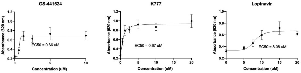


 EN
 SK