The number of type 2 severe acute respiratory syndrome (SARS-CoV-2) coronavirus infections continues to rise globally, and hospital physicians are currently preparing therapies for FDA-approved therapies as treatment options for SARS-CoV-2. We have been researching anti-cancer therapies for feline infectious peritonitis (FIP) for several years. [1,2], a coronavirus disease with almost 100% mortality in felines. Feline enteric coronavirus (FECV), which is common in many asymptomatic cats, mutates into virulent and lethal FIP coronavirus [3]. We believe that our experimental research in the treatment of FIP may be relevant and applicable to the latest results of in vitro SARS-CoV-2 tests. [4] in the absence of large-scale laboratory and human clinical studies. The FIP coronavirus protease inhibitor (GC376) has been successful in treating FIP in a subset of felines; however, in cases of neurological impairment, the protease inhibitor was unable to prevent the progression of central nervous system (CNS) disease, resulting in neurological FIP and subsequent euthanasia. [5]. The polymerase inhibitor GS-441524 has already shown significant activity in a feline clinical study against FIP [1], but treatment for neurological impairment has yet to be demonstrated. Remdesivir, a prodrug of GS-441524, has high hopes for the treatment of SARS-CoV-2. [6], but is not currently FDA approved and is only available in intravenous form. There is an urgent need for therapies against SARS-CoV-2, with good oral bioavailability and FDA approved, suitable for organs that express SARS-CoV-2 target angiotensin-converting enzyme 2 (ACE2) and can synergize with remedivr. Although detailed experimental results will be reported elsewhere (unpublished data from BGM Laboratory), we believe that our research could help physicians with treatment options in addition to supportive care.
Type 1 severe acute respiratory syndrome (SARS-CoV-1) and SARS-CoV-2 coronavirus target ACE2 as a receptor [7]which is expressed in humans in the lungs, heart, gastrointestinal tract and CNS [8]. SARS-CoV-1 is known to penetrate the CNS via the olfactory nerve and olfactory bulb [9], like other coronaviruses [10]. Anosmia (loss of smell) often occurs in patients with coronavirus disease 2019 (COVID-19), a disease caused by SARS-CoV-2, suggesting that SARS-CoV-2 uses the same route after infection. Almost all beta-coronaviruses enter the CNS [10] and SARS-CoV-1 and SARS-CoV-2 have the same ACE2 receptor. It is also reasonable to assume that a massive brainstem infection in experimental animals after nasal exposure to SARS-CoV-1 [9] can also occur with SARS-CoV-2, which may contribute to sudden respiratory failure, as seen in some patients [10]. It is unclear whether SARS-CoV-2 CNS penetration may also occur in patients with recent blood-brain barrier (BBB) damage following a stroke or other stroke. As we have shown in cats, the consequences of CNS penetration underline the need for a multi-purpose organ-friendly strategy that suppresses SARS-CoV-2 in both the periphery and the brain.
We found that nelfinavir and amodiaquine show in vitro anti-FIP activity that is comparable to chloroquine and is superior to ribavirin, penciclovir, favipiravir and naphamostat against SARS-CoV-2. [4]. Amodiaquin, like chloroquine and hydroxychloroquine, is a CNS-penetrating 4-aminoquinoline antimalarial drug that inhibits hemozoin production in parasites but has been withdrawn from the US market, although it is still available in other countries. Amodiaquine is known to have some antiviral activity and derivatives have been developed to inhibit Ebola virus infection. [11]. Pharmacogenomics has shown that the presence of the CYP450 2C8 * 2 allele contributes significantly to the toxicity of amodiaquine [12]. Suitable monitoring parameters include complete blood counts with differential and liver function tests, as agranulocytosis and hepatotoxicity are serious side effects, with nausea, vomiting and pruritus being mild side effects. Amodiaquine / artesunate is available for the treatment of malaria; is cost effective and available outside the US. This is the third study of 4-aminoquinoline and its coronavirus activity and complements clinical research from China. [13,14]. In addition, it is well known that 4-aminoquinolines penetrate the BBB and have been investigated for broad-spectrum antiviral activity against various viral infections, including Zika viruses. [15], Dengue [16] and Ebola [17]. It may also be useful for those patients who suffer from SARS-CoV-2 in the brainstem. Antiviral mechanisms of chloroquine action may involve altering the release of endosomal RNA [15], altering autophagically dependent viral replication [15] and inhibition of ACE2 glycosylation [18].
Nelfinavir is an older human immunodeficiency virus (HIV) protease inhibitor capable of inhibiting HIV-1 and, to a lesser extent, HIV-2 protease [19], but it is no longer the first choice of treatment. However, it has a spectrum of activity that includes SARS-CoV-1 [20] also FIP coronavirus [21], is orally bioavailable and can reach a plasma concentration of 7.3 mg / l at a dose of 3000 mg twice daily [ 22]. For the treatment of SARS-CoV-1 [23] Other protease inhibitors, including the combination of lopinavir and ritonavir, have been used to treat SARS-CoV-2 in Singapore. [24] and China [25]. However, there are problems with prescribed dose toxicity [24] as well as efficacy in the monotherapy regimen. [25]. The hypothesis of the use of older antiretroviral agents with higher toxicity but potentially wider antiviral activity spectrum is not new. Experimental study of nelfinavir suppressing FIP coronavirus [21] however, it provides additional data to suggest that nelfinavir could be considered as an alternative to SARS-CoV-2. Suitable nelfinavir monitoring parameters include echocardiogram for QT prolongation and torsades de pointes, as well as diarrhea, fatigue (10–20%), lipodystrophy and hyperglycaemia.
These in vitro activities against FIP coronavirus do not replace clinical data and studies, but may provide guidance for therapeutic off-label strategies. Mutation of FECV to FIP coronavirus may provide a paradigm for considering the relationship between different SARS-CoV-2 strains. Nelfinavir, chloroquine, and hydroxychloroquine are FDA-approved, orally bioavailable, and commercially available, and there is at least data on their effect against SARS-CoV-1 or SARS-CoV-2 in vitro. Nelfinavir may be an alternative to lopinavir / ritonavir. Amodiaquine, hydroxychloroquine and chloroquine show the ability to penetrate the CNS. Amodiaquine may be an alternative to chloroquine in areas where it is available. These agents may offer clinicians an additional therapeutic strategy beyond supportive care as monotherapy or in combination.
References
Murphy BG, Perron M, Murakami E, Bauer K, Park Y, Eckstrand C. The nucleoside analog GS-441524 strongly inhibits feline infectious peritonitis (FIP) virus in tissue culture and experimental cat infection studies. Vet Microbiol. 2018; 219: 226–233. doi: 10.1016 / j.vetmic.2018.04.026. [PMC free article] [PubMed] [CrossRef] [Google Scholar]
Pedersen NC, Eckstrand C, Liu H, Leutenegger C, Murphy B. Levels of feline infectious peritonitis virus in blood, effusions, and various tissues and the role of lymphopenia in disease outcome following experimental infection. Vet Microbiol. 2015; 175: 157–166. doi: 10.1016 / j.vetmic.2014. [PMC free article] [PubMed] [CrossRef] [Google Scholar]
Licitra BN, Millet JK, Regan AD, Hamilton BS, Rinaldi VD, Duhamel GE. Mutation in spike protein cleavage site and pathogenesis of feline coronavirus. Emerg Infect Dis. 2013; 19: 1066–1073. doi: 10.3201 / eid1907.121094. [PMC free article] [PubMed] [CrossRef] [Google Scholar]
Wang M, Cao R, Zhang L, Yang X, Liu J, Xu M. Remdesivir and chloroquine effectively inhibit the recently emerged novel coronavirus (2019-nCoV) in vitro. Cell Res. 2020; 30: 269-271. doi: 10.1038 / s41422-020-0282-0. [PMC free article] [PubMed] [CrossRef] [Google Scholar]
Pedersen NC, Kim Y, Liu H, Galasiti Kankanamalage AC, Eckstrand C, Groutas WC. Efficacy of a 3C-like protease inhibitor in treating various forms of acquired feline infectious peritonitis. J Feline Med Surg. 2018; 20: 378–392. doi: 10.1177 / 1098612X17729626. [PMC free article] [PubMed] [CrossRef] [Google Scholar]
Ko WC, Rolain JM, Lee NY, Chen PL, Huang CT, Lee PI. Arguments in favor of remdesivir for treating SARS-CoV-2 infections. Int J Antimicrob Agents. 2020 doi: 10.1016 / j.ijantimicag.2020.105933. [PMC free article] [PubMed] [CrossRef] [Google Scholar]
Qiu Y, Zhao YB, Wang Q, Li JY, Zhou ZJ, Liao CH. Predicting the angiotensin converting enzyme 2 (ACE2) utilizing capability as the receptor of SARS-CoV-2. Microbes Infect. 2020 Mar 19 doi: 10.1016 / j.micinf.2020.03.003. [Epub ahead of print] [PMC free article] [PubMed] [CrossRef] [Google Scholar]
Hamming I, Timens W, Bulthuis ML, Lely AT, Navis G, van Goor H. Tissue distribution of ACE2 protein, the functional receptor for SARS coronavirus. A first step in understanding SARS pathogenesis. J Pathol. 2004; 203: 631–637. doi: 10.1002 / path.1570. [PMC free article] [PubMed] [CrossRef] [Google Scholar]
Netland J, Meyerholz DK, Moore S, Cassell M, Perlman S. Severe acute respiratory syndrome coronavirus infection causes neuronal death in the absence of encephalitis in mice transgenic for human ACE2. J Virol. 2008; 82: 7264–7275. doi: 10.1128 / JVI.00737-08. [PMC free article] [PubMed] [CrossRef] [Google Scholar]
Li YC, Bai WZ, Hashikawa T. The neuroinvasive potential of SARS-CoV2 may play a role in the respiratory failure of COVID-19 patients. J Med Virol. 2020 Feb 27 doi: 10.1002 / jmv.25728. [Epub ahead of print] [PMC free article] [PubMed] [CrossRef] [Google Scholar]
Sakurai Y, Sakakibara N, Toyama M, Baba M, Davey RA. Novel amodiaquine derivatives potently inhibit Ebola virus infection. Antiviral Res. 2018; 160: 175–182. doi: 10.1016 / j.antiviral.2018.10.025. [PMC free article] [PubMed] [CrossRef] [Google Scholar]
Gil JP, Gil Berglund E. CYP2C8 and antimalaria drug efficacy. Pharmacogenomics. 2007; 8: 187–198. doi: 10.2217 / 14622416.8.2.187. [PMC free article] [PubMed] [CrossRef] [Google Scholar]
Yao X, Ye F, Zhang M, Cui C, Huang B, Niu P. In vitro antiviral activity and projection of optimized dosing design of hydroxychloroquine for the treatment of severe acute respiratory syndrome coronavirus 2 (SARS-CoV-2) Clin Infect Dis. 2020 Mar 9 doi: 10.1093 / cid / ciaa237. [Epub ahead of print] [PMC free article] [PubMed] [CrossRef] [Google Scholar]
Gao J, Tian Z, Yang X. Breakthrough: Chloroquine phosphate has shown apparent efficacy in treatment of COVID-19 associated pneumonia in clinical studies. Biosci Trends. 2020; 14: 72–73. doi: 10.5582 / bst.2020.01047. [PubMed] [CrossRef] [Google Scholar]
Zhang S, Yi C, Li C, Zhang F, Peng J, Wang Q. Chloroquine inhibits endosomal viral RNA release and autophagy-dependent viral replication and effectively prevents maternal to fetal transmission of Zika virus. Antiviral Res. 2019; 169 doi: 10.1016 / j.antiviral.2019.104547. [PubMed] [CrossRef] [Google Scholar]
Al-Bari MAA. Targeting endosomal acidification by chloroquine analogs as a promising strategy for the treatment of emerging viral diseases. Pharmacol Res Perspect. 2017; 5: e00293. doi: 10.1002 / prp2.293. [PMC free article] [PubMed] [CrossRef] [Google Scholar]
Du X, Zuo X, Meng F, Wu F, Zhao X, Li C. Combinatorial screening of a panel of FDA-approved drugs identifies several candidates with anti-Ebola activities. Biochem Biophys Res Commun. 2020; 522: 862–868. doi: 10.1016 / j.bbrc.2019.11.065. [PubMed] [CrossRef] [Google Scholar]
Vincent MJ, Bergeron E, Benjannet S, Erickson BR, Rollin PE, Ksiazek TG. Chloroquine is a potent inhibitor of SARS coronavirus infection and spread. Virol J. 2005; 2: 69. doi: 10.1186 / 1743-422X-2-69. [PMC free article] [PubMed] [CrossRef] [Google Scholar]
Witvrouw M, Pannecouque C, Switzer WM, Folks TM, De Clercq E, Heneine W. Susceptibility of HIV-2, SIV and SHIV to various anti-HIV-1 compounds: implications for treatment and postexposure prophylaxis. Antivir Ther. 2004; 9: 57–65. [PubMed] [Google Scholar]
Yamamoto N, Yang R, Yoshinaka Y, Amari S, Nakano T, Cinatl J. HIV protease inhibitor nelfinavir inhibits replication of SARS-associated coronavirus. Biochem Biophys Res Commun. 2004; 318: 719–725. doi: 10.1016 / j.bbrc.2004.04.083. [PMC free article] [PubMed] [CrossRef] [Google Scholar]
Hsieh LE, Lin CN, Su BL, Jan TR, Chen CM, Wang CH. Synergistic antiviral effect of Galanthus nivalis agglutinin and nelfinavir against feline coronavirus. Antiviral Res. 2010; 88: 25–30. doi: 10.1016 / j.antiviral.2010.06.010. [PMC free article] [PubMed] [CrossRef] [Google Scholar]
Pan J, Mott M, Xi B, Hepner E, Guan M, Fousek K. Phase I study of nelfinavir in liposarcoma. Cancer Chemother Pharmacol. 2012; 70: 791–799. doi: 10.1007 / s00280-012-1961-4. [PMC free article] [PubMed] [CrossRef] [Google Scholar]
Yao TT, Qian JD, Zhu WY, Wang Y, Wang GQ. A systematic review of lopinavir therapy for SARS coronavirus and MERS coronavirus — a possible reference for coronavirus disease-19 treatment option. J Med Virol. 2020 Feb 27 doi: 10.1002 / jmv.25729. [Epub ahead of print] [PMC free article] [PubMed] [CrossRef] [Google Scholar]
Young BE, Ong SWX, Kalimuddin S, Low JG, Tan SY, Loh J. Epidemiologic features and clinical course of patients infected with SARS-CoV-2 in Singapore. JAMA. 2020 Mar 3 doi: 10.1001 / jama.2020.3204. [Epub ahead of print] [PMC free article] [PubMed] [CrossRef] [Google Scholar]
Cao B, Wang Y, Wen D, Liu W, Wang J, Fan G. A trial of lopinavir – ritonavir in adults hospitalized with severe Covid-19. N Engl J Med. 2020 Mar 18 doi: 10.1056 / NEJMoa2001282. [Epub ahead of print] [PMC free article] [PubMed] [CrossRef] [Google Scholar]
Original article on dvm360.com, published 18.3.2020.
Researchers at Colorado State University want to stop the feline enteric coronavirus before it has a chance to mutate to the feline infectious peritonitis virus.
A new candidate in the fight against feline infectious peritonitis (FIP) may soon enter the scene. Researchers at Colorado State University (CSU), funded by the Morris Animal Foundation, are developing an oral vaccine to beat the disease by targeting feline enteric coronavirus (FECV) - a highly contagious and common virus that can accidentally mutate to FIPV.
“Vaccination attempts that have been made in the past have targeted FIP, but that’s too late,” Gregg Dean, DVM, PhD, chair of the Department of Microbiology, Immunology and Pathology in the College of Veterinary Medicine and Biomedical Sciences, told dvm360. “We know that enteric coronavirus replicates at an amazing rate. So it’s likely that it will mutate into the virus that causes FIP—so there’s more going on inside the cat than is being transmitted between cats. If we can control that replication, we might be able to prevent the mutant from emerging.”
It's all about the gut
As a first step toward understanding what a vaccine would have to do to stimulate a protective immune response against FECV, Dr. Dean and his team studied cats that had been naturally infected with the disease, specifically looking at their mucosal immune responses. “Enteric virus is quite restricted in where it replicates, and that is, as we know, in the intestinal tract,” he explained. “So understanding the immune response at that site is critically important.”
The vaccine design reflects this insight. The vaccine will be administered orally and includes the bacterium Lactobacillus acidophilus, a probiotic found in some foods and in the gastrointestinal tracts of many animals. “The role of probiotics is essential,” said Dr. Dean. “Some probiotics stimulate the immune system naturally, and by combining the antigens from feline coronavirus with the natural immunostimulatory activity of the probiotic, we hope to induce a protective and lasting immune response.” He explained that the probiotic’s effect is enhanced by the fact that it is alive and replicating. “It also produces antigens – in our case, feline coronavirus antigens – so it is very economical,” continued Dr. Dean. “We don’t have to use expensive processes to produce large quantities of antigen. We are using the natural activity of the bacteria.”
Dr. Dean and his team plan to begin the first trials of the FECV vaccine this fall. “We’re going to give the vaccine to cats in a colony environment where enteric coronavirus naturally circulates. We’re going to take new kittens, vaccinate them, and then see if they get FECV,” he said. “Almost every cat, and probably every cat in that colony, is infected.”
Disease recognition
Meanwhile, Dr. Dean is leading another study funded by the Morris Animal Foundation with a different but related goal: to develop a new diagnostic test for FIP that is accessible, inexpensive, and rapid. “As we move closer to real, viable treatment options for FIP, an accurate diagnostic test will be an important part of that decision-making process,” he explained. “Early diagnosis will be important as long as effective treatment is available.”
Instead of focusing on the cat’s immune response or looking for the virus itself, Dr. Dean and his team are looking for specific biomarkers that are unique to FIP infection. They have identified 18 proteins that appear to be common in cats with FIP, and they are in the process of validating them. If the test is successful, all that will be needed is a blood sample from the patient. “We still have a lot of work to do, but it’s a top priority for us,” he said.
We have a lot of work to do
With these exciting prospects for cat health on the horizon, Dr. Dean praised the past and present work of his veterinary colleagues: "There are a lot of people who work hard to solve these important problems for cats, and it is the excellent work of this community that leads us all to a common goal."
Sarah Mouton Dowdy, a former dvm360.com editor, is a freelance writer and editor in Kansas City, Missouri.
Please note that these answers were provided by Dr. Pedersen individual cat owner. Consider them as a useful guide and not as individual veterinary advice. In no case do they replace standard veterinary care.
General questions about GS-441524 injection
What if my cat doesn't respond to GS immediately or her behavior doesn't change after a few weeks on the drug?
All we can do is watch what is happening over the next few days and hope for the best. If increased pain develops at the injection site, it is possible that this is the cause of the symptoms we are seeing. If the place is sensitive to touch, it goes without saying that it bothers the cat. It is possible that the situation will worsen and an open wound will form in the painful area, or it will improve quickly. Even if no one wants severe reactions at the injection site, injection is still a better choice.
My cat is experiencing “ups and downs” during treatment. Sometimes she seems full of energy, other times she is lethargic. Is this normal?
The most dramatic improvement usually occurs in the first week, or the first two, after which the process slows down. Do these cycles show that the cat is less active after the injection and more active a few hours after the injection? During our tests, we noticed that some cats seemed a little depressed and depressed after the injection. A higher dose worsens the situation. If it is a relapse, the reaction is even more pronounced, but if it is really just a reaction to the drugs, we will have to wait and see what happens after the end of treatment.
What should I do if my cat feels increased pain at the injection site from using GS?Would lidocaine or anything else help with the pain?
We are working to change the diluent for the drug to reduce stinging and pain, but it will probably take several more months and it will only solve the problem with the diluent and the effect of the drug itself.Applying lidocaine cream to the post-injection area is like locking the barn door after the horses get out.We tried to use it at the injection sites before treatment, but the owners who tried it were not satisfied with the results and stopped using it.
Is it safe / appropriate to continue administering FIO (feline interferon omega) concurrently with GS?
It won't help, it won't hurt either.
Should my cat continue in pre-zone with GS?
Not. You can stop prednisolone after a few days.A cat with FIP should respond very quickly to treatment and no other medication is needed at this time unless clearly indicated.
What are the chances that after a cat is cured of FIP, it will become infected again with a coronavirus that mutates again to FIPV?Should I treat a cat that secretes coronavirus?
Although theoretically possible, we have not yet seen the FIP-cured cats develop the disease later.Therefore, I would focus on curing your FIP positive cat.Do not treat a cat that only secretes enteric coronavirus.This cat has a very low risk of developing FIP and treatment with enteric coronavirus carriers will only promote the occurrence of drug-resistant strains.
Should I vaccinate or neuter my cat during GS?
Vaccination or castration is not required during treatment.Healing the cat is paramount. There will be plenty of time after treatment to resolve the issue of castration.Personally, I am not a strong supporter of FVRCP vaccines (polyvalent vaccines) or the need to vaccinate cats against rabies (unless required by state or local legislation) or administering FeLV to cats that are at low risk for infection.If we treat a cat with FIP, it should be treated in accordance with standard professional care, locality (rabies) and your practice.In short, treat the cat like any other cat, but have it cured first.
Questions about what will happen after the end of GS treatment
My cat has been with GS for several weeks and she seems to be doing well, but the titer of coronavirus antibodies is still high.Does that mean she's still sick?Should I be worried?
We do not see the coronavirus antibody titer value for you, so I cannot answer your question.However, we would expect it to take many months for permanent remission to occur.The fact that the titer is still high at this early stage does not worry us, but it may be something that should be monitored to catch a possible relapse. But we are optimistic.Symptoms indicate complete remission, and just as importantly, the cat has gained weight and figure in recent weeks.These were also favorable symptoms in our other cases, which did not suffer from a neurological disease.The high titer of the coronavirus also reflects the intense response of the immune system to the FIP virus.It is desirable for this titer to remain high for a while and then slowly return to a much lower titer.
What if your cat relapses after you stop taking GS? How do I find out? (Question about a neurological cat)
We hope that your cat will remain healthy after the end of treatment, but we must also prepare for a possible relapse.Relapses can occur within 1-84 days, based on our experience with other cats we have treated.The clinical signs of relapse will be evident in neurological cases, and will be essentially a recurrence of the same problems.They get a fever, the cat stops eating, becomes less active and the neurological symptoms she had before return.It is important to document what is happening.You can also measure her temperature every day for several weeks and then every other day until we reach day 84.During this period, it will be important to monitor the cat's level of activity and weight.
What thermometer should I use to measure the temperature of a cat?
I prefer digital rectal thermometers, which are characterized by high measurement speed.Do you measure temperature during rest and not after a certain time of physical activity?Temperatures may vary. One is higher, one is higher.
I noticed that after the GS was discontinued, the cat's temperature rose sharply. Does that mean the cat has relapsed?
Cats and other small animals are not as consistent in their systemic temperature as humans, and the fluctuations you see are not uncommon.The problem is that the more you worry, the more you measure the temperature and the more of these fluctuations you will see.For this reason, we are not very afraid of these fluctuations. It is much worse when temperatures are consistently higher than 39.5 ° C.It is important to note the overall condition of the cat, its appetite, activity levels and clinical signs. In short, fluctuations like these are fine unless they become permanent and are associated with loss of appetite and activity.If your cat is otherwise well and there is no evidence of a persistent fever, we recommend that the cat be only at home, where it is possible to protect it from unnecessary stress and monitor it closely.
Questions about laboratory results during or after GS treatment
My cat is being treated with GS and her lymphocyte count is high.That's all right?
The lymphocyte count is only slightly higher than normal, but in reality it is good.A low lymphocyte count would indicate that the infection is active and severe, while a high number would indicate that it elicits an immune response.
My cat has been on GS for several weeks and laboratory reports have increased creatine kinase and phosphorus.Is this a problem?
It doesn't have to mean anything if urea and creatinine are in the normal range.High phosphorus content may not mean anything yet. I recommend checking the values in a month.
We are concerned about the seemingly rapid increase in urea and creatinine in the blood.Do you have any other recommendations, or do you just need to supplement your fluids / diet and hope that the progression of kidney disease will slow down, as it is a young cat and it seems that the FIP has been successfully cured?
Yes, there is evidence of kidney damage and given the recent change and the fact that your cat is being treated this way, I think your cat may have suffered mild kidney damage.We only observed kidney damage that was milder than this in one of the more than 30 cats we treated for a total of 12-36 weeks and even at higher doses than you used.However, this type of medicine is known to be toxic to the kidneys in some people treated for HIV / AIDS or hepatitis C. This damage is reversible if you stop treatment early enough, and I assume you have already stopped treatment.If not, and you are at the end of 12 weeks or close, quit it.It does not reduce the chance of healing.Make sure the cat has access to water, and if her appetite is affected, feed her a moist diet that she likes with added water.Cats love wet food with juice and you can still mix a little extra water.There is a commercial and domestic diet for cats with kidney disease, but it is not very tasty and does not cure the kidneys, but rather protects the remaining kidney tissue.I would also make sure that your veterinarian does a urine test to find out what the specific gravity is - the lower it is, the greater the kidney damage.The more water cats drink and the more they urinate, the greater the kidney damage.It is necessary to determine whether the urine contains any signs of bacterial infection in the urine sediment or after cultivation - if they are found, antibiotics should be used immediately.
Questions regarding the oral form of GS
Some companies produce GS in tablet form.Is this form safe / effective?
Yes, at least one company in China also produces the oral form GS-441524, which it claims will work just like injections. This is not surprising, since the modifications required to convert injectable drugs into an oral form are well known.All drugs for HIV / AIDS and hepatitis C were also transformed into an oral form.However, like many others, you may find it more difficult to give tablets to cats than to inject.They usually adapt to the injections over a short period of time, but if they don't agree with the pills, it still gets worse and worse.Therefore, if you go this route, do not invest much in it until you are sure that you will be able to give the tablets to your cat.
Companies that make the oral form recommend splitting the dose and giving it twice a day.Why?
I do not know anything more about the oral form of GS, so I cannot provide a reliable answer to this question.However, there must be a reason why this Chinese manufacturer tells people to give GS twice a day and not once a day.Maybe it's because putting it all at once is too much of a burden for the cat system.It does not make sense to think that the medicine is excreted from the blood more quickly when given orally, which is another possible reason why they want to split the dose.It is important to me that these people are pushing hard for the oral form of the medicine, but have not provided any preliminary information on the success or tolerability, nor have any clinical trials been conducted.
Is there a risk of alternating injection and oral forms of GS?
It would probably not be appropriate to alternate these forms on a daily basis. But there is probably no problem with the transition from one form to another.The diluted drug in injection form usually contains 15-16.5 mg / ml and the dose for cats is 4 mg / kg, SC.
The problems associated with adverse sexual behavior in intact cats and males during GS treatment have caught my attention. The questions often come from countries where castration is either postponed or not part of common practice. There are also frequent questions about routine vaccinations. There are concerns that stress during castration / sterilization or vaccination may affect the outcome of GS-441524 treatment. I am convinced that such fears are exaggerated. If a cat is treated and in remission, or is considered cured, it can be neutered, but preferably in the least stressful way. Cats can be neutered and returned to their homes quickly and efficiently on the day of neutering with a minimum of preoperative, surgical and postoperative treatments and restrictions (eg caging, e-collars). Such surgeries will be less stressful for cats (and owners whose behavior is reflected back on their cats) than intolerable sexual behavior alone.
I am not in favor of hormonal treatment to prevent inappropriate sexual behavior in cats, and I think that effective castration will be less stressful in the long run than such prevention. Therefore, if a permanent change in this behavior is required, surgical castration is always better than chemical castration.
Is it better to neuter cats if they still have some part of the treatment in front of them, provided they look healthy and blood test results are normal? Yes, castration during treatment makes sense. However, we do not recommend unreasonable resumption of treatment during surgical castration during the 12-week observation period after the end of treatment, the so-called post-treatment. Castration is unlikely to change the fate of a cat with FIP, as the cure for FIP is given by the very cessation of treatment. The observation period is not intended to further improve, but only to confirm the cure.
Some owners of cured cats do not want their castration for breeding purposes. We know that genetic and environmental factors also affect FIP, which has led to the recommendation that purebred cats that give birth to kittens with FIP should not be used for breeding. This should be even more true for cats that have been cured of the FIP.
When it comes to vaccines, many already know that I am not a big supporter of vaccines for cats after reaching adulthood, nor of boosted vaccines in the first year. I also think that rabies vaccines are not justified for normal use in cats, neither in terms of public health nor in cats. Nevertheless, I am aware that these views are not generally accepted and that laws in some states require rabies vaccinations against cats. In any case, we did not notice any consequences of vaccination in any of our cured cats. However, this is not something I would recommend for cats in treatment. The immune system of these cats is responsible for other things than responding to the vaccine.
When are drugs other than GS-441524 indicated for the treatment of FIP?- NC Pedersen
There are often questions about the need for complementary medicines to GS-441524. Initially, supportive (symptomatic) treatment may be needed to keep cats alive long enough for the antiviral effect to take effect. Drugs often used in this early stage usually include antibiotics (doxycycline / clindamycin), analgesics (opioids, gabapentin), anti-inflammatory drugs (corticosteroids, NSAIDS), immunostimulants (interferons, non-specific immunostimulants) and fluids. I have tried to avoid excessive use of these drugs, except for their temporary use, and only if there are legitimate reasons for their use, especially in severely ill cats during the first few days. The most important goal of FIP treatment is to stop the replication of the virus in macrophages, which immediately stops the production of numerous inflammatory and immunosuppressive cytokines that cause the symptoms of FIP. While some drugs, such as corticosteroids (prednisolone) or NSAIDS (meloxicam), can inhibit inflammatory cytokines, the only drugs that completely inhibit these harmful cytokines are antivirals, such as GS-441524 or GC376. These drugs lead to a dramatic improvement in fever, activity, appetite, etc. within 24-48 hours. This improvement will be much more pronounced than with any other medicine. Therefore, unless there are reasons to take other medicines, they should be discontinued as soon as there is a significant and stable improvement in FIP symptoms. I'm not even a supporter of the supplements that cats often take during treatment. B12 only treats B12 deficiency, not FIP anemia. This also applies to a wide range of nutritional supplements and many types of special cat food. Follow the proven commercial brands of cat food supplemented with animal meat.
Mutian, the first Chinese company to sell GS-441524 on an unapproved market, recently advertised that using its oral formulation for one (or more?) Days would stop the fecal excretion of FeCV in healthy cats. Some veterinary researchers also take this approach. The reason is to prevent the formation of a mutant virus causing FIP (FIPV), and thus to prevent FIP. Although this approach is at first sight attractive, it is a very incorrect introduction of GS441524 in cats. Until now, GS441524 has only been recommended for the treatment of cats suffering from FIP. There are many problems associated with this approach.
In his advertisements, Mutian suggests that FeCV can be easily eliminated by a simple administration of oral GS441524. However, it does not talk about possible research that led to this approach. There is currently no published field research to demonstrate whether chronic FeCV excretion can be permanently cured by healthy cats. 40% or more individuals in any population of multiple cats may be positive for this virus. Naturally occurring FeCV infection appears at an early age and is not associated with any significant symptoms of the disease. Virus shedding takes weeks, months, and in some cases indefinitely, but in most cats it eventually stops building immunity. However, once immunity is stopped, antibody levels fall and cats become susceptible to infection again. This cycle of primary and secondary infections occurs in many cats throughout their lives. GS441524 treatment is very unlikely to result in more permanent immunity than is seen in nature. It would not be important if FeCV was short-lived in the environment and would not be easy to transfer to clothing, rubbish, hair and other items. FeCV survives in the environment for up to 2 weeks and transmission by contaminated objects is well documented. Therefore, although it is possible to treat FeCV in a particular cat, the virus easily returns to the population. This has also been documented in the experience of isolating mothers and prematurely weaning their kittens, which is a slightly similar strategy to preventing FeCV infection.
Although our current knowledge of FeCV infection calls this approach into question, there may be more compelling reasons not to treat healthy cats with GS441525 or any other safe and effective antiviral drugs in the future. It is unlikely that the excretion of FeCV can be stopped for a significant period by a single or short-term treatment, as stated, and this will undoubtedly require a longer period. This approach will be extremely expensive if applied to a large number of cats in a kennel, shelter or rescue station.
The most important reason to avoid this FIP prevention approach is the problem of drug resistance. We already know that some cats with naturally occurring FIP are infected with one or more GS441524-resistant FIPV strains. Our field experiment with 31 cats found that one was infected with a virus that was highly resistant. There is laboratory research confirming that resistance can develop very easily in cell cultures. Drug resistance is also well known in viral diseases such as HIV / AIDS and in people infected with the hepatitis C virus. The fastest way to achieve resistance to any microbes, including viruses, is to overuse drugs where they are not needed. The problem of antimicrobial drug resistance is sufficiently confirmed by our experience with bacteria in animals and viruses in humans. There is no doubt that overuse of drugs such as GS441524 in large groups of healthy cats will lead to a positive selection of FeCV strains that are partially or completely drug resistant. This resistance is also manifested in the mutant FIP-causing FeCV virus (FIPV), which precludes the use of GS441524 in a larger number of cats with FIP. Unfortunately, veterinary medicine does not have the resources of human medicine or financial incentives to discover, test and obtain multiple drug licenses in order to circumvent the acquired drug resistance.
I urge cat owners and scientists to refuse to use safe and effective antivirals in this way. I have nothing to do with voracious drug manufacturers who prefer profit to serious science, common sense and the fears of cat owners suffering from FIP now or in the future. There is a saying, "Just because we can do something doesn't mean we should." NC Pedersen
Is it possible that GS-441524 will lose its effectiveness, leading to relapses?
In particular, GS-441524 is extremely stable in the highly acidic solution used by its suppliers. We stored it in the fridge for months. The more likely reasons for relapses are inadequate treatment (poor injection, poor medicine), inability to get enough medicine in places with inflammation (such as the brain), or drug resistance (we saw this in 1 cat in both our GC376 and GS studies). -441524).
Are secondary infections such as toxoplasma or infectious anemia associated with relapse?
This is very unlikely for secondary infections that lead to relapse. Cats are the ultimate host for Toxoplasma gondii and the infection is rarely systemic. In fact, the systemic form is so unusual compared to FIP that there is no good clinical reason for its routine testing in cats suspected of having FIP. In addition, toxoplasmosis would be much more likely to occur in outdoor cats that hunt than in shelter or indoor cats that are fed commercial feed. Infectious feline anemia, which is caused by a common and largely overt blood infection with one or more types of hemotropic mycoplasma or hemoplasm, could break into clinical form upon immunosuppression by another infectious agent. However, I cannot recall the case of FIP, which was complicated by these organisms. However, feline leukemia virus, and not coronavirus, is strongly associated with hemotropic mycoplasma. Nevertheless, I suspect that this is one of the reasons why so many veterinarians include doxycycline in the cocktail of drugs they used to "treat" the FIP.
Should cats receive Virbagen omega after GS treatment to strengthen the immune system?
Published double-blind, placebo-controlled studies of feline interferon omega in FIP have shown that it has no therapeutic benefit. I also perceive this with other immunomodulators such as PI.
Should I try to eliminate feline enteric coronavirus (FECV) from my cat's environment after the cat is no longer treated with GS-441524?
This virus is so ubiquitous, difficult to eliminate, and easily reinfected that I have come to the conclusion that attempting to eliminate it will be very costly and largely unproductive. Although it is possible to eliminate FECV in a small group of cats that are kept in strict isolation and by faecal testing, in reality it is easier said than done. In the end, even if you succeed with great effort and cost, what will you do then ?! The only way to maintain the situation is to observe an extremely strict quarantine of cats and people moving in and out of your premises, which greatly complicates the ability to maintain a functional shelter or rescue or kennel.
How useful is the AGP test to find out if my cat is cured?
The AGP test is popular in Europe, but I can't figure out why. The test measures what we call the "phase reactant" and is a measure of inflammation in the body. It is completely non-specific and has no added value compared to routine examinations, such as general health, weight loss, fever, abnormalities in white blood cell parameters, albumin and globulin levels, A: G ratio, etc.
Summary: Carol Johnson DVM, PhD. A Heather Lorimer Ph.D. For clarification, additional information from literature articles by Dr. Pedersena.
Feline Infectios Peritonitis (FIP) is one of the most complex infectious diseases and is undoubtedly one of the worst diseases imaginable. It is caused by an RNA virus from the coronavirus family Nidovirales. Other RNA viruses that the reader may be familiar with include the Ebola virus, influenza virus, AIDS, and rhinoviruses. Coronaviruses are named after protrusions that look like a crown (or corona) when viewed under an electron microscope. Coronaviruses can cause disease in almost every animal species, but as such, coronaviruses are species-specific: feline coronaviruses do not infect humans, dogs, or other animals.
Feline enteric coronavirus (FECV, also referred to in the literature as FCoV) usually remains in the intestinal tract and infects the upper layer of cells lining the small intestine before settling in the large intestine. FECV can cause mild diarrhea and vomiting, but is not considered a serious pathogen in the intestinal tract.
An FECV-infected cat can shed the virus in large quantities and excretion can continue for months. Cats are obligatory carnivores and, as a result, have a very short digestive tract. This may play a role in high virus shedding. Immunity to FECV is usually temporary and previously immune animals may be reinfected. This is one of the reasons why vaccination prevention is difficult, if not impossible.
FECV specific mutations they allow him to leave the intestinal tract, where he infects cells of the immune system called macrophages, which usually help fight infection. This mutated version of FECV is called FIPV (feline infectious peritonitis virus). Infected macrophages spread the disease in the cat's body in a similar way as tuberculosis (TB) bacteria spread in humans or animals. FIPV-associated mutations occur in three parts of the FECV genome, but a particular mutation may be unique to each cat. As many as 20% FECV infections can lead to subclinical macrophage infection, but relatively few cats develop FIP. Similarly, up to 40% of the world's human population is infected with tuberculosis bacteria, but few people have developed complete tuberculosis. Unlike TB, FIPV is not transmitted to other cats; the transmission takes place via an unmutated FECV.
FIP is a population-related disease high-density cats, where kittens are part of the equation. Kittens are most susceptible to developing FIP and are usually infected with FECV at about 9 weeks of age. In general, FIP can also occur in a dense urban or rural population of free-range cats. In the United States, FIP is more common in cats and kittens from conventional, high-density shelters and feline shelters or rescue stations where kittens may be exposed to large viral loads. Another source of FIP are kennels. Not only are a number of cats concentrated in farms, but there may also be genetic predispositions that may also play a role (up to 50% in some cases). Although the genetic risk is clear, genetic analysis shows that susceptibility to FIP is likely to involve a large number of genes. As a result, inbreeding is associated with susceptibility to FIP, but genetic testing for this susceptibility is not currently possible. Overall, FIP occurs in about 0.3% cats, but can occur in up to 1-5% (or more) cats in high-density sites, such as kennels or rescue stations.
The FIP is on the rise probably due to the increasing number of rescued cats in which kittens can be fed from bottles, weaned prematurely and exposed to large amounts of FECV.
There is a dry and wet form of FIP, but there may be combined forms and it is possible to change from one form to another. In general, the moist form, characterized by effusion, which leads to enlargement of the abdomen or fluid in the lungs, along with other symptoms, has a rapid course and can kill the cat very quickly (in this case, it is often death due to euthanasia). The dry form can last for months to about a year, sometimes longer.
Risk factors for FIP development they include the prevalence of cats that shed FECV, the intensity of virus shedding, the number of cats aged 4 to 29 months (most sensitive) and genetic predisposition (in bred cats). FIP often occurs in young cats after a stressful event, such as castration or sterilization. In these cases, the cat may already have FIPV-infected macrophages in the lymph nodes, and stress allows FIP to break out.
FIP can also develop in older cats. It typically happens that one (or more) cats in a multi-cat household die due to old age and the owner feels that the remaining cat needs a new friend, so he gets the kitten out of the shelter. The older cat has long since lost immunity to FECV and is now susceptible to infection. Due to older age, an older cat may have a worse immune response than a younger animal and is more likely to develop FIP.
Dr. Pedersen is of the opinion that FIP can usually be diagnosed relatively easily. Kittens or young cats with a viscous, slimy, yellow-colored fluid in the abdomen or thoracic cavity are likely to have FIP. The dry form of FIP may be more difficult to diagnose, but in young cats it is usually a combination of symptoms associated with chronic ill health, including weight loss, cyclic fever, characteristic blood count (anemia, decreased albumin, increased globulin, low albumin to globulin ratio, increase in absolute neutrophil count, decrease in absolute lymphocyte count, increase in bilirubin, etc.) and coronavirus titre ≥ 1: 3200, which helps to guide the diagnosis of FIP. Dry FIP may present with neurological symptoms such as convulsions or ocular lesions. Autopsy of most cases confirms characteristic lesions and leads to characteristic histological findings. Tissue or histological specimens can be further tested by various techniques, such as polymerase chain reaction (PCR), immunofluorescence (IFA) (frozen tissue only) or immunohistochemistry (IHC) (formalin-fixed tissue), but Dr. Pedersen considers these tests to be confirmatory. He believes that in almost all cases it is possible to diagnose FIP by routine examination. Some diagnostic tools lead to false negative results. For example, PCR, a commonly used diagnostic test, has about 30% false negative results.
Traditional treatment does not work. Immunosuppressants such as corticosteroids may make the cat feel better, but the course of the disease will not change. Biological agents do not work. Vaccines do not work because the kitten is usually infected with FECV before vaccination and because the immunity is only temporary. The most common cause of death in cats with FIP is euthanasia due to loss of quality of life as the disease progresses relentlessly. Although the wet form of FIP is often very fast, some cats may live longer with supportive care than one might think (weeks or months). Pulmonary fluid should be aspirated, but abdominal fluid should generally not be aspirated. Dry cats can stay alive for months or more. Spontaneous remissions may occur, but usually all cats eventually succumb to FIP.
Hope comes in the form of new antivirals. RNA viruses share many of the same types of genes and therefore present similar goals in drug development. This means that drugs designed to block one type of RNA virus may prove useful in the treatment of another type. Two potential drugs that may be effective in the treatment of FIP are protease inhibitors and nucleoside analogs (NUCs), which specifically target viral enzymes. RNA viruses often form one very large protein, which is cleaved by very specific proteases into the individual viral proteins needed to assemble new viruses. Drugs that inhibit specific proteases have been developed as antiviral drugs for various viruses. NUCs used to prevent HIV genome replication (reverse transcriptase inhibitors) in AIDS patients may also block RNA-dependent RNA polymerases that replicate the coronavirus genome. Cats, like all mammals, do not have an RNA-dependent RNA polymerase, so it is an enzyme specific for a given virus. Protease inhibitors act in the late phase of cell infection, while NUCs act in the early phase.
GC376, a protease inhibitor, was the first drug of its kind to be studied in cats. Dr. Pedersen worked with a team of veterinarians and chemists at Kansas and Wichita State Universities to treat cats experimentally infected with FIP, and later naturally infected cats. The treatment included 20 naturally infected symptomatic cats with FIP. Thirteen eventually died, some relapsing after remission and death due to neurological FIP. Seven cats survived and now appear to be free of disease, one to 1 year after treatment. A study in symptomatic, naturally infected cats determined a safe dose of drugs and determined an optimal treatment time of 12 weeks. However, treatment of cats with neurological symptoms was not successful, probably because the drug does not cross the blood-brain barrier. The drugs had few adverse effects; one of the most pronounced was the inhibition of adult tooth formation, which is a known side effect of this group of drugs. The limiting factor in the study was the final amount of drug produced.
EVO984 is a nucleoside analogue - reverse transcriptase inhibitor developed by Gilead Sciences. NUCs may have some advantage over protease inhibitors because they act in the early stages of virus infection of the cell. Gilead has provided several NUCs for which Dr. Pedersen tested their effectiveness against FIPV in vitro, followed by a pharmacokinetic study and finally a study in cats with artificially induced FIP. UC Davis needed to obtain extensive documentation and knowledge before treatment could be done on client pets with naturally developed FIP, and this study is underway right now. The drug appears to be safe, has been able to reverse the symptoms of FIP, including effusion, and some cats have successfully entered remission. Like GC376, EVO984 is not effective on neurological FIP because it does not cross the blood-brain barrier well. Although this study has only been running for a few months, it looks more promising than GC376. The test has only lasted 12 weeks, but all 24 cats that took part are currently alive.
Translator's note: EVO984 later became known under a new name, which is known to almost everyone who treated a cat with FIP. EVO984 is nothing but GS441524. In addition, GS441524 has been shown to cross the blood-brain barrier at increased doses and thus to treat neurological forms of FIP.
Next steps: While the studies appear promising, many unanswered questions remain. Dr. Pedersen believes that if his study is successful, it will likely need to be confirmed by a second group. It has been pointed out that additional funding may be needed through the Winn Feline Foundation’s Bria Fund, which has invited contributions for future FIP research. Gilead is excited about the preliminary results, but the company develops drugs for human use and does not have an animal health division. Gilead has indicated to Dr. Pedersen that if the results continue to be promising, they may pursue it or seek a partner that specializes in animal health products. However, given the overall prevalence of FIP worldwide, the drug may not be attractive enough for large pharmaceutical companies. However, Dr. Pedersen pointed out that there may be a way to find a way around it using provisions of the FDA’s Minor Use and Minor Species Animal Health Act of 2004.
Questions and Answers:
The question was asked why more cats could not be treated in the study. The drugs are experimental and have limited supplies. Dr. Pedersen feels that he only needs about 20 cats to draw conclusions, as this is usually a deadly disease. He also said that the desperate owners sometimes offered him a large amount of money so that their cat could be included in the study. He said it was very sad, but strict criteria had to be followed to be included in the study and it was not possible to meet everyone.
Asked about the genetic predisposition and whether it is possible to select purebred cats resistant to FIP, Dr. Pedersen shared his experience with randomly selected breeding cats experimentally infected with the highly lethal FIPV laboratory strain. Despite infection, about 20% cats did not develop FIP. When these cats were housed together, their kittens did not develop FIP in only about 10%. When the second generation of surviving cats were bred together, FIP developed in all their offspring. Dr. Pedersen believes that these results support the concept that the most resistant cats are crossbred cats, where many immune system genes are heterozygous (having two different versions of each gene), so that a cat's immune system can respond and attack a wider range of pathogenic targets. He believes that when cats are bred, the cat's immune system becomes more homozygous (they have two copies of the same version of each gene), which reduces the variety of targets to which the immune system can respond. Because mutations that change FECV to FIPV vary, cats with a broader ability to respond to small viral changes are likely to be better protected. Breeders should minimize the use of males that have fathered kittens that have died of FIP. Why males? Because individual breeding males generally produce more offspring than breeding females, they therefore have a greater impact on the next generation of cats.
The aim of this study was to evaluate acute phase protein (APP) measurements as a diagnostic tool to distinguish between feline infectious peritonitis (FIP) and other diseases in cats with body cavity effusions.
Methods
Cats with pleural, abdominal or pericardial effusion have been included prospectively. Cats were classified as FIP positive or negative based on immunohistochemistry (if available) or a sophisticated statistical method using machine learning methodologies using concepts from game theory. FIP-free cats were further divided into three subgroups: heart disease, neoplasia and other diseases. Serum amyloid A (SAA), haptoglobin (Hp) and α1-acid glycoprotein (AGP) were measured in serum and exudate using previously validated tests in cats.
The results
Serum and effusion samples from 88 and 88, respectively, were available for APP measurement. 67 cats. Serum and effusion APP concentrations were significantly different in cats with and without FIP (P <0.001 for all three APPs). The best APP to distinguish between cats without FIP was AGP in effusion; the cut-off value of 1550 µg / ml had a sensitivity and specificity of 93% for the diagnosis of FIP.
Conclusions and significance
AGP has been found, especially when measured in effusion, to be useful in distinguishing between FIP and other diseases, while SAA and Hp are not. The concentration of all three APPs in some diseases (e.g. septic processes, disseminated neoplasia) was as high as in cats with FIP; so none of them can be recommended as a single diagnostic test for FIP.
Introduction
Feline infectious peritonitis (FIP) is a fatal infectious disease that can occur in two clinically distinct forms, the more common effluent (wet) form and the granulomatous (dry) form.1-3 Abdominal distension and dyspnoea are common in cats with effusive FIP. Ascites or pleural effusion caused by FIP is important to distinguish from other potential causes such as heart disease, neoplasia or septic effusion.4,5 Although several diagnostic tests have been developed for the diagnosis of FIP, differentiation between FIP and diseases with similar clinical manifestations remains a challenge. Although recent advances in the development of antiviral drugs may change the outcome of the disease in the future, 6 there is currently no widely available effective treatment for FIP (translator's note - as we know, treatment already exists - the article is from 2016). It is essential to make a correct diagnosis, as the consequences are currently fatal. In most cases, a combination of several diagnostic tests is necessary.3
Because FIP is an inflammatory condition, an increase in acute phase protein (APP) concentrations can be expected. APPs are produced by hepatocytes as part of an acute phase response, which is an early and non-specific but very complex response of the body to various injuries (infection, trauma, necrosis, malignant growth, etc.). 7-9 Depending on the extent of their response to triggers, APPs can be classified as major (10- to 100-fold increase), mean (two- to ten-fold increase), and minor (10- to 100-fold increase). <dvojnásobné zvýšenie). U mačky sú hlavnými APP sérový amyloid A (SAA) a α1-kyslý glykoproteín (AGP); haptoglobín (Hp) je priemerný APP.10
Several studies have examined the diagnostic potential of APP in the diagnosis of FIP, as well as the possible role of APP in the pathogenesis of FIP. 11–17 These researches had certain disadvantages that limited the implementation of the results in practice. First, APP in cats with FIP was compared with APP in healthy cats or cats exposed to feline coronavirus (FCoV) (based on FCoV positive titer) instead of comparison with cats potentially suffering from FIP, based on similar clinical manifestations.11,13 Second, it was not clear whether effusion or serum was used to measure APP.12 In addition, the sample size was too small to draw meaningful conclusions, as only four cats without FIP and eight cats with FIP were included in one study.
The aim of this study was to evaluate the ability of APP (measured in serum and effusion) to distinguish FIP from other diseases.
Materials and methods
The study prospectively included cats with pleural, abdominal, or pericardial effusions or a combination thereof, which were examined at a small animal clinic at Justus-Liebig University in Giessen, Germany, and Tierklinik Hofheim, Germany, for a period of two years. Abdomino, thora- or pericardiocentesis were performed as routine diagnostic procedures to examine the nature of the disease process in each cat. Cats from which less than 5 ml of effusion could be obtained were excluded from this study.
Routine serum hematology and biochemistry, tests for antibodies to feline immunodeficiency virus (FIV) and feline leukemia virus (FeLV) antigen (SNAP FIV / FeLV Combo Test; IDEXX Laboratories) as well as standard laboratory analysis (total content) were performed on all cats. protein, albumin and total number of nucleated cells) and cytological examination of the effusion. Residual serum and effluent samples were stored for up to 24 months at -80 ° C until APP was analyzed.
In connection with FIP, several diagnostic tests have been performed in other studies, namely the Rivalt effusion test, the anti-FCoV antibody test in serum and effusion, immunofluorescence staining of the FCoV antigen in effusion macrophages, PCR in EDTA blood and effusion. 18 The rival's test was performed in a central laboratory at a small animal clinic, as mentioned above.19 Direct and indirect detection of FCoV was performed at the Justus-Liebig University Institute of Veterinary Virology in Giessen, with the exception of immunofluorescence staining of FCoV antigen in effusion macrophages, which was performed in an external laboratory (Landesbetrieb Hessisches Landeslabor, Giessen). Anti-FCoV antibody titers were determined by indirect immunofluorescence assay using a methodology similar to the previous study.20 Embedded PCR with reverse transcriptase from EDTA blood and effusion was performed by the method of Herrewegh et al. 21 Immunofluorescence staining of FCoV antigen in effusion macrophages was performed by a method identical to that used by Parodi et al. 22 Additional diagnostic procedures (thoracic and / or abdominal radiography, abdominal ultrasonography, CT, echocardiography, cytology, histopathology, chest or abdomen surgical examination) were performed depending on the medical condition. Cats that died or were killed during hospitalization were subjected to a post-mortem inspection at the Justus-Liebig Institute for Veterinary Pathology, Giessen University, subject to the owner's consent. Pathohistological examination included immunohistochemistry as the gold standard for the diagnosis of FIP according to the method described by Kipar et al. 23
The final diagnosis (FIP vs. non-FIP) in the previous study was made based on the results of immunohistochemistry, if possible.18 The remaining cats, for which no immunohistochemistry was available, either because the cats were discharged from the clinic or the owners refused postmortem examination, were classified as FIP-positive or FIP-negative using a sophisticated statistical method.24 This method combines machine learning methodology with partial supervision using concepts from cooperative game theory using the results of several diagnostic tests in individuals whose actual state of health is unknown.24 The method takes into account the accuracy of diagnostics and the clinical significance of individual tests and their combinations for the determination of the final diagnosis. Briefly, 29/100 cats included in the previous study underwent post mortem examination (including immunohistochemistry).18 and 11/29 cats were diagnosed with FIP. Results of several diagnostic tests (Rivalta effusion test, anti-FCoV antibody test in serum and effusion, immunofluorescent staining of FCoV antigen in effluent macrophages, PCR in EDTA blood and effusion) from 16/29 cats for which FIP / non-FIP status was known on the basis of immunohistochemistry, were used to teach the device. The remaining 13/29 cats were used as test samples. Using this statistical method, the FIP / non-FIP status of the remaining 71 cats in the previous study was then evaluated.
Cats without FIP were further divided according to the results of all diagnostic tests into three subgroups: (1) heart disease - cats in which echocardiography diagnosed effusion cardiomyopathy; (2) neoplasia - cats in which it has been possible to diagnose a tumor causing effusion on the basis of cytological and / or histological examination; and (3) others - cats with effusion caused by a disease other than FIP, heart disease or tumor.
The cats included in this study represent a subset of the population from previous research.18 The classification of cats into the FIP / non-FIP category, as well as the further classification of cats without FIP into three subgroups (heart disease, neoplasia, etc.), was performed in the same manner as in this study.
Measurement of acute phase proteins
Serum and effluent samples were thawed at room temperature before measuring APP. The presence of hemolysis was assessed visually.
Haptoglobin and SAA were measured with an ABX Pentra 400 automated analyzer (Axon Lab) using reagents from the Phase Range Haptoglobin kit (second generation) (Tridelta Development) and the LZ Test 'Eiken' SAA (Eiken Chemical). Both tests were used to measure APP in cats. 11,25,26 AGP was measured by the manual method using reagents from the SRID Assay Kit (Phidel Feline α1 Acid Glycoprotein) (Tridelta Development). This test has also been validated and used in cats. 13,14
Statistical analysis
The data were tested for normality using the D'Agostino and Pearson summary normality test and found not to be normally distributed. Numeric values are therefore given as median and range. APPs in cats with and without FIP were compared using the Mann-Whitney U-test, while the four groups (depending on the final diagnosis) were compared using the Kruskal-Wallis test, followed by Dunn's comparison. Receiver operating characteristic curves (ROC) and area under the curve (AUC) were calculated for each parameter tested. Statistical analysis was performed using commercial software (GraphPad Prism version 6). Statistical significance was defined as P ⩽0.05.
The results
Serum samples were available for APP measurement from 88 cats. In 67/88 cats, APP was also measured in effusion. The median age of the 88 cats was 8.3 years (range 0.3-17.9 years); the age of the four cats was unknown. Thirty (34%) cats were females (four intact, 26 sterilized) and 57 (65%) males (seven intact, 50 neutered); in one (1%) cat, gender was not recorded. The most common breed was the domestic shorthair (63 cats [72%]); Another 11 breeds were recorded. Thirty-seven (42%) cats had ascites, 44 (50%) cats had pleural effusion, five (6%) cats had ascites and pleural effusion, and two (2%) cats had pericardial effusion. Of the 88 cats, 20 (23%) cats had FIP (nine cats confirmed this by immunohistochemistry; the remaining 11 cats were found statistically) and 68 (77%) cats had other diseases (16 cats were diagnosed by histopathology). Of these, 22 (25%) cats had heart disease, 24 (27%) had tumor and 22 (25%) cats had other diseases. In the second group, the diagnosis was made in all but two cats. Cats were diagnosed with one of the following diseases: septic peritonitis or pyothorax (n = 10), sepsis (with chest effusion classified as transudate based on cell number and total protein; n = 1), idiopathic chylothorax (n = 3), kidney disease ( n = 2), hepatic amyloidosis (n = 1), cholangiohepatitis (n = 1), permethrin intoxication (n = 1) and trauma (n = 1).
Two cats were positive for FIV, two for FeLV and one was positive for both FIV and FeLV. The underlying diseases responsible for effusion in these cats were FIP, septic peritonitis, carcinoma, lymphoma and trauma.
Three serum samples and 12 effusion samples were markedly hemolytic.
Serum and effusion concentrations of the three APPs were significantly different in cats with and without FIP (P <0.001 for all three APPs) (Table 1).
FIP
Non-FIP
P value
Serum
Hp (mg / ml)
2.0 (2.0-9.0)
1.8 (0-2.0)
<0.001
AGP (μg / ml)
2900 (960-5040)
690 (120-4500)
<0.001
SAA (μg / ml)
98.5(1.3-163.4)
7.6(0.1-163.8)
<0.001
Effusion
Hp (mg / ml)
2.2(0.1-9.3)
0.8(0.1-2.5)
<0.001
AGP (μg / ml)
2570(1300-5760)
480 (190-3800)
<0.001
SAA (μg / ml)
80.4(0.1-207.4)
0.1 (0.1-182.7)
<0.001
Table 1 Serum haptoglobin (Hp), α1-acid glycoprotein (AGP) and serum amyloid A (SAA) concentrations (20 cats with FIP and 68 cats without FIP) and effusion (14 cats with FIP and 53 cats without FIP)
There was a significant difference in serum and effusive concentrations of all three APP concentrations between cats with FIP, heart disease, neoplasias, and others (Figures 1a – ca 2a, b), with the sole exception of serum SAA, which did not differ between cats with FIP and other diseases (Figure 2c).
Figure 1 (a) Haptoglobin, (b) α1-acid glycoprotein (AGP) and (c) serum amyloid A (SAA) concentration in the fusion of cats with feline infectious peritonitis (FIP; n = 14), heart disease (n = 17)), neoplasia (n = 21) and other diseases (n = 15). The boxes represent the 25th and 75th quartiles, the horizontal line represents the median. The protrusions represent the range of data. Asterisks represent levels of significance (*** P <0.001, ** P <0.01, * P <0.05) when comparing the group with heart disease, neoplasia and other diseases with the FIP group
Figure 2 (a) Haptoglobin, (b) α1-acid glycoprotein (AGP) and (c) serum amyloid A (SAA) in the serum of cats with feline infectious peritonitis (FIP; n = 20), heart disease (n = 22)), neoplasia (n = 24) and other diseases (n = 22). The boxes represent the 25th and 75th quartiles, the median is expressed by a horizontal line. The protrusions represent the range of data. Asterisks represent levels of significance (*** P <0.001, ** P <0.01, * P <0.05) when comparing the group with heart disease, neoplasia and other diseases with the FIP group
The ROC curves for the three APPs in the diagnosis of FIP are shown for the serum in Figure 3 and for the effusion in Figure 4.
Figure 3 Curves of the operating characteristics of the acceptor of three acute phase proteins, haptoglobin (Hp), α1-acid glycoprotein (AGP) and serum amyloid A (SAA) in the serum of cats with infectious peritonitis in cats (FIP; n = 20) and cats without FIP (n = 68) )
Figure 4 Curves of the operating characteristics of the receiver of the three acute phase proteins haptoglobin (Hp), α1-acid glycoprotein (AGP) and serum amyloid A (SAA) from the effusion of cats with infectious peritonitis in cats (FIP; n = 14) and cats without FIP (n = 53)
Table 2 shows the AUC for each APP, including the best cut-off values based on the ratio of the best probability to its sensitivity and specificity.
AUC
Cut-off
Sensitivity (%)
Specificity (%)
Serum
Hp
0.777
2.0 mg / ml
55
82
AGP
0.899
2260 μg / ml
85
90
SAA
0.800
97.3 μg / ml
55
87
Effusion
Hp
0.870
2.1 mg / ml
79
87
AGP
0.950
1550 μg / ml
93
93
SAA
0.885
43.6 μg / ml
71
91
Table 2 Area under the curve (AUC), optimal cut-off values and sensitivity and specificity of the three acute phase proteins in serum and effusion
AGP in effusion has been shown to be the best marker for distinguishing FIP from other diseases; the cut-off value of 1550 μg / ml had a sensitivity and specificity of 93% for the diagnosis of FIP.
Discussion
This is the first study to evaluate all three important APPs in cats, measured in serum and effusion, as a diagnostic tool to distinguish FIP from other diseases. The best APP to distinguish cats with or without FIP was AGP in effusion. The AUC of the ROC curve for this analyte was 0.95; the cut-off value of 1550 μg / ml had a sensitivity and specificity of 93% for the diagnosis of FIP. The cut-off values for the tested parameters were chosen preferentially to obtain high specificity, as a false positive result could be potentially fatal for the cat. In the present study, only four cats with non-FIP diseases had effusion AGP concentrations higher than 1550 μg / ml and would therefore be falsely diagnosed with FIP. Of these cats, three had septic effusion and one had metastatic pancreatic carcinoma, endocarditis, and purulent bronchopneumonia. This cat had ascites, and although no tumor cells were found on cytology, the cause was most likely metastatic pancreatic cancer and the cat was classified as a "tumor group." However, this cat also tested positive for FIV and FeLV, which may have affected APP levels. As FIV or FeLV contributed to APP concentrations in the remaining four cats with FIV and / or FeLV, it is not possible to determine using current methods.
In all APPs tested in serum and effusion, some cats with septic processes and several cats with disseminated neoplasms had APP levels as high as cats with FIP. However, cats with heart disease had low APP, with the sole exception of one cat with endocarditis with an AGP concentration at an effusion of 1500 μg / ml. This cat also showed signs of bronchopneumonia at post-mortem examination, which probably contributed to the high concentration of AGP. However, this cat was classified as a "heart disease" because AGP was measured in ascites, which was considered a secondary consequence of heart failure based on post-mortem and cytological findings (modified transudate). Interestingly, in all APPs when measuring APP in effusion, there was less overlap between cats with FIP and cats with septic or neoplasia. The cause of this finding is currently unknown.
Based on these findings, a proposed diagnostic algorithm could be useful in clinical practice. In cats with body effusions, AGP in effusions should be determined. If AGP levels are high, FIP or septic or disseminated neoplastic disease should be considered. Septic effusion can be easily identified by the results of hematology and cytological examination of the effusion. If there is no evidence of a septic process, additional FIP diagnostic tests should be performed. The main disadvantage of this diagnostic algorithm is the poor availability of AGP testing compared to other methods, including PCR. However, this could be improved in the future by introducing tests that can be used with automated analyzers.27
Several studies have found elevated APP levels in cats with FIP; 11–14, however, high levels of APP have also been reported in a variety of other inflammatory and non-inflammatory conditions, such as neoplasia. 25,26,28–30 Two studies in cats with FIP evaluated only AGP, 13,14 and one study evaluated both AGP and Hp.12 All three APPs have also been the subject of other research; however, the results were only compared with those in healthy cats.11 In one study, the ROC curve for serum AGP showed an AUC of 0.850,13, which is similar to the value found in the current research (0.899). However, clinically healthy cats were used to calculate the ROC in this study, which prevented direct comparison of results.13 For sensitivity AGP, excellent sensitivity and specificity have been reported for the diagnosis of FIP, each with 100% specificity using a cut-off value of 1.5 mg / ml (ie 1500 /g / ml).14 However, only eight cats with FIP and four cats with other diseases were included and these results should be interpreted with caution. Using the same cut-off value, AGP had a sensitivity of 85% and a specificity of 100% in another study.12 This excellent specificity can be attributed to the fact that no cats with septic effusion were included.12 Septic cats, according to this study, have AGP values as high as cats with FIP, which reduces the specificity of this parameter.
There are several limitations in this study. First, immunohistochemistry, the gold standard method for diagnosing FIP, could only be performed in 11 cats for confirmation and in 18 cats for FIP exclusion (cats suffering from diseases other than FIP) because post-mortem examination was performed only in this subgroup of cats.18 However, in the latest comprehensive review of FIP diagnosis, histological examination and immunohistochemistry were not considered necessary requirements for FIP diagnosis.31 Instead, the typical characteristics, anamnesis, and clinical signs of FIP have proven to be important diagnostic tools. However, the limitations of every laboratory method, including immunohistochemistry, have also emerged.31 Other authors based the diagnosis of FIP on the findings of cytological examination of the effusion in combination with other laboratory parameters and a typical history for cases where histology was not available.32,33 To overcome the problem of the absence of post-mortem examinations, a highly reliable and sophisticated statistical method combining the results of several tests was used to classify FIP positive or FIP negative cats.24
The assays used to measure APP were not specifically designed to measure these analytes in effusion. However, according to the manufacturer's instructions, the AGP tests in this study can be used to determine the concentration of AGP in feline serum or other samples. A similar test was previously used to measure AGP in effusion.12 Long-term storage may have affected APP concentrations in our samples, as some were stored for up to 2 years at -80 ° C prior to analysis.
It is known that some laboratory methods are affected by the presence of hemolysis, lipemia and bilirubinemia in the analyzed samples. We still do not have much information about the effect of these substances on the measurement of APP in dogs and cats.10 To our knowledge, no specific measures have been published for the immunoturbidimetric assay used to measure the concentration of SAA or single radial immunodiffusion used to measure the concentration of AGP in cats in this study. When measuring the Hp concentration, haemolysis could be of concern, as free hemoglobin could bind to haptoglobin in the sample, which in turn could lead to falsely reduced Hp concentrations. 34 Because hemolytic samples were not excluded from the analysis, their use could affect the results and could be responsible for the limited usefulness of Hp in the diagnosis of FIP.
In conclusion, it is important to emphasize that the findings of this study are only applicable to cats with effusions in body cavities, which may suffer from the effusive (wet) form of FIP. Further studies are needed to evaluate APP in cats with granulomatous (dry) FIP.
Conclusions
Although AGP has been found to be a useful diagnostic tool for distinguishing between FIP and other diseases causing effusion, the contribution of SAA and Hp has not been sufficient in this regard. Measurement of AGP in effusion provided the highest diagnostic benefit among APPs tested in both serum and effusion. However, because some AGP overlap has been found in cats with FIP and septic disease or disseminated neoplasia, AGP cannot be used as the sole test for FIP.
Thanks
We would like to thank Sabina Zielinska for technical support and all colleagues from the Clinic of Small Animals and Dr. Christiane Stengel from Tierklinik Hofheim, Germany, for sampling.
Conflict of interests The authors did not indicate any potential conflict of interest in connection with the research, authorship or publication of this article.
Financing The authors have not received any financial support for the research, authorship and / or publication of this article.
Part of this study was presented as an oral abstract presentation at the 22nd Annual ECVIM-CA Congress in 2012 in Maastricht, the Netherlands.
Received: June 16, 2016
References
1.
Wolfe, RG, Griesemer, LA. Feline infectious peritonitis. Pathol Vet 1966; 3: 255–270. Google Scholar | SAGE Journals
2.
Montali, RJ, Strandberg, JD. Extraperitoneal lesions in feline infectious peritonitis. Vet Pathol 1972; 9: 109–121. Google Scholar | SAGE Journals | ISI
3.
Pedersen, NC. A review of feline infectious peritonitis virus infection: 1963–2008. J Feline Med Surg 2009; 11: 225–258. Google Scholar | SAGE Journals | ISI
4.
Tasker, S, Gunn-Moore, D. Differential diagnosis of ascites in cats. In Pract 2000; 22: 472–479. Google Scholar | Crossref
5.
Beatty, J, Barrs, V. Pleural effusion in the cat: a practical approach to determining aetiology. J Feline Med Surg 2010; 12: 693–707. Google Scholar | SAGE Journals | ISI
6.
Kim, Y, Liu, H, Galasiti Kankanamalage, AC. Reversal of the progression of fatal coronavirus infection in cats by a broad-spectrum coronavirus protease inhibitor. PLoS Pathog 2016; 12: e1005531. Google Scholar | Crossref | Medline | ISI
7.
Kushner, I. The phenomenon of the acute phase response. Ann NY Acad Sci 1982; 389: 39–48. Google Scholar | Crossref | Medline | ISI
8.
Baumann, H, Gauldie, J. The acute phase response. Immunol Today 1994; 15: 74–80. Google Scholar | Crossref | Medline
9.
Koj, A. Initiation of acute phase response and synthesis of cytokines. Biochim Biophys Acta 1996; 1317: 84–94. Google Scholar | Crossref | Medline | ISI
10.
Cerón, JJ, Eckersall, PD, Martínez-Subiela, S. Acute phase proteins in dogs and cats: current knowledge and future perspectives. Vet Clin Path 2005; 34: 85–99. Google Scholar | Crossref | Medline | ISI
11.
Giordano, A, Spagnolo, V, Colombo, A. Changes in some acute phase protein and immunoglobulin concentrations in cats affected by feline infectious peritonitis or exposed to feline coronavirus infection. Vet J 2004; 167: 38–44. Google Scholar | Crossref | Medline | ISI
12.
Duthie, S, Eckersall, PD, Addie, DD. Value of α1-acid glycoprotein in the diagnosis of feline infectious peritonitis. Vet Rec 1997; 141: 299–303. Google Scholar | Crossref | Medline | ISI
13.
Paltrinieri, S, Alessia, G, Vito, T. Critical assessment of the diagnostic value of feline α1-acid glycoprotein for feline infectious peritonitis using the likelihood ratios approach. J Vet Diagn Invest 2007; 19: 266–272. Google Scholar | SAGE Journals | ISI
14.
Giori, L, Giordano, A, Giudice, C. Performances of different diagnostic tests for feline infectious peritonitis in challenging clinical cases. J Small Anim Pract 2011; 52: 152–157. Google Scholar | Crossref | Medline | ISI
15.
Paltrinieri, S, Metzger, C, Battilani, M. Serum α1-acid glycoprotein (AGP) concentration in non-symptomatic cats with feline coronavirus (FCoV) infection. J Feline Med Surg 2007; 9: 271–277. Google Scholar | SAGE Journals | ISI
16.
Paltrinieri, S, Gelain, ME, Ceciliani, F. Association between faecal shedding of feline coronavirus and serum α1-acid glycoprotein sialylation. J Feline Med Surg 2008; 10: 514–518. Google Scholar | SAGE Journals | ISI
17.
Ceciliani, F, Grossi, C, Giordano, A. Decreased sialylation of the acute phase protein α1-acid glycoprotein in feline infectious peritonitis (FIP). Vet Immunol Immunopathol 2004; 99: 229–236. Google Scholar | Crossref | Medline | ISI
18.
Held, S. Accuracy diagnostic tests for Feline Infectious Peritonitis (FIP) in cats with a body height test. Dissertation, Justus-Liebig University Giessen, 2013. Google Scholar
19.
Hirschberger, J. Körperhöhlenergüsse. In: Kraft, W, Dürr, UM (eds). Clinical laboratory diagnostics in animal health. 6th ed. Stuttgart: Schattauer, 2005, p 243. Google Scholar
20.
Pratelli, A. Comparison of serologic techniques for the detection of antibodies against feline coronaviruses. J Vet Diagn Invest 2008; 20: 45–50. Google Scholar | SAGE Journals | ISI
21.
Herrewegh, AAPM, DeGroot, RJ, Cepica, A. Detection of feline coronavirus RNA in feces, tissues, and body fluids of naturally infected cats by reverse transcriptase PCR. J Clin Microbiol 1995; 33: 684–689. Google Scholar | Crossref | Medline | ISI
22.
Parodi, MC, Cammarata, G, Paltrinieri, S. Using direct immunofluorescence to detect coronaviruses in peritoneal and pleural effusions. J Small Anim Pract 1993; 34: 609–613. Google Scholar | Crossref | ISI
23.
Kipar, A, Bellmann, S, Gunn-Moore, DA. Histopathological alterations of lymphatic tissues in cats without feline infectious peritonitis after long-term exposure to FIP virus. Vet Microbiol 1999; 69: 131–137. Google Scholar | Crossref | Medline | ISI
24.
Pfannschmidt, K, Hüllermeier, E, Held, S. Evaluating tests in medical diagnosis: combining machine learning with game-theoretical concepts. Proceedings IPMU-2016 International Conference on Information Processing and Management of Uncertainty in Knowledge Based Systems; 2016 Jun 20–24; Eindhoven, The Netherlands. Springer, 2016, pp 450–461. DOI: 10.1007 / 978-3-319-40596-4 38. Google Scholar | Crossref
25.
Hansen, AE, Schaap, MK, Kjelgaard-Hansen, M. Evaluation of a commercially available human serum amyloid A (SAA) turbidimetric immunoassay for determination of feline SAA concentration. Vet Res Commun 2006; 30: 863–872. Google Scholar | Crossref | Medline | ISI
26.
Tamamoto, T, Ohno, K, Ohmi, A. Verification of measurement of the feline serum amyloid A (SAA) concentration by human SAA turbidimetric immunoassay and its clinical application. J Vet Med Sci 2008; 70: 1247–1252. Google Scholar | Crossref | Medline
27.
Bence, LM, Addie, DD, Eckersall, PD. An immunoturbidimetric assay for rapid quantitative measurement of feline alpha-1-acid glycoprotein in serum and peritoneal fluid. Vet Clin Pathol 2005; 34: 335–340. Google Scholar | Crossref | Medline | ISI
28.
Kajikawa, T, Furuta, A, Onishi, T. Changes in concentrations of serum amyloid A protein, α1-acid glycoprotein, haptoglobin, and C-reactive protein in feline sera due to induced inflammation and surgery. Vet Immunol Immunopathol 1999; 68: 91–98. Google Scholar | Crossref | Medline | ISI
29.
Sasaki, K, Ma, Z, Khatlani, TS. Evaluation of feline serum amyloid A (SAA) as an inflammatory marker. J Vet Med Sci 2003; 65: 545–548. Google Scholar | Crossref | Medline | ISI
30.
Selting, KA, Ogilvie, GK, Lana, SE. Serum alpha 1-acid glycoprotein concentrations in healthy and tumor bearing cats. J Vet Intern Med 2000; 14: 503–506. Google Scholar | Medline | ISI
31.
Pedersen, NC. An update on feline infectious peritonitis: diagnostics and therapeutics. Vet J 2014; 201: 133–141. Google Scholar | Crossref | Medline | ISI
32.
Gamble, DA, Lobbiani, A, Gramegna, M. Development of a nested PCR assay for detection of feline infectious peritonitis virus in clinical specimens. J Clin Microbiol 1997; 35: 673–675. Google Scholar | Crossref | Medline | ISI
33.
Ishida, T, Shibanai, A, Tanaka, S. Use of recombinant feline interferon and glucocorticoid in the treatment of feline infectious peritonitis. J Feline Med Surg 2004; 6: 107–109. Google Scholar | SAGE Journals | ISI
34.
Eckersall, PD, Duthie, S, Safi, S. An automated biochemical assay for haptoglobin: prevention of interference from albumin. Comp Haematol Int 1999; 9: 117–124. Google Scholar | Crossref
I would like to summarize the research that Hsieh and his colleagues did in the article.1 The researchers hypothesized that interferon-gamma is involved in protecting against FIP, a reasonable assumption based on previous research by other scientists. They sequenced the feline interferon-gamma gene (fIFNG) and identified differences between cats (i.e., genetic polymorphisms) at three different positions, which they designated 401, 408, and 428B. These polymorphisms included a single nucleotide polymorphism (SNP) at each of these positions. These SNPs were not located in the parts of the genes that code for the fIFNG protein (i.e., exons) but rather in the regions that surround the exons (i.e., introns). Intron regions are known to contain both nonsense sequences and genetic elements that regulate the level of protein expression by the gene. They then collected DNA from 66 healthy cats and 60 cats with FIP. They found no relationship between the differences in SNPs at positions +401 and +408, but there was a relationship with disease status for the SNP at position +428B. I quote their work – “of all SNPs tested; only fIFNG + 428C/T was shown to be significantly associated with infection outcome. At position +428, there was a higher frequency of the CT genotype in asymptomatic control cats (19.5 %) than in cats with FIP (6.3 %) and the data showed a significant correlation with disease resistance (p = 0.03) (Table 2).” It is noteworthy that among the 66 healthy and 60 cats with FIP, only cats that had CC or CT were present; no cats that had TT were present in either population. The table showing the relevant findings is given below:
Healthy cats
FIP cats
+428 SNPs
(% of cats)
(% of cats)
CC
66 (80.5)
60 (93.8)
CT
16 (19.5)
4 (6.3)
TT
0 (0.0)
0 (0.0)
The authors concluded that the difference between 16 healthy cats with CT (19.5 % healthy cats) was significantly different from the 4 FIP cats with CT (6.3 % cats with FIP). Using the odds ratio (OR), they calculated that the odds of CC in FIP cats were 3.6 times higher than in CT cats, and concluded that the T SNP is dominant over C. There were two problems with this conclusion, however. First, no cat in their study was TT, and given the fact that 20 cats in both populations carried the T SNP (20/126=15.8 %), one would expect 9 % (0.158 x 0.158=.092), or 11 cats out of 126 cats to be TT. This suggests that either TT is embryonic lethal or, more likely, the populations studied were not a random representation of all cats. Statistical comparisons are only valid when the populations are randomly selected, and special care should be taken when the significance level approaches the minimum P value of P ≤ 0.05 and the number of cases and controls is small. The null value for TT cats in the population also made it impossible to assess whether cats containing two copies of the “protective allele” also had significantly higher resistance than cats that were CC. One would expect that if T is dominant and CT is protective, TT would also be protective. Knowledge of the incidence of cats that are TT at +428 is crucial to the use of the test, because the only way to breed to the CT genotype, if it is the only genotype associated with resistance to FIP, is to maintain CC cats in the population. If TT cats are shown to have a similar degree of resistance to FIP as CT cats, then it is theoretically possible to breed for the T allele alone (CT and TT) assuming that the T allele is indeed associated with resistance. Therefore, the value of the test depends on the prevalence of the CC, CT and TT genotypes in the populations in which it will be used. It is likely that some breeds will be homozygous for the common C allele and therefore will not be covered by the test. Although unlikely, some breeds may have a high prevalence of T and again assuming that both CT and TT are protective, testing may not be cost-effective. If the prevalence of T is low, positive selection for T carries a risk of inbreeding (see discussion below). The bottom line is that breeders should not pay for this test until they have the information they need to use it properly in their breeding programs.
The authors had the biggest problem explaining the biological significance of the T SNP polymorphism. Most significant genetic mutations are found in exons (protein coding regions) and not in introns (non-protein coding regions). To provide biological relevance, the authors had to demonstrate that an intron mutation caused an increase in fIFNG production. The usual way to do this would be to isolate a normal gene with a CC SNP and an alternative gene with a CT SNP and show that there was a difference in the expression level of fIFNG between the two genes in a test system (ie in-vitro). The simplest test would be to identify a large group of cats that are CC, CT or TT, isolate their blood lymphocytes, stimulate them to produce IFNG, and then compare the levels of either specific RNA (by qRT-PCR) or the actual protein by ELISA or other assay. If their observations are correct, cat lymphocytes that are CT or TT should express more fIFNG than cats that are CC. Instead of this complex experiment, the authors decided to only measure fIFNG levels in the plasma of cats with FIP, arguing that cats with FIP and CT SNPs would have higher plasma levels of fIFNG than cats with FIP and CC SNPs. They found that 12/12 cats with FIP with CC SNP had very low levels of fIFNG, while all three cats with FIP with CT mutation had high levels. The conclusion was that the CT mutation allows for higher expression of fIFNG. There are several serious problems with this conclusion. First, it is paradoxical to conclude that a CT mutation confers resistance to FIP and that this resistance is associated with increased fIFNG expression, but then to prove this using plasma from CT cats that have suffered from FIP. The second problem is that there were only three cats in the CT group and 12 cats in the CC group, which makes any statistical comparison irrelevant. Finally, there is no good evidence that cats develop FIP because they are unable to produce sufficient levels of fIFNG. Several FIP studies have measured cytokine expression in cats with FIP.3 These studies were more concise when they showed an increase in FIFNG in the serum of cats with FIP and an increased production of blood lymphocytes in cats with FIP after stimulation. In fact, the general conclusion is that IFNG is stimulated rather than inhibited in cats with FIP.
Up to this point, I assume that this research is justified and that further research is needed. The problem is to make a huge leap from some weak research findings to practical applications. There is already ample evidence that FIP resistance cannot be attributed to a single polymorphism in a single gene. Our attempts to find the only gene responsible for FIP resistance in field studies with Burmese cats4 and randomly bred cats in our own experimental breeding program5 did not allow us to identify a single genetic marker of FIP resistance / susceptibility that we would consider significant. In fact, the only genetic risk factor we were able to identify for FIP resistance was kinship crossing itself.6 Breeding resistant cats with resistant cats did not increase the resistance of their offspring, but rather made them even more susceptible. This is exactly what you would expect from a genetic trait that includes many different genes and gene pathways. The more you try to select a single resistance factor, the more you inadvertently limit the role of multiple genes and gene pathways. Interestingly, the current recommendation is still the best, ie to avoid inbreeding and not keep cats that have fathered kittens with FIP. This recommendation is based on the theory that such cats carry a greater proportion of susceptibility factors than cats that do not produce kittens that have developed FIP and will produce kittens with an even higher proportion of risk factors when bred together. Based on these findings, selection for resistance to FIP using a single genetic marker is more likely to favor inbreeding and, in fact, increase the incidence of FIP. Finally, it should be recalled that heredity explains only 50 % susceptibilities to FIP, 7 the remainder being attributed to epigenetic (genetic changes that occur after birth) and environmental factors.
Although a simple genetic test that would significantly reduce the risk of FIP does not seem realistic, there is reason to continue to look for genetic explanations for why some cats may appear to be resistant to FIP and others succumb to it, and why some cats develop a wet form of FIP and in others a chronic dry form. These studies should focus on farms where FIP exists, where pedigrees are known and where genetic traits can be easily traced. However, the cost of such research should not be at the expense of breeders who order the test. The normal procedure for placing a genetic test for a disease trait on the market is to verify it before it is offered to the public. I don't think this test has been sufficiently researched.
Cited literature
Hsieh LE, Chueh LL. Identification and genotyping of feline infectious peritonitis-associated single nucleotide polymorphisms in the feline interferon-γ gene. Vet Res. 2014, 45:57.
Pedersen NC. A review of feline infectious peritonitis virus infection: 1963-2008. J Feline Med Surg. 2009, 11 (4): 225-58.
Pedersen NC. An update on feline infectious peritonitis: virology and immunopathogenesis. Vet J. 2014, 201 (2): 123-32
Golovko L, Lyons LA, Liu H, Sørensen A, Wehnert S, Pedersen NC. Genetic susceptibility to feline infectious peritonitis in Birman cats. Virus Res. 2013, 175 (1): 58-63.
Pedersen NC, Liu H, Gandolfi B, Lyons LA. The influence of age and genetics onnatural resistance to experimentally induced feline infectious peritonitis. Vet Immunol Immunopathol. 2014, 162 (1-2): 33-40.
Pedersen NC, et al. Immunity to feline infectious peritonitis virus infection is diminished rather than enhanced by positive selection for a resistant phenotype over three generations.Manuscript in preparation.
Foley JE, Pedersen NC: Inheritance of susceptibility of feline infectious peritonitis in purebred catteries. Feline Practice, 1996, 24 (1): 14-22
Ascitic feline coronavirus (FCoV) RNA was examined in 854 cats suspected of having feline infectious peritonitis (FIP) by RT-PCR. Positivity was significantly higher in purebreds (62.2%) than in hybrids (34.8%) (P <0.0001). Among purebred breeds, the positives of the Norwegian Forest Cat (92.3%) and the Scottish Fold cat (77.6%) were significantly higher than the average of purebred breeds (P = 0.0274 and 0.0251, respectively). Positivity was significantly higher in males (51.5%) than in cats (35.7%) (P <0.0001), whereas the prevalence of anti-FCoV antibodies generally showed no gender difference, suggesting that FIP in males infected with FCoV is more common, so males are more likely to break FIP. Genotyping was performed on 377 gene positive samples. Type I (83.3%) was detected much more frequently than type II (10.6%) (P <0.0001), similar to previous serological and genetic surveys.
See for more information original article. Among other things, it is mentioned that the incidence of FIP shows seasonal changes, and most often occurs in the autumn and winter months, while in summer the incidence is significantly lower.


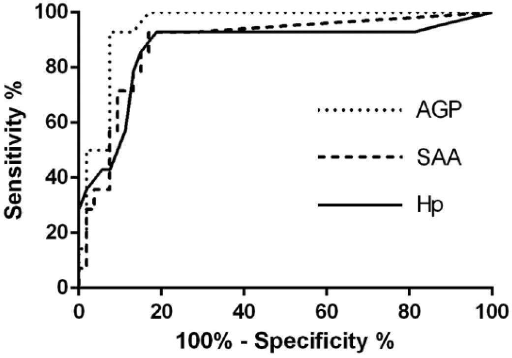


 EN
 SK