There is currently no evidence to suggest that humans infected with humans play a role in the spread of COVID-19. (*)
COVID-19 is a disease caused by a new type of coronavirus SARS-CoV-2. As the number of new cases grows worldwide, there is a growing concern for human and animal welfare, and important questions arise regarding prevention and best practices in the event of a positive diagnosis. In addition, another major doubt has arisen among pet owners: can dogs and cats also become infected, or can they transmit the virus?
So far, there are only a few known cases of pet infections around the world, including two dogs in Hong Kong and two cats in Belgium. Both dogs showed no signs of disease, but one cat had respiratory and digestive disorders. In addition, he had US Department of Agriculture The Malaysian Tiger also tested positive at the Bronx Zoo in New York.
Coronaviruses are a large group of viruses that are quite common in animals. There are four genera of the family Coronaviridae (Alphacoronavirus, Betacoronavirsus, Gammacoronavirus and Deltacoronavirus), which cause different diseases in different species of animals. The WHO emphasizes that people are occasionally infected with these viruses, which can spread to other people. For example SARS-CoV has been associated with civets and MERS-CoV is transmitted by single-humped camels.
Relationship with animals
Possible animal sources of COVID-19 have not yet been confirmed. Although the OIE claims that current evidence suggests that the COVID-19 virus came from an animal source, investigations are underway to find that source (including relevant species) and to determine the possible role of the animal reservoir in the disease.
To understand these differences, we spoke to our veterinarians. Hervé Poulet, the global leader in vaccines for pets and horses, shares the view that, despite the current lack of evidence that pets can transmit COVID-19, the OIE emphasizes that people who have the disease should avoid being very close. contact with your pets. Furthermore, they should follow good hygienic practices when handling and caring for them - this is a behavior that we should always follow.
"Cats and dogs can also be affected by their specific coronavirus, for example canine enteric coronavirus (CCoV), which belongs to the genus Alphacoronavirus and causes mild enteritis in dogs and infects cells in the gut, ”says Hervé.
Jean-Philippe Tronel, global technical director for pet vaccines, notes that vaccination against this animal disease already exists and is present in polyvalent vaccines for dogs in North America and some countries in Asia and Latin America. In addition, in the context of felines, there is a feline coronavirus (FCoV) that can cause a serious feline disease known as feline infectious peritonitis. The vaccine against it is available in some countries in Europe, Asia and North America.
It's not the same coronavirus
In contrast, people may be positive for presence beta-coronaviruses - such as SARS-CoV-2, responsible for the current pandemic; SARS-CoV, which originally caused Severe Acute Respiratory Syndrome in China in 2002 (SARS); a MERS-CoV, responsible for Middle East Respiratory Syndrome (MERS), which appeared in 2012 in Saudi Arabia. It is important to emphasize that there is no evidence of human-to-animal transmission in both diseases.
"Given these differences, the treatment options for different types of coronavirus are certainly not the same. Among the growing panic over the COVID-19 pandemic, there have been reports that people are seeking vaccines against canine and feline coronavirus as a desperate - and wrong - way to immunize. For this reason, it is important to emphasize that these vaccines are exclusively for dogs or cats. There are no clinical trials to ensure safety and efficacy in humans, ”says Hervé.
Last but not least, Karin Botteon, a veterinary consultant in the pet segment in Brazil, emphasizes that in case of any doubts, more than ever, it is crucial to always seek the advice of a veterinarian during this pandemic period. It is essential to strengthen the need for preventive medicine for pets, including proper adherence to vaccination programs.
"Because most countries in the world are currently facing a series of restraints, it is very important that pet owners contact their veterinarians to obtain correct and professional information on best practices and boards of directors for their own country or even region. Remember: when animals are healthy, people are also healthier, "concludes Karin.
(*) The situation regarding COVID-19 is constantly evolving. Research into all aspects is taking place around the world, including the mode of infection, and there is no way to go as far as possible to obtain as much information as possible. From what we know so far, there is no scientific knowledge about any transmission of the disease from pets to humans.
We follow all related research results and recommendations of international experts, especially the World Organization for Animal Health (OIE) and the World Health Organization (WHO). We will share these updates when they become available.
Translator's Note: There is only 63% similarity between SARS-CoV-2 and FCoV, and even so, it is limited to only about 1/4 of the genome.
Our savannah kittens were diagnosed with fatal feline infectious peritonitis. Luna's story of surviving the disease could only be realized thanks to ongoing research.
Meet Luna
What is FIP? (Our cat, Luna, underwent clinical trials of a new drug at FIP at the University of California, Davis). All photos are the property of Debra Roberts
Our first pet: a beautiful savannah cat
After a thorough survey and education to learn as much as possible about savannah cats, and after choosing a renowned breeder, Luna became a member of our family in March 2017 at the age of 10 weeks. When I picked her up in Boston, she was brave, active, playful, and at first sight brimming with good health. Luna was extremely friendly, fun, quirky, loving, with a strong personality. She exceeded all our expectations regarding the savannah breed. She quickly invaded our home and our hearts as our first darling and exceptional family member.
Luna was to receive her second combination vaccine and undergo a veterinary examination. On April 13, 2017, she received a vaccine that is part of a routine protocol for cats. After receiving the vaccine, Luna refused to eat, slept constantly and was hot to the touch. Her husband and I thought her middle stomach was enlarged, but when I mentioned it to the vet during the visit, she said it was a "normal kitten's belly."
The only abnormal finding during the examination was cardiac murmurs III. degrees that were thought to be benign. The plan was to re-examine the murmur in a few months and consider a possible cardiological examination if the condition did not improve.
Luna with an enlarged belly
Luna's diagnosis FIP: At the right time in the right place
The stomach continued to grow and on April 21 we took Luna to the emergency clinic. I looked for the causes of abdominal distension (expansion of the abdomen) in kittens and I also read about infectious peritonitis in cats (FIP). I tried to push the FIP out of my mind; after all, it is not such a common disease. I thought in the worst case, it could be a parasite.
During the examination, the veterinarian sucked fluid from her abdomen and brought a tube with her sample into the room. She was sure that due to the appearance of fluid, anorexia, lethargy and fever of 40.6 ° C, Luna has a moist form of FIP. She told us about the gloomy prognosis, and that Luna had only a few days ahead of her, or maybe two weeks of life. She suggested that we return a few days to consult an internist and discuss some life-extension options and hear a second opinion, or undergo euthanasia if her condition worsened over the weekend.
So on April 24, we met with an expert to hear his independent opinion. Lune underwent an echocardiogram, underwent X-ray and ultrasound examinations, and other blood tests to confirm the diagnosis of FIP. It took a long and exhausting three days. We refused to accept the reality of the loss of our new child. We started looking for all the ways we could save her. FIP is 100% lethal, there is no cure. How is it possible?
There must still be a way to save her. As a nurse in a large teaching hospital, I encounter clinical trials and research. I thought, "I'm sure someone is working for the FIP." During the very long and exhausting weekend of waiting for a specialist, I called and sent e-mails to every major veterinary school I was able to find on the Internet.
Luna's trip to UC Davis for life-saving medicine
To my great surprise, Michael Bannasch of UCD, Dr. Niels Pedersen's assistant, called me on April 23. Dr. Pedersen is a veterinarian, professor and scientist who has been treating FIP since the 1960s. Michael told me about researching and testing a hitherto unapproved drug. We discussed Luna's diagnosis and examinations, and Michael asked me to email him all of Luna's records and test results. On April 24, I received another phone call during which we talked about further tests and the possibility of flying with Luna to Sacramento - if we can get approval for her participation in the experiment.
If successful, Luna would be the first private cat in the world with a naturally acquired FIP to be tested for a new drug. She had high hopes, she was at the perfect age to respond to the new antiviral. Luna would stay at the clinic for 5 to 10 days for drug testing and monitoring.
Time was running out, her condition was getting worse. She had a high fever. Dr. Pedersen told us to stop palliative care (feline interferon omega and prednisone) and to arrive as soon as possible.
We arrived in Sacramento on April 28, seven days after being diagnosed with FIP. Dr. Pedersen acquainted us in detail with the treatment protocol, showed us the graphs, explained the mechanism of action of the drug to stop the replication of the virus. Then we went to the room where Luna was to stay for the next 5-10 days. At that time, she received her first injection and at the same time underwent a thorough examination.
Luna's stay at UC Davis with Dr. Niels Pedersen
First day of testing.
The experimental antiviral drug known as EVO984 / GS-441524 took effect immediately
Luna responded extremely quickly to a drug called 'EVO984' (later called GS-441524). We noticed an immediate reduction in fever and an improvement after the first injection. Luna reacted so well that we could go home on the sixth day. She was to receive injections for at least another 11 weeks. It was like bringing home a healthy, new kitten, a kitten without fever, malaise and an enlarged belly.
At home, I continued to give daily antiviral injections for a total of 12 weeks. Dr. Pedersen guided us throughout the treatment and advised us when to have the local veterinarian repeat the blood tests. Dr. Pedersen evaluated the results at the same time as regular weight checks to allow the dose to be adjusted accordingly.
By the way, this was the second drug testing at FIP since 2016, which took place at UCD. The first drug, GC376, was able to bring five out of 20 cats that had not relapsed to successful remission. Luna's drug, EVO984 / GS-441524, was even more successful, with approximately 26 cats achieving complete remission, which later changed to "cured."
To read more about FIP drug testing at UCD and the current status of the above antivirals, I recommend the following articles:
Important note: Both researches on UCD are already completed.
Left: large belly, dull, pale coat, piloerection (bristly coat). Wednesday: five days after starting treatment. Right: Abdominal distension is almost gone. Debra RobertsOne year without FIP. Debra Roberts
Happy ending for Luna and hope for cats diagnosed with FIP
On July 23, 2017, Luna received the last injection of EVO984 / GS-441524. She is doing well, showing no signs of illness and her laboratory results are still great. She loves camping and our adventure trips with a backpack on her back. She walked up to 13 miles, either with a harness or on a backpack. You don't see the cat on a leash on the sidewalks or in the camps every day!
Luna has been without FIP for almost four years. He lives a normal and healthy life and enjoys his three savannah friends - Titan, Calypso and Phoebe, which we brought home in October 2017 and November 2018.
Steve Dale, an animal behaviorist and well-known radio personality, interviewed me about Luna's participation in testing a new drug on the program "Steve Dale's Pet World", "In radio interview (Flash Player required) so you can hear Luna's story.
Luna loves camping and hiking with her human companions
The original article is followed by questions and answers…
MOSCOW (Reuters) - Russian drugmaker Pharmasyntez has asked the Kremlin to allow it to produce a generic version of the US drug COVID-19 remdesivir, which is used to treat US President Donald Trump, despite not having a patent, the company director said on Monday.
Remdesivir is not available in Russia, said Pharmasyntez CEO Vikram Punia, but the generic version cannot be produced and distributed without the consent of the patent holder, the American company Gilead Sciences GILD.O.
In July, Pharmasyntez asked the American company for approval in the form of a voluntary license, but did not receive any response, Punia said.
The Siberian drugmaker is now asking the Kremlin to activate a compulsory licensing process based on national security concerns, giving it the right to produce a generic Remdeform without the consent of Gilead.
"This drug could save many lives. The longer this drug is unavailable, the more people we will lose," Punia said.
The law has never been applied to a pharmaceutical product in Russia, Punia said after the daily Vedomosti reported on a letter from his company to the Kremlin on Monday.
It was not possible to contact Gilead on Monday outside normal business hours to comment on the situation. It has already granted voluntary licenses to manufacturers in 127 countries, mainly in low-income countries or in countries that have other significant barriers to accessing healthcare.
Russia recorded a record high 18,665 new cases of coronavirus on Sunday, bringing the total number since the start of the pandemic to 1,655,038.
Remdesivir was granted emergency use by the US Food and Drug Administration in May after government-initiated tests have shown that it shortens hospital stays for patients with COVID-19. He was one of the drugs Trump received during his treatment for this disease.
The FDA officially approved the drug this month, despite the recent results of a study sponsored by the World Health Organization, which showed that remdesivir did not improve patients.
According to a record in the registry, Pharmasyntez has completed clinical trials of a generic drug on 300 patients with COVID-19 in 23 Russian hospitals.
Punia said the company could sell its drug at a significantly lower price of around 540$ for 6 vials. Five days of remedivirus treatment from Gilead, sold under the Veklury brand, was priced at 3120$.
This is the first report of successful treatment of a case of non-fusive FIP uveitis using the oral form of the adenosine analog and feline interferon omega, and alpha-1 acid glycoprotein (AGP) as an indicator of recovery. A 2-year-old neutered Norwegian Forest Cat has been affected by uveitis, keratic clots, mesenteric lymphadenopathy and weight loss. He was hypergammaglobulinemic and had non-regenerative anemia. Feline coronavirus (FCoV) RNA was detected by thin-needle aspiration of mesenteric lymph nodes using polymerase chain reaction reverse transcription (RT-PCR) - non-fusive FIP was diagnosed. Prednisolone acetate eye drops were administered three times daily for 2 weeks. Treatment with an oral form of an adenosine analogue (Mutian) was started. After 50 days of Mutian treatment, the cat gained more than one kilogram, the globulin level decreased from 77 to 51 g / l, and the hematocrit increased from 22 to 35%; uveitis subsided and vision improved. Serum AGP levels decreased from 3100 to 400 μg / ml (within normal limits). Symmetric dimethylarginine (SDMA) was above normal at 28 μg / dl, declining to 14 μg / dl at the end of treatment; It is not known whether the increase in SDMA was due to FIP lesions in the kidney or Mutian. After cessation of Mutian treatment, low doses of the oral form of recombinant feline interferon omega were started - recovery of the cat continued.
Feline coronavirus (FCoV) is a highly infectious enteric virus that causes subclinical infection or diarrhea in most infected cats. [1], but in about 10% it causes potentially fatal immune-mediated monocyte-associated granulomatous vasculitis [2], known as feline infectious peritonitis (FIP). FCoV is an alpha coronavirus with positive polarity, a member of the Coronaviridae family of the Nidovirales family. [3].
Although treatment of FIP with an injectable nucleoside analog has been described in the past [4] and cure of enteric FCoV infection by oral form of adenosine analog (Mutian, Nantong Biotechnology, China) [5], this is the first reported case of recovery of a cat with a systemic FCoV infection - i.e. FIP - using Mutian.
2. Case study
Skywise was a 2-year-old neutered male Norwegian Forest Cat from a household with five cats; he developed uveitis in the remaining eye (in the right, he lost his left eye as a kitten) and occasional diarrhea. Its iris was unnaturally colored and grainy fat precipitates were visible (Figure 1a).
Figure 1 Cat's eye before (a) and (b) after treatment with Mutian and topical steroids. The cat had uveitis and keratic clots (a). Right (b): the same eye after 7 days of systemic prednisolone, 2 weeks of topical prednisolone and 7 weeks of Mutian treatment, showing almost complete resolution of uveitis.
His medical history included bringing two 11-month-old Norwegian Forest Cats (Link and Zelda) into the home three months ago and the onset of uveitis five days after revaccination (Leucofeligen, Virbac, France). Skywise was tested for FCoV antibodies and was found to have a very high titer above 10,240 (Idexx Laboratories, Wetherby, UK) three weeks before presentation due to the contact cat (Paddy) being persistently pyrexic and malaise. . Paddy was negative for FeLV p27 antigens and FIV antibodies (Idexx Laboratories, Wetherby, UK), but his anti-FCoV antibody titer was found to be very high (Table 1), so all other domestic cats were tested for FCoV antibodies to decided whether they should be isolated from Paddy, but since they were also found to have high FCoV antibody titers (Table 1), segregation was not necessary.
Day −29
Day −19
Day 7
Day 41
Skywise
2 years - Norwegian Forest Cat (NFC) FIP
> 10,240
CT 30
Neg
Paddy
2 years - NFC, persistent pyrexia, lethargy
> 10,240
CT 18
Neg
Oliver
8 years old - Domestic shorthair cat
> 10,240
CT 20
Neg
Link
1 year - NFC
> 10,240
CT 20
Neg
Zelda
1 year - NFC
640
Neg
Neg
Table 1
Feline coronavirus (FCoV) antibody titer and qRT-PCR C resultsT.
CT - Cycle threshold for FCoV qRT-PCR performed on a stool sample. The days are relative to the first presumed diagnosis of feline infectious peritonitis (FIP) at Skywise on day 0 (April 18, 2020); ie. its FCoV antibody titer was tested 19 days before the diagnosis of FIP. Mutian treatment was started for Skywise on day 6 and for other FCoV infected cats on day 18 for 5 days.
At presentation, the patient's body weight was 2.89 kg (Figure 2) and the fitness score was 2/9. Ultrasound confirmed the absence of abdominal or thoracic effusion, but mesenteric lymphadenopathy was present. Mesenteric lymph node (MLN) aspirate was collected by ultrasound-guided thin-needle aspiration biopsy (FNA) and tested for FCoV reverse transcription polymerase chain reaction (qRT-PCR) (Idexx Laboratories, Wetherby, UK). The assay was positive for the M1058L mutation and negative for the S1060A mutation [6].
Figure 2 Weight before, during and after FIP recovery and timeline. The weight of the cat is shown in the graph above: Day 0 represents the day of presentation with uveitis at the clinic. Revaccination was performed 5 days before the onset of uveitis, but the cat's weight was not recorded before that day, so it is not known whether she lost weight before revaccination or whether there was weight loss after revaccination; ie. whether the FIP was pre-vaccinated or whether the FIP was due to revaccination itself. The cat lost 30 g of weight during systemic treatment with prednisolone (days 0 to 6), then only topical steroids were administered. Mutian treatment was between days 6 and 56; after starting Mutian treatment, the cat's weight increased rapidly and weight gain continued even after Mutian was replaced by feline interferon omega, until the cat's weight reached about 4.35 kg.
The blood results are shown in Table 2. Hematology revealed mild to moderate non-regenerative anemia with hematocrit (HCT) 22%, lymphocyte count 2.07 × 10⁹/ la by eosinopenia below the detection level (reported as 0.0 × 10⁹/ l). Biochemical analysis of the blood showed hyperglobulinemia (77.2 g / l) and an albumin to globulin (A: G) ratio of 0.31. Bilirubin was 11 micromoles per liter (μmol / l) (0.64 mg / dl). Alpha-1 acid glygoprotein (AGP) was increased to 3100 μg / ml (Idexx Laboratories, Wetherby, UK) (normal level is ≤ 500 μg / ml [7]).
AGP
TP
Alb
Glob
A: G
Bilirubin
Hct
Lymphocytes
ALT
AP
GGT
SDMA
Creat
Urea
Ref. range
<500 μg / mL
57-89 g / L
22-40 g / L
28-51 g / L
> 0.8 *
0–15 μmol / L
30–52%
0.92–6.88 × 10⁹
12-130 U / L
14-111 U / L
0–4 U / L
0-14 μg / dL
71–212 μmol / L
5.7–12.9 mmol / L
Day 0
ND
111
20
91
0.22
11
25.7
0.95
64
ND
ND
ND
ND
ND
Day 2
3100
103
24
77
0.31
ND
22.0
2.07
ND
ND
ND
ND
ND
ND
Day 18
700
83
26
57
0.46
6
34.4
3.61
27
32
0
ND
88
9.6
Day 41
400
80
29
51
0.57
5
35.1
3.85
67
40
0
28
93
8.3
Day 62
400
78
30
48
0.60
5
35.2
2.64
61
40
0
19
106
6.5
Day 90
300
78
31
47
0.70
6
40.0
4.15
81
40
0
14
113
10.0
Day 153
400
67
34
33
1.03
1
35.2
4.49
81
46
2
ND **
98
7.8
Table 2
Laboratory data related to FIP treatment monitoring.
* A: G above 0.8 has a strong negative predictive value for FIP [8]. ** SDMA was not repeated because urine analysis was normal. AGP - alpha-1 acid glycoprotein; TP - total protein; Alb - albumin; Glob - globulin; A: G - ratio of albumin to globulin; Hct - hematocrit; ALT - alanine aminotransferase; AP - alkaline phosphatase; GGT - gamma glutamyltransferase; SDMA - symmetric dimethylarginine; Creat - creatinine; ND = not determined.
Only parameters that have been monitored repeatedly are shown in this table; others for which few (or no) results were available (eg aspartate aminotransferase) or which were not relevant to the procedure in this case are omitted. FCoV antibody titers and faecal FCoV qRT-PCR results are shown in Table 1. Day 0 is the day of FIP diagnosis in the first cat; Mutian was administered between days 6 and 56, after which the cat was treated with 100,000 units of the oral form of feline interferon omega.
Systemic treatment was initiated with prednisolone 5 mg for uveitis (Table 3). The dose was reduced to 2.5 mg sid, then stopped after 7 days and replaced with topical prednisolone-acetate 1% eye suspension (Pred Forte®, Allergan, Dublin, Ireland) applied every 8 hours for two weeks. On the sixth day after the presentation, treatment with an oral adenosine analogue began (Mutian 200, Nantong Biotechnology, Nantong, China). [5] at a dose of 8 mg / kg every 24 hours in divided doses: this was twice the normal dose, in an effort to ensure penetration into the eyeball. S-adenosyl-L-methionine supplementation was recommended to support the liver during treatment with Mutian. The dose of Mutian was reduced to 6 mg / kg on day 25, but by then the weight of the cat had increased, so the same number of capsules per day was required.
Treatment
Day
Ophthalmoscopic findings
Systemic prednisolone
0
Day of presentation to the primary veterinarian.
1
Eye examined using a standard ophthalmoscope. Cornea unchanged, anterior chamber slightly cloudy, but sedation would be required for a detailed examination. Cat blind and stressed.
2
Blood samples and aspirates of mesenteric lymph nodes with a thin needle taken under sedation.
Topically prednisolone acetate 1% tid
4
Keratin (protein) precipitated on the lens, which partially covers the fundus. Pupillary light reflex present but reduced.
Mutian 8mg / kg for 19 days, then 6mg / kg
6
The cat's caregiver said he thought the cat could see.
17
The cat's caregiver said the cat was chasing a bird.
18
Uveitis recedes, anterior chamber clear. Ceramic precipitates as before.
41
Uveitis has largely subsided, keratic precipitates still present. The fundus was visible, the chorioretinitis had subsided, and the cat's high temperament made detailed examination impossible. Excellent visual sensitivity.
Table 3 Timeline of treatment and resolution of uveitis.
To evaluate the susceptibility of the FCoV strain to the antiviral drug used, faecal samples were subjected to an FCoV qRT-PCR test (Veterinary Diagnostic Services, University of Glasgow, Scotland) [5]; the result was weakly positive in the threshold cycle (CT) 30, then negative, indicating that the virus was sensitive to the antiviral (Table 1). The faeces of three of the other four domestic cats were also positive for FCoV RNA (Table 1).
After starting Mutian treatment, the cat's weight increased rapidly, while it decreased by 30 g (2.89-2.86 kg) during prednisolone treatment (Figure 2). After 40 days of Mutian, the cat gained one kilogram (Figure 2). The dose of Mutian was adjusted during treatment in accordance with weight gain.
Blood tests were repeated on day 13 of Mutian treatment (18 days after diagnosis) (Table 2). Most importantly, the anemia reversed, HCT increased from 22.0 to 34.4%, and bilirubin decreased from 11 to 6 μmol / L. Globulin decreased to 57 g / l and albumin increased by 2 g / l, so the albumin: globulin ratio increased from 0.31 to 0.46, which was still a small but still improvement (Figure 3). Clinical examination revealed that iris color had returned to normal (Figure 1, Table 3) and vision improved; the caregiver reported an increase in activity - playing with other cats - and that the cat was trying to catch a bird. His droppings regained their normal consistency and frequency.
Figure 3 Timeline of treatment in relation to globulin albumin levels, showing a decrease in globulin (and thus total protein) levels, an increase in albumin and an albumin to globulin ratio. Improvement in these parameters continued after discontinuation of Mutian and feline interferon omega therapy.
The dose of Mutian was reduced to 6 mg / kg on day 20 and treatment with Mutian was stopped after 7 weeks for two reasons: AGP levels returned to normal (400 μg / ml - normal is <500 μg / ml), and because SDMA rose to 28 μg / dl (reference range is below 14 μg / dl). Recombinant feline interferon omega (rFeIFN-ω; Virbagen Omega, Virbac, France) started at 100,000 units every 24 hours orally. SDMA decreased to 19 μg / dl on day 62 (one week after the end of Mutian treatment) and to 14 μg / dl on day 90, which was 35 days after the end of Mutian treatment (Table 2). Urine analysis performed at days 62, 90, and 153 revealed no abnormalities, and the specific gravity was 1,050, 1,055, and 1,050, respectively. Weight gain continued after stopping Mutian treatment and using feline interferon omega (Figure 1), albeit at a slower rate (average 9g / day instead of 23g / day).
Cohabiting cats were successfully treated with Mutian at a dose of 4 mg / kg for five days to prevent coronavirus secretion and thus recurrent recovery of the recovered cat; the faeces of all five cats were confirmed as negative by RT-PCR (Table 1).
3. Discussion
The successful treatment of FIP with antiviral drugs was first published by Dr. Pedersen [4,9]. Cat owners can obtain various anti-FCoV drugs, including Mutian, an oral form of adenosine analog, over the Internet. However, such medicines are not licensed for veterinary use, which puts treating veterinarians in a dilemma: they cannot prescribe or supply such treatment, but they can help clients who choose to take these medicines by providing diagnostic and routine supportive care. It is particularly useful to make an accurate diagnosis that will prevent the treatment of about 40% cats that would otherwise be misdiagnosed (data not shown). Our view is that it would be more appropriate for cat owners to use such drugs under proper veterinary supervision, even if these products are unlicensed. The purpose of this case study is to report what has worked for us in monitoring a case of non-fusible FIP treated with Mutian followed by feline interferon.
Diagnosis of non-fusive FIP is challenging because the symptoms are diverse and the list of differential diagnoses is often lengthy. Dunbar et al. 2019 [10], reported that detection of FCoV RNA from Mesenteric Lymph Node (MLN) FNA (Fine Needle Aspiration) is 96% specific for FIP diagnostics. Unfortunately, the lab initially erroneously performed the FCoV RT-PCR test on a blood sample from day 2, instead of the MLN FNA as required - and the test turned negative. FCoV RT-PCR on blood samples is not useful in the diagnosis of FIP because most cats with FIP are not viremic at the time of clinical manifestations. [11] and also because about 5% cats do not test positive for FIP [12,13] because FCoV-infected cats undergo transient viremia [12,13,14,15]. FCoV RNA was found in feline MLN FNA and revealed to be positive for the M1058L mutation, which was further evidence to support the diagnosis of FIP [6,11]; although substitution with methionine for leucine at position 1058 in the FCoV spike protein was found in 89% tissue samples from 14 cats without FIP, suggesting that the mutation indicates systemic spread of FCoV from the gut rather than a virus with the potential to cause FIP [16].
Despite laboratory delays in confirming the diagnosis of FIP, we initiated Mutian treatment based on history, clinical signs, blood scores, and elevated AGP, which had previously been shown to be very specific for distinguishing FIP from other diseases with similar manifestations. [17]. There was an urgency to start treatment because the FIP male staging score was 6, indicating death within two weeks. [18]. The HCT of the cat decreased rapidly (from 25.7 to 22% in two days), which proved to be an indicator of imminent near death: Tsai et al., (2011) [18] found that HCT levels drop dramatically 2 weeks before death and bilirubin increases one week before death.
The history of the cat was typical of many cases of FIP: two new 11-month-old purebred cats were introduced into the household, which were probably the source of a recent coronavirus infection; the cat was vaccinated five days before the onset of uveitis. Introducing new cats and vaccinations are stressful for cats - it is known that stress causes FIP in cats infected with FCoV [19] and Riemer et al., (2016) [20] found that vaccination was a suspected stress factor in 6.9 % cases of FIP. Unfortunately, the weight of the cat between the introduction of new cats and vaccination was not recorded, so it is not known whether he lost weight during revaccination (ie was subclinically ill with FIP) or whether weight loss started later. This case nevertheless illustrates that vaccination of FCoV-infected cats may pose a risk to the development of FIP, and that optimal vaccination should be performed after the cat has stopped shedding the virus, which occurs within a few months for most FCoV type 1 infections. [21 ].
Clinical signs of weight loss, appetite, diarrhea and uveitis were also consistent with the diagnosis of non-fusive FIP. FIP is a major cause of uveitis in young cats - ophthalmologists from the North Carolina Veterinary School diagnosed FIP in 19 of 120 cats (15.8%) with uveitis [22]. Uveitis was reported in 17 of 59 (29%) cats with non-fusive FIP [23].
Although treatment with an injectable nucleoside analogue has been documented previously in the treatment of FIP [4], to our knowledge, oral treatment of FIP with an adenosine analogue has not been previously reported, although it has been used to treat FCoV infection in asymptomatic or diarrhea cats. [5]. The patient was given twice the normal therapeutic dose of FIP (ie, 8 mg / kg vs. 4 mg / kg) to potentially increase the concentration of the drug in the eye and to rule out any virus that might be present in the brain because only 20% of the absorbed drug passes through the blood-brain barrier (T Xue, personal communication). The response to treatment was rapid, dramatic and extremely positive. Although we tried to reduce the dose of Mutian as soon as possible in the event of possible side effects, in practice the amount of medication did not change significantly because the cat gained weight at the same time. Pedersen et al., (2019) [4] stated that the simplest indicator of response to treatment was weight gain, which was staggering in this case, as the cat gained 1.1 kg during the first 50 days of treatment. In contrast, he lost 30g during the week on prednisolone, before starting treatment with Mutian.
In two studies, cats with FIP treated with corticosteroids had a median survival of 7.5 days [24] and 8 days [25], which is significantly shorter than the 21 - day average reported by Tsai et al., 2011 [18] (although the potential adverse effects of biopsies on survival need to be considered in the two studies and many deaths were due to secondary bacterial infection due to extensive corticosteroid-induced immunosuppression [25]). In addition, prednisolone treatment was previously found to shorten survival in FIP cats that were co-treated with polyprenyl immunostimulant. [23]. Therefore, in our case, systemic corticosteroids were replaced by topical corticosteroids for the treatment of uveitis.
We do not know if it was Mutian, systemic and topical steroids, or a combination of all treatments that affected the overall cure of uveitis. The cat owner reported a partial return of vision on day 6, which was the day Mutian treatment was started, suggesting that steroids were responsible for return vision (Table 3). Legendre et al., (2017) [23] reported progress in 3 of 17 cats with ocular FIP treated with polyprenyl immunostimulant (PI). Their initial symptoms included anterior uveitis (all three cats), clots of ceramic origin (one cat), iris discoloration (one cat) and anisocoria (one cat). In two cats, anterior uveitis significantly improved or resolved after 2 months of PI treatment without corticosteroids. In the third cat, uveitis did not improve; the cat received topical ocular corticosteroids at the same time as PI and the eyes were enucleated [23]. Whether or not Mutian alone can affect the treatment of FIP-associated uveitis without current topical corticosteroids remains to be seen. Systemic corticosteroids appear to have little or no significance in these cases.
No clinical side effects were observed during treatment with Mutian and the biochemical parameters of the liver and kidney were within the reference range, with the exception of plasma symmetric dimethylarginine (SDMA), which increased (Table 2). SDMA is an early indicator of kidney disease [26,27]. There are three possible explanations for increased SDMA: first, it may be due to FIP lesions in the kidneys. Azotemia is more likely in non-effusive than effusive FIP [20], but as far as we know, no study has been conducted to determine whether SDMA is rising in cats with FIP - veterinary pathologists speculate that transient FCoV infection of the kidneys may be the cause of interstitial nephritis observed in many older cats (W Jarrett and S Toth, personal communication ). Second, predisposition to the breed may be a factor - Birm cats have been found to have elevated SDMA levels. [28], but no study has been performed on SDMA levels in Norwegian Forest Cats and in cats, SDMA levels have fallen to normal levels, suggesting that the increased SDMA levels did not appear to be related to his breed. Third, increased SDMA may have been a side effect of Mutian treatment - SDMA decreased within seven days after the end of Mutian treatment and returned to normal within 35 days after the end of Mutian treatment. However, because we did not test it earlier during the disease, we do not know if it was increased before the deployment of Mutian. During treatment, cat creatinine and urea were always within normal limits, although a progressive increase in creatinine was observed (Table 2), which will be closely monitored in the future.
Because the recommended time of administration of GS-441524 was 84 days [4], cat owners using other antivirals have been treating their cats with FIP for the same length of time. The optimal duration of FIP oral Mutian treatment has not been established. However, this case illustrates that treatment can be stopped after a much shorter cycle if laboratory indicators for FIP return to normal. We believe that in our case, complete recovery occurred after 50 days of treatment for the following reasons - uveitis subsided, the cat recovered from anemia, lymphopenia reversed, bilirubin decreased and AGP decreased permanently. Nevertheless, we did not want to leave the cat completely without antiviral coverage, so a low oral dose of rFeIFN-omega was used as described above. [29]. This drug has antiviral and immunomodulatory properties and was an optional treatment for FIP with meloxicam prior to the onset of specific antiviral drugs against FCoV. [30]. The cat continued to gain weight with rFeIFN-omega, although more slowly than before, stabilized at 4.35 kg after 100 days (Figure 2) and hematological parameters continued to improve (Table 2, Figure 3).
FIP relapse did not occur in this - or in other - cases whose treatment we monitored. We attribute our success primarily to assessing the effectiveness of an antiviral agent against the FCoV strain in an infected cat by monitoring the amount of virus excreted in the stool (by qRT-PCR) to ensure that the viral load decreased in response to the antiviral agent; second, preventing FIP-cured re-infection by identifying other sources of viruses in the same household and stopping FCoV shedding by Mutian [5]; third, short-term treatment with a dose that eliminated the virus from the brain; fourth, after treatment with a nucleoside analogue (or protease inhibitor) by initiating a low dose of oral feline interferon until the FCoV antibody titer has decreased - an indicator that no coronavirus remains in the body.
Skywise and his roommates are alive and well 6 months after diagnosis.
4. Conclusions
To our knowledge, this is the first published case of curing a non-fusive FIP cat using an oral adenosine analogue, followed by a recombinant feline interferon, and AGP used as a recovery marker from FIP. To protect the liver and monitor liver enzymes, we recommend taking SAMe with Mutian. SDMA levels in future cases following this FIP treatment protocol require further investigation. We suggest that systemic corticosteroids be contraindicated in the treatment of FIP for non-palliative purposes.
Acknowledgement
We would like to thank the reviewers for their time in thoroughly reviewing our work and for their helpful comments: Table 1 and Table 3 were added thanks to their comments.
Author's contributions
Conceptualization, DDA and JC-R .; methodology, MF, OJ and DDA .; validation, OJ, DDA, JC-R. and MF .; formal analysis, MF and DDA .; investigation, MF and JC-R .; sources, JC-R. and MF .; data management, DDA, JC-R. and MF .; writing - preparation of the original proposal, DDA; writing - reviews and edits, OJ, DDA, JC-R. and MF .; visualization, DDA and JC-R .; supervision, DDA and JC-R .; project administration, JC-R., MF. financing acquisition, JC-R. and DDA All authors have read and agreed to the published version of the manuscript.
Financing
Cat treatment and lab tests were funded by JC-RDDA Thank you subscribers www.catvirus.com for support during this study and we are very grateful to the Angelica Fund donor for the payment of publication fees.
References
Addie DD, Toth S., Murray GD, Jarrett O. The risk of feline infectious peritonitis in cats naturally infected with feline coronavirus. Am. J. Vet. Res. 1995;56: 429–434. [PubMed] [Google Scholar]
Kipar A., May H., Menger S., Weber M., Leukert W., Reinacher M. Morphologic features and development of granulomatous vasculitis in feline infectious peritonitis. Vet. Pathol. 2005;42: 321–330. doi: 10.1354 / vp.42-3-321. [PubMed] [CrossRef] [Google Scholar]
De Groot RJ, Baker SC, Baric R., Enjuanes L., Gorbalenya AE, Holmes KV, Perlman S., Poon L., Rottier PJM, Talbot PJ, et al. Family Coronaviridae. In: Adams MJ, Carstens EB, Lefkowitz EJ, editors. Ninth Report International Committee on Taxonomy of Viruses; King AMQ. Elsevier; Amsterdam, The Netherlands: 2012. pp. 806–828. [Google Scholar]
Pedersen NC, Perron M., Bannasch M., Montgomery E., Murakami E., Liepnieks M., Liu H. Efficacy and safety of the nucleoside analog GS-441524 for the treatment of cats with naturally occurring feline infectious peritonitis. J. Feline Med. Surg. 2019;21: 271–281. doi: 10.1177 / 1098612X19825701. [PMC free article] [PubMed] [CrossRef] [Google Scholar]
Duthie S., Eckersall PD, Addie DD, Lawrence CE, Jarrett O. Value of alpha1-acid glycoprotein in the diagnosis of feline infectious peritonitis. Vet. Rec. 1997;141: 299–303. doi: 10.1136 / vr.141.12.299. [PubMed] [CrossRef] [Google Scholar]
Shelly SM, Scarlett-Kranz J., Blue JT Protein electrophoresis on effusions from cats as a diagnostic test for feline infectious peritonitis. J. Am. Anim. Hosp. Assoc. 1988;24: 495–500. [Google Scholar]
Pedersen NC, Kim Y., Liu H., Galasiti KAC, Eckstrand C., Groutas WC, Bannasch M., Meadows JM, Chang KO Efficacy of a 3C-like protease inhibitor in treating various forms of acquired feline infectious peritonitis. J. Feline Med. Surg. 2018;20: 378–392. doi: 10.1177 / 1098612X17729626. [PMC free article] [PubMed] [CrossRef] [Google Scholar]
Dunbar D., Kwok W., Graham E., Armitage A., Irvine R., Johnston P., McDonald M., Montgomery D., Nicolson L., Robertson E., et al. Diagnosis of non-effusive feline infectious peritonitis by reverse transcriptase quantitative polymerase chain reaction from mesenteric lymph node fine needle aspirates. J. Feline Med. Surg. 2019;21: 910–921. doi: 10.1177 / 1098612X18809165. [PubMed] [CrossRef] [Google Scholar]
Felten S., Leutenegger CM, Balzer HJ, Pantchev N., Matiasek K., Wess G., Egberink H., Hartmann K. Sensitivity and specificity of a real-time reverse transcriptase polymerase chain reaction detecting feline coronavirus mutations in effusion and serum / plasma of cats to diagnose feline infectious peritonitis. BMC Vet. Res. 2017;13: 228. doi: 10.1186 / s12917-017-1147-8. [PMC free article] [PubMed] [CrossRef] [Google Scholar]
Fish EJ, Diniz PPV, Juan YC, Bossong F., Collisson EW, Drechsler Y., Kaltenboeck B. Cross-sectional quantitative RT-PCR study of feline coronavirus viremia and replication in peripheral blood of healthy shelter cats in Southern California. J. Feline Med. Surg. 2018;20: 295–301. doi: 10.1177 / 1098612X17705227. [PubMed] [CrossRef] [Google Scholar]
Simons FA, Vennema H., Rofina JE, Pol JM, Horzinek MC, Rottier PJ, Egberink HF A mRNA PCR for the diagnosis of feline infectious peritonitis. J. Virol. Methods. 2005;124: 111–116. doi: 10.1016 / j.jviromet.2004.11.012. [PMC free article] [PubMed] [CrossRef] [Google Scholar]
Herrewegh AAPM, de Groot RJ, Cepica A., Egberink HF, Horzinek MC, Rottier PJM Detection of feline coronavirus RNA in feces, tissue, and body fluids of naturally infected cats by reverse transcriptase PCR. J. Clin. Microbiol. 1995;33: 684–689. doi: 10.1128 / JCM.33.3.684-689.1995. [PMC free article] [PubMed] [CrossRef] [Google Scholar]
Tasker S. Diagnosis of feline infectious peritonitis: Update on evidence supporting available tests. J. Feline Med. Surg. 2018;20: 228–243. doi: 10.1177 / 1098612X18758592. [PubMed] [CrossRef] [Google Scholar]
Porter E., Tasker S., Day MJ, Harley R., Kipar A., Siddell SG, Helps CR Amino acid changes in the spike protein of feline coronavirus correlate with systemic spread of virus from the intestine and not with feline infectious peritonitis. Vet. Res. 2014;45: 49. doi: 10.1186 / 1297-9716-45-49. [PMC free article] [PubMed] [CrossRef] [Google Scholar]
Giori L., Giordano A., Giudice C., Grieco V., Paltrinieri S. Performances of different diagnostic tests for feline infectious peritonitis in challenging clinical cases. J. Small Anim. Pract. 2011;52: 152–157. doi: 10.1111 / j.1748-5827.2011.01042.x. [PMC free article] [PubMed] [CrossRef] [Google Scholar]
Tsai HY, Chueh LL, Lin CN, Su BL Clinicopathological findings and disease staging of feline infectious peritonitis: 51 cases from 2003 to 2009 in Taiwan. J. Feline Med. Surg. 2011;13: 74–80. doi: 10.1016 / j.jfms.2010.09.014. [PMC free article] [PubMed] [CrossRef] [Google Scholar]
Rohrer C., Suter PF, Lutz H. The diagnosis of feline infectious peritonitis (FIP): A retrospective and prospective study. Kleinterpraxis. 1993;38: 379–389. [Google Scholar]
Riemer F., Kuehner KA, Ritz S., Sauter-Louis C., Hartmann K. Clinical and laboratory features of cats with feline infectious peritonitis – A retrospective study of 231 confirmed cases (2000–2010) J. Feline Med. Surg. 2016;18: 348–356. doi: 10.1177 / 1098612X15586209. [PubMed] [CrossRef] [Google Scholar]
Addie DD, Jarrett JO Use of a reverse-transcriptase polymerase chain reaction for monitoring feline coronavirus shedding by healthy cats. Vet. Rec. 2001;148: 649–653. doi: 10.1136 / vr.148.21.649. [PubMed] [CrossRef] [Google Scholar]
Jinks MR, English RV, Gilger BC Causes of endogenous uveitis in cats presented to referral clinics in North Carolina. Vet. Ophthalmol. 2016;19(Suppl. S1): 30–37. doi: 10.1111 / vop.12324. [PMC free article] [PubMed] [CrossRef] [Google Scholar]
Legendre AM, Kuritz T., Galyon G., Baylor VM, Heidel RE Polyprenyl Immunostimulant Treatment of Cats with Presumptive Non-Effusive Feline Infectious Peritonitis in a Field Study. Front. Vet. Sci. 2017;4: 7. doi: 10.3389 / fvets.2017.00007. [PMC free article] [PubMed] [CrossRef] [Google Scholar]
Fischer Y., Ritz S., Weber K., Sauter-Louis C., Hartmann K. Randomized, placebo controlled study of the effect of propentofylline on survival time and quality of life of cats with feline infectious peritonitis. J. Vet. Intern. Med. 2011;25: 1270–1276. doi: 10.1111 / j.1939-1676.2011.00806.x. [PMC free article] [PubMed] [CrossRef] [Google Scholar]
Ritz S., Egberink H., Hartmann K. Effect of feline interferon-omega on the survival time and quality of life of cats with feline infectious peritonitis. J. Vet. Intern. Med. 2007;21: 1193–1197. doi: 10.1111 / j.1939-1676.2007.tb01937.x. [PMC free article] [PubMed] [CrossRef] [Google Scholar]
Jepson RE, Syme HM, Vallance C., Elliott J. Plasma asymmetric dimethylarginine, symmetric dimethylarginine, l-arginine, and nitrite / nitrate concentrations in cats with chronic kidney disease and hypertension. J. Vet. Intern. Med. 2008;22: 317–324. doi: 10.1111 / j.1939-1676.2008.0075.x. [PubMed] [CrossRef] [Google Scholar]
Relford R., Robertson J., Clements C. Symmetric Dimethylarginine: Improving the Diagnosis and Staging of Chronic Kidney Disease in Small Animals. Vet. Clin. U.S. Small Anim. Pract. 2016;46: 941–960. doi: 10.1016 / j.cvsm.2016.06.010. [PubMed] [CrossRef] [Google Scholar]
Paltrinieri S., Giraldi M., Prolo A., Scarpa P., Piseddu E., Beccati M., Graziani B., Bo S. Serum symmetric dimethylarginine and creatinine in Birman cats compared with cats of other breeds. J. Feline Med. Surg. 2018;20: 905–912. doi: 10.1177 / 1098612X17734066. [PubMed] [CrossRef] [Google Scholar]
Gil S., Leal RO, McGahie D., Sepúlveda N., Duarte A., Niza MM, Tavares L. Oral Recombinant Feline Interferon-Omega as an alternative immune modulation therapy in FIV positive cats, clinical and laboratory evaluation. Res. Vet. Sci. 2014;96: 79–85. doi: 10.1016 / j.rvsc.2013.11.007. [PMC free article] [PubMed] [CrossRef] [Google Scholar]
Addie D. Feline coronavirus infections. In: Green C., editor. Infectious Diseases of the Dog and Cat. Elsevier; Maryland Heights, MO, USA: 2012. pp. 92–108. [Google Scholar]
Diane D Addie, Sheryl Curran, Flora Bellini, Ben Crowe, Emily Sheehan, Lesya Ukrainchuk, Nicola Decaro
Abstract
Feline coronavirus (FCoV) is common in domestic cats living in groups. About 10% infected cats can potentially develop a fatal disease, feline infectious peritonitis (FIP). The virus is transmitted by the faecal-oral route. Mutian® Xraphconn (Mutian X) is a product for the treatment of FIP, but it is also used to stop coronavirus secretion, although there are no clear guidelines for its use. The aim of this study was to determine the minimum dose and duration of treatment required to reliably eliminate the virus in the faeces of asymptomatic, virus-secreting cats. In five households with several cats, Mutian X tablets received 29 cats naturally infected with FCoV, and actively shedding the virus in the feces. Virus shedding was monitored by a quantitative reverse transcription polymerase chain reaction (RT-qPCR) controlled on fecal inhibitors to ensure susceptibility. Mutian X administered orally eliminated the virus in 29 cats; although four cats required re-therapy to stop virus shedding. A dose of 4 mg / kg every 24 hours for four days was found to be the optimal treatment protocol: 2 mg / kg cleared only 80% cats. Therapeutic use of a sensitive RT-qPCR assay was necessary to ensure that virus was eliminated, as failure of even one cat could result in re-infection of other cats. Pre-treatment virus shedding records provided retrospective control: significantly more cats stopped shedding virus after Mutian X than recovered from infection during the control period (p <0.00001). This is the first report of successful elimination of FCoV excretion by the faecal route in chronically infected cats.
Keywords: Antivirals; Faecal shedding of the virus; Feline coronavirus (FCoV); Feline Infectious Peritonitis (FIP); Itraconazole; Mutian X tablets
Thousands of cat owners are resorting to black market medicine to cure their beloved pets from the FIP.
Cats face their own pandemic: a coronavirus strain that is estimated to kill tens of thousands of cats worldwide each year.
Feline infectious coronavirus, also known as FIPV, is not a novel virus and is a different strain from the virus that causes COVID-19 in humans. So far, it has been a death sentence for cats that develop the most severe forms of infection.
Most cats encounter feline coronavirus at some point in their lives, but show almost no signs of disease. However, in some cats, the virus mutates - and causes fatal FIP.
"I didn't even know about FIP until now, and 24 hours later I was already holding the drugs in my hands," Amy Shafer told Eyewitness News.
Her 13-year-old cat, Mr. Charlie Bear, was on the verge of death at the end of March this year, in the grip of the FIP.
"Vomiting, lethargy, decreased appetite and fever," says Amy. "He stopped playing with toys, he didn't eat as much as before."
Veterinarian Amy said there was no hope and that "she could bring him to bed in a day or two."
She desperately wanted to save the cat, which she says helped her in a difficult divorce. Amy and her friend Christophe Liglet got a tip on the Facebook group FIP Warriors.
"So they're like a secret group, someone has to tell you about them," Amy says.
Later that night, Amy and Christophe went to Torrance to meet a couple they had contacted through the FIP Warriors who had several vials of FIP medicine. Amy handed over $ 441 in cash for an illegal, presumably life-saving drug.
"It felt like a drug deal," Christophe recalls with a laugh. "I thought I'd call the cops in five minutes if he didn't come out."
Amy says the results were almost instantaneous. The fever subsided, he started to gain weight and now he plays with toys and climbs on his cat tree. Charlie Bear is about halfway through the 84-day treatment protocol, which Amy estimates will cost eight to ten thousand dollars.
"I'm sure my family thinks I'm crazy, but they would do the same," Amy says. "Charlie Bear is the best, he's like a little man.
50 YEARS OF RESEARCH LEADS TO A POTENTIAL HEALINGBE
Dr. Niels Pedersen, Professor Emeritus at UC Davis, has dedicated his 50-year career to studying FIP and seeking treatment.
"One percent of all cats die at FIP," says Dr. Pedersen for Eyewitness News. "It's a cat pandemic."
Pedersen and his team at UC Davis found the drug in a substance known as GS-441524, or GS for short. It blocks the virus's ability to replicate.
"We experimented with 31 cats and we were able to cure 25 of them," says Pedersen.
"It's amazing," says Dr. Brian Murphy, who has since taken over the feline coronavirus program at UC Davis. "It's a miracle for me because we found this medicine, like a needle in a haystack."
But there is a problem. GS-441524 is almost identical to Remdesivir, which is currently much talked about as a drug for COVID-19, and which is manufactured by Gilead, California. Gilead initially hoped that Remdesivir would work to treat people with Ebola.
Gilead invented and owned patents for GS-441524 and GS-5734 (Remdesivir), and his researchers are co-authors of the UC Davis studies, which demonstrate the efficacy of the drug in the treatment of FIP. So far, however, Gilead has refused to license GS-441524 for the treatment of cats.
Gilead did not respond to repeated requests for comments on the team, but last year told the Veterinary Information Network that the company was suspending the licensing of GS-441524 to other parties for commercial development until Remdesivir had FDA approval.
“It’s very frustrating because we were faced with the fact that we had this huge discovery, a major breakthrough,” Pedersen says. “We had a drug that could safely and effectively cure the majority of cats suffering from FIP. It was something I had dreamed about for 50 years.”
UC Davis is currently testing a similar drug for FIP - GC376. Another California company, Anivive, has licensed this drug. Company Anivive seeks FDA approval use this drug in cats and hopes to begin clinical trials at UC Davis this year.
Murphy of UC Davis claims that his team receives e-mails every week from cat owners who are desperately trying to get their cats into clinical trials.
"We are understanding and empathetic about their predicament," says Murphy. "But by the time we get to the tests, their cat will be dead."
Without legal access to the drug, thousands of cat owners have turned to the black market. Medicines containing GS-441524 are manufactured by various companies in China.
"The need is so intense and so great that people will find a way to get medication," says Pedersen. "I warned Gilead of that."
CAT OWNERS AND VETERINARIANS ARE IN A DIFFICULT SITUATION
On the FIP Warriors Facebook page, cat owners share before and after photos, advice and support on how to get and use the medicine. Since its inception, the group has grown to more than 24,000 members.
The FDA informs Eyewitness News that any unauthorized drug is considered illegal, but the FDA is unlikely to target cat owners.
"In general, the FDA usually focuses its law enforcement activities on the manufacturer and / or seller of an unapproved product, not the end user," the FDA spokesman said.
"Whenever you use unapproved drugs, you need to watch out for the buyer," says Pedersen, who notes that 100% treatment is not guaranteed and some cats relapse after treatment.
Saving the lives of loved ones with illegal drugs puts veterinarians who want to help in a legal and ethical trap.
"I don't buy the product, I can't prescribe it," said an anonymous LA veterinarian who agreed to an anonymous interview with Eyewitness News. "It's a black market."
The veterinarian estimates that so far she has treated 18 cats with this drug and all but two have survived.
“They’re beaming, happy, with no signs of illness,” she says. “They’re happy at home with their owners, or their rescuers. And that was amazing to me because I’d never seen that before. Before, FIP was always a death sentence.”
She is aware that the administration of the medicine could potentially jeopardize her veterinary license.
"It's a risk," he says. "And if these pets didn't succeed and their lives didn't improve, or their quality and length of life didn't improve, I wouldn't do it."
Koi is a one-year-old rescued cat that came with the FIP in mid-April. Her owner, a musician with the stage name "Tillie", calls Koi her "little punk girl."
"It's scary because it's a lot of money," says Tillie. "You really have no guarantee."
Tillie says she couldn't stand the idea of losing Koi after losing another beloved cat to cancer last fall.
“I would say within two days we could know it was working,” Tillie says of the black market drug. “It’s a completely different cat, running around, wanting to get everywhere.”
Tillie knows this is against the law, but says the potential life-saving Koi is worth it.
"It would be really cruel to persecute people trying to save their animals," says Tillie. "We've gotten into a position where we have to decide whether to let our animal die or do everything in our power to save it, and I do."
Do you have a tip? Email ABC7 investigative producer Lisa.Bartley@abc.com
Amy Shafer and Tillie raise money to cover the cost of treating their cats.
Peter J. Dickinson, Michael Bannasch, Sara M. Thomasy, Vishal D. Murthy, Karen M. Vernau, Molly Liepnieks, Elizabeth Montgomery, Kelly E. Knickelbein, Brian Murphy, Niels C. Pedersen
Abstract
Feline infectious peritonitis (FIP) is caused by a mutant feline enteric coronavirus biotype. The resulting FIP virus (FIPV) in the case of the non-fusion form commonly causes pathologies of the central nervous system (CNS) and the eyes. More than 95% cats with FIP succumb to the disease several days to months after diagnosis, despite various historically used treatments. Recently developed antiviral drugs have shown promise in the treatment of non-neurological FIPs, but data on the treatment of neurological cases of FIP are limited. Four cases of naturally occurring FIP with CNS involvement were treated with the antiviral nucleoside analog GS-441524 (5-10 mg / kg) for at least 12 weeks. Cats were monitored continuously by physical, neurological and ophthalmological examinations. One cat underwent magnetic resonance imaging (MRI), cerebrospinal fluid (CSF) analysis, including FCoV titer, RT-PCR, and ophthalmic examination using Fourier optical coherence tomography and in vivo confocal microscopy (IVCM). All cats responded positively to treatment. The three cats are still alive (528, 516 and 354 days after the start of treatment) with normal results of physical and neurological examinations. One cat was euthanized 216 days from the start of treatment after relapses after primary and secondary treatment. In 1 case, the cure of the disease was determined on the basis of normalization of MRI and CSF findings and examination of cranial and caudal segment disease using ocular imaging methods. Treatment with GS-441524 shows its clinical efficacy and may lead to clearance and long-term regression of neurological FIP. The doses required for CNS disease may be higher than the doses used for non-neurological FIPs.
The experimental treatments were approved by the Institutional Committee on the Care and Use of Animals and the Clinical Trials Evaluation Committee of the Veterinary Medical University School of the University of California, Davis. GS-441524 was provided by Gilead Sciences Inc. (Foster City, CA), as previously described. 1, 2 The clinical diagnosis of FIP was based on a combination of characteristic information, medical history, disease symptoms, laboratory test results including hyperglobulinemia, decreased albumin: globulin (AG) ratio and FCoV antibody titers (indirect immunofluorescence test [IFA]), Fuller Laboratories, Fullerton, California) 3 and response to virus-specific treatment. The status of feline leukemia virus (FeLV) and feline immunodeficiency virus (FIV) was determined for FeLV antigen and FIV antibody by ELISA (IDEXX, Westbrook, Maine). In one case, repeated advanced diagnostic testing was performed, including MRI, CSF analysis, CSF FCoV RT ‐ PCR (Real-time PCR Research and Diagnostics Core Facility, UC Davis, Davis, CA) and serology, FD ‐ OCT and IVCM.
2. Case 1
A neutered male of an 8-month-old blue Siamese cat obtained as a kitten from a rescue group. History of several months of lethargy and decreased appetite and monthly progressive pelvic limb ataxia confirmed by neurological examination. The cat weighed 3.0 kg, which was 1 kg less than the female sibling. Serum biochemistry abnormalities included increased total protein concentration (8.9 g / dl; reference interval 6.3-8.8 g / dl) with an AG ratio of 0.53 (albumin, 3.1 g / dl; reference interval 2, 6-3.9 g / dl; globulin), 5.8 g / dl; reference interval, 3.0-5.9 g / dl). Tests for FeLV and FIV antibody titers and Toxoplasma gondii IgM and IgG (Protatek, Mesa, Arizona) were negative and the FCoV antibody titer was positive at 1:12 800. OU). Ultrasound of the abdomen showed circumferential hyperechoic lines at the renal corticomedullary junctions, linear hyperechoic lines parallel to the luminal surface of the jejunum and ileum, and enlarged colic and mesenteric lymph nodes. The cat was treated with 5 mg / kg GS-441524 SC once daily for 14 weeks. Appetite and activity, including jumping on elevated surfaces, improved within 4 days. The total serum protein concentration after the end of treatment was 7.8 g / dl with an AG ratio of 0.77. Neurological examination was normal and body weight was 5.1 kg. The cat remains clinically normal at the time of writing 528 days after the start of treatment.
3. Case 2
The annual neutered domestic shorthair cat born to the rescued feral cat had a 3-month history of anterior uveitis and a weekly history of lethargy, altered behavior, tail twitching, generalized seizures, decreased appetite, and pelvic dysphagia and ataxia. The cat weighed 3.7 kg. At the neurological examination, the cat was dulled by generalized ataxia, decreased postural responses in the left thoracic and right pelvic limbs, decreased physiological OU nystagmus, and decreased nasal perception on the right side. Approach response was reduced in both eyes, anisocoria (mydriasis in the left eye [oculus sinister, OS]) was present with reduced direct and consensus pupillary light reflex (PLR) with OS illumination. Ophthalmic examination revealed anterior uveitis of the OU with retinal detachment of the OS and retinal vasculitis of the OU. The cat weighed 3.7 kg and serum biochemistry abnormalities included an increased total protein concentration (8.6 g / dl; reference interval, 6.6-8.4 g / dl) with an AG ratio of 0.48 (albumin, 2.8 g reference interval, 2.2-4.6 g / dl; globulin, 5.8 g / dl; reference interval, 2.8-5.4 g / dl), increased total bilirubin concentration (1.8 mg / dl; reference interval, 0.0-0.2 mg / dl) and increased AST activity (128 IU / dl) L; reference interval, 17-58 IU / L). The feline leukemia virus, FIV and heartworm antigen test (SNAP Feline Triple Test, IDEXX, Westbrook, Minnesota) was negative. Abdominal palpation showed enlarged mesenteric lymph nodes. The cat was treated with 5 mg / kg GS-441524 SC, once daily for 14 weeks. The body weight at the end of treatment was 5.9 kg. The level of mentation and activity improved significantly 48 hours after the start of treatment. After 3 weeks of treatment, neurological and ophthalmic symptoms were unobservable except for mild intermittent anisocoria and chorioethinal scars OU. Previously reported abnormalities in serum biochemistry resolved with a total serum protein concentration of 8.1 g / dl and an AG ratio of 0.77. Three weeks after the end of the treatment, the cat weighed 6.4 kg, physical and neurological examinations were found and the total serum protein concentration was 7.0 mg / dl with an AG ratio of 0.84. The cat remains clinically normal at the time of writing 516 days after the start of treatment.
4. Case 3
The 18-month-old neutered domestic shorthair cat obtained from the shelter had a 3-month history of eye disease, a 3-week progressive history of lethargy and anorexia, and a several-day history of progressive limb paresis. The cat was treated with prednisolone-acetate 1% OU eye drops, every 6 hours for 5 days before presentation. At the neurological examination, the cat showed inappropriate behavior and hypersensitivity to cranial palpation. The cat was non-ambulatory paraparetetic with reduced pelvic limb reflexes. The blink reflex lacked OU with anisocoria (pupil of medium extent in the right eye [oculus dexter, OD], mydriasis OS). The pupillary light reflex lacked OD due to posterior synechiae and absent OS with direct or indirect illumination. Reflections for OU glare and vision were present and the cat showed reduced vision under photopic conditions. Ophthalmic examination revealed uveitis and OU hyperviscosity syndrome. The cat weighed 2.6 kg. Serum biochemistry abnormalities included increased total protein concentration (11.7 g / dl; reference interval 6.3-8.8 g / dl) with an AG ratio of 0.2 (albumin, 2.0 g / dl; reference interval 2, 6-3.9 g / dl; globulin), 9.7 g / dl; reference interval, 3.0-5.9 g / dl). Tests for FeLV and FIV were negative and the FCoV antibody titer was positive at 1: 6400. Ultrasound of the abdomen showed hepatosplenomegaly, small kidneys with indistinct corticomedullary junctions, and enlarged mesenteric lymph nodes. The cat was treated with 5 mg / kg GS-441524 SC, once daily for 15 weeks. After 1 month of treatment, uveitis improved, but is still present and the cat was outpatient paraparetetic with normal segmental reflexes. The cat weighed 3.3 kg and the AG ratio was 0.55. After 2 months of treatment, mild signs of active anterior ODitis were present, but there was only a slight improvement in outpatient paraparesis. Body weight increased to 3.7 kg and the AG ratio was 0.67. After 15 weeks of treatment, there were only minimal signs of uveitis OD and ambulatory paraparesis, which was static during the previous 4 weeks, improved. The cat weighed 4.0 kg and the AG ratio was 0.76. After cessation of treatment, lethargy, anorexia and anisocoration returned within 36 hours. Treatment was resumed at 5 mg / kg GS-441524 SC once daily and symptoms resolved within 24 hours. The signs remained static during the 12 weeks of the second round of treatment, but the reduced activity reappeared after the end of treatment. The cat was euthanized in part due to increased resistance to drug administration. Histopathological evaluation after autopsy showed multifocal chronic dysfunctional meningitis, encephalomyelitis and ventriculitis, lymphocyte, histiocytic uveitis and choroiditis OU and interstitial nephritis. Positive coronavirus immunohistochemical immunoreactivity (FIP antibody - V3-70, Custom Monoclonals International, Sacramento, CA) was identified within histiocytes associated with lesions in the brain, kidney and eye.
5. Case 4
A seven-month-old sterilized female domestic shorthair cat adopted from a shelter had a three-week history of lethargy and anorexia and a two-week history of ataxia and crouching. At the neurological examination, the cat had an ataxic gait that was worse in the pelvic limbs. Postural responses were reduced in the pelvic limbs. Anisocoria (midrange OD, miotic OS) was present with incomplete PLR OUs. Blink reflexes, dazzling reflexes and vision were present by the OU. The cat weighed 2.7 kg. Brain magnetic resonance imaging has shown multifocal T2-weighted hyperintensity in the entire parenchyma, most severely in the midbrain and thalamus. Post-contrast T1-weighted images showed diffuse thickening and thickening of the meninges and brainstem with marked ventriculomegaly (Figure 1). Cerebrospinal fluid collected from the cerebellomedular cistern was xantochromic with a total nucleated cell count (TNCC) of 888 / μL (reference interval, <3 cells / μL) and a total CSF protein concentration of 1790 mg / dl (reference interval, <25 mg / dl). Serum and CSF titers of FCoV antibodies were positive at> 1:20 480 and real-time TaqMan RT-PCR for FCoV in CSF was positive with a threshold cycle (Ct) value of 18.87. Tests for FeLV and FIV were negative. Serum biochemistry and CBC abnormalities included a total protein concentration of 8.5 g / dl (reference interval, 6.6-8.4 g / dl), an AG ratio of 0.37 (albumin 2.3 g / dl; reference interval 2.2 -4.6 g / dl, globulin 6.2 g / dl, reference interval 2.8-5.4 g / dl), total bilirubin concentration 0.5 mg / dl (reference interval 0.0-0.2 mg / dl), anemia (hematocrit, 25.8%; reference interval, 30 -50%) and lymphopenia (835 / μL; reference interval, 1000-7000 / μL). Ultrasound of the abdomen showed hyperechoic kidneys and retroperitoneal fat, several enlarged lymph nodes, and mild peritoneal effusion. An ophthalmological examination of the FD ‐ OCT and IVCM revealed anterior uveitis with keratinous clots present in the OU at the back; ocular hypertension (25 mmHg OD, 11 mmHg OS) and chorioretinitis were also identified by OD (Figure 2). The cat was treated with 5 mg / kg GS-441524 SC, once daily for 4 weeks and prednisolone acetate 1% eye drops OU q8h and dorzolamide 2% eye drops OD q8h during the first 3 weeks of GS-441524 treatment. Activity and mentation improved within 24 hours after onset. treatment. After 4 weeks, the ophthalmic disease improved significantly (Figure 2), but ataxia was still present and the cat lost 0.2 kg of body weight (Figure 3). Serum total protein concentration was steadily increased (8.6 g / dl) with an improved AG ratio of 0.72; lymphopenia and anemia resolved. Due to lack of weight gain and persistent neurological deficits, the dose of GS-441524 was increased to 8 mg / kg SC, once daily for another 10 weeks (a total of 14 weeks). The cat also received a two-week course of prednisolone 1 mg / kg PO every 24 hours. Increased activity and willingness to jump on elevated surfaces were observed within 24 hours, and 1 week after the end of GS-441524 treatment, neurological examination was negative and no active ophthalmic disease was detected. Body weight was increased to 3 kg and total serum protein concentration was normal (7.6 g / dl) with an AG ratio of 0.8. Repeated magnetic resonance imaging (Figure 1) showed minimal increase in meningeal contrast, but ventriculomegaly increased. Repeated CSF RT ‐ PCR for FCoV RNA was negative and TNF in CSF decreased compared to the previous count, but still high at 224 / μL. Due to the evidence from the cerebrospinal fluid analysis that the infection was still active, the dose of GS-441524 was further increased to 10 mg / kg SC, once daily for another 5 weeks (a total of 19 weeks). The cat remained clinically normal with increased activity during this period and body weight increased to 4.7 kg (Figure 3). Immediately after the end of treatment, neurological and ophthalmological examinations remained unchanged and repeated MRI was found in addition to persistent ventriculomegaly. Repeated CSF analysis showed a continued decrease in TNCC (8 cells / μL) and total protein concentration (85 mg / dl), negative RT ‐ PCR for FCoV RNA, and a reduced 1: 128 FCoV antibody titer. Approximately 8 months after the start of treatment and 3 months after the end of treatment, the MRI did not change from the previous image, except for less severe ventriculomegaly. Cerebrospinal fluid analysis showed TNCC 6 cells / μL, total protein concentration 52 mg / dl, negative RT ‐ PCR for FCoV RNA and static antibody titer FCoV 1: 128. Serum total protein concentration was 7.1 g / dl with AG 0 ratio , 97. No signs of active inflammation were observed on ophthalmology, although focal areas of retinal dilution were identified. The cat remains clinically normal at the time of writing 354 days after the start of treatment.
Figure 1 Sequential magnetic resonance imaging from case 4. The lines represent selected postcontrast (gadolinium) T1 weighted transverse images of the brain obtained in one imaging sequence. Routine cerebrospinal fluid analysis at the time of imaging is shown in white for each imaging time point: TNCC = total number of nucleated cells in CSF (cells / μl); TP = total protein CSF (mg / dl); N = neutrophils, SM = small mononuclear cells, LM = large mononuclear cells. The characteristic neutrophil pleocytosis resolved during treatment. Additional CSF analyzes related to FCoV detection are presented in yellow for each time point: PCR = FCoV RT-PCR result [positive (+) or negative (-)]; Dilution ratio = FCoV antibody titer in cerebrospinal fluid. For each imaging sequence, the time points and doses of GS-441524 provided prior to imaging are described. The initial marked increase in meningeal pain contrast resolves after treatment with GS-441524 and does not recur after treatment. Ventriculomegaly, which is present after the initial response to treatment, slowly resolved upon subsequent imaging. The reduction in abnormalities in the CSF analysis findings in parallel corresponded to reduced abnormalities in MR imaging.
Figure 2 Sequential multimodal imaging of cranial and caudal segments from case 4. On the presentation of (A, B) predilation and (G, H) post-dilatation photographs of cranial segments showing mild diffuse corneal edema, pigmented ceramic clots, rubeosis iridis, detail of cloudy iris with flashes in aqueous humor and incomplete dilatation of the OU; dyskorrhea with incomplete pupillary dilatation due to caudal synechia OS (H) was also observed. Ceramic clots were also visualized by OS biomicroscopy with slit lamp (V), corneal FD-OCT (M) and endothelial IVCM (X, arrows); increased corneal thickness was also observed with FD-OCT (X). Imaging of the retina and choroid by FD ‐ OCT revealed a cellular infiltrate in the choroid (P, arrow), which was visible as a hyporeflective lesion with infrared photography (S). 0.8 months after the start of GS-441524 treatment, photographs of pre- (C, D) and post-dilatation (I, J) cranial segments of the clear cornea and cranial ventricle OU, isocoria, reduced rubeosis iridis and complete dilatation of the pupil. A significant decrease in pigmented ceramic precipitates was observed in slit lamp (W), corneal FD ‐ OCT (N) and IVCM endothelial (Y, arrow) biomicroscopy. Normal retinal and choroid morphology is observed in FD ‐ OCT (Q), although hyporeflective lesions remain on infrared imaging (T). At 7.6 months, pre- (E, F) and post-dilatation (K, L) photographs of the cranial segment showed clear corneas and cranial chambers of the OU, isocorrium, normal iris morphology, and post-inflammatory pigment on OS lens capsules. Corneal FD-OCT (O) lacks ceramic precipitates. In FD ‐ OCT and infrared imaging, dilution of the dorsal peripheral retina (R, arrow) was present with loss of normal stratification but no cellular infiltrate or retinal detachment (U).
Figure 3 Case 4 body weight development plotted with time-initiated GS-441524 treatment. Drug dose changes are indicated by red arrows. After increasing the dose of the drug above the initial dose of 5 mg / kg, an increase in body weight was observed and was accompanied by a remission of clinical signs.
6. Discussion
Infectious feline peritonitis is a major cause of mortality in young cats and a common cause of neurological disease. Several experimental treatments have not shown consistent efficacy against FIP, and cats are euthanized or die several days to months after the development of clinical disease, especially when FIP affects the CNS. Fortunately, drugs aimed at replicating RNA viruses in important human diseases, such as human immunodeficiency virus (HIV), hepatitis C and Ebola, have provided a model for treating viral diseases in other species, such as FIP. GS-441524 is a 1'-cyano-substituted adenine C-nucleoside ribose analog that inhibits viral RNA synthesis. GS-441524 and the 3C-Like viral protease inhibitor have demonstrated efficacy against FIPV in experimentally induced and naturally occurring FIP. However, preliminary studies suggest that treatment of ocular and CNS forms of FIP may be more complex due to limited drug passage across the blood-blood and blood-brain barriers. A high rate of relapse of FIP with CNS involvement has been observed in protease inhibitor-based therapy, while GS-441524 has become more promising in the treatment of ocular and neurological FIP. An initial clinical study with GS-441524 on naturally occurring, non-neurological FIP used doses of 2 mg / kg, which appeared to be insufficient for cats that developed neurological symptoms during treatment. However, 2 cats that developed neurological disease at this dose appeared to respond to 4 mg / kg. 4 cases of neurological FIP from our study were treated with a dose of 5 mg / kg, with duration of treatment and subsequent dose increase based on clinical reactions. A dose of 5 mg / kg, SC, once daily for 12 to 14 weeks was sufficient to treat 2 less severe neurological cases of FIP (cases 1, 2), but repeated cycles of 5 mg / kg in the most severely clinically affected cat (case 3 ) only resulted in improvement in clinical symptoms with rapid clinical regression after cessation of treatment. This therapeutic failure resulted in a gradual increase in dose from 5 to 10 mg / kg in case 4. The in vitro 50% effective concentration (EC50) for GS-441524 to prevent viral cytopathic effects was reported at 0.8 μM with complete inhibition of viral replication at 10 μM and partial inhibition at 1 μM. 1 Limited pharmacokinetic studies in cats from the same study showed that CSF concentrations of GS-441524 were approximately 20% in plasma and a dose of 10 mg / kg resulted in CSF concentrations of 0.8 to 2.7 μM. These data correspond to the limited efficacy associated with doses of 5 mg / kg in cases 3 and 4 and the apparent efficacy associated with increasing the dose to 8 to 10 mg / kg in cases 4. To further define the optimal dose of GS-441524 in cats with neurological FIP, extended pharmacokinetic studies in healthy and affected cats with intact and impaired blood-brain barrier function. As in previous reports, limited adverse reactions associated with long-term use of GS-441524 have been observed. Local skin reactions and discomfort after SC injection were the only clinically relevant adverse reactions, but this was a major factor influencing the euthanasia decision. 3. Although responses to treatment were measurable by MRI, CSF analysis, and eye imaging, clinical responses to treatment at the appropriate dose were equally useful, with rapid improvements in mentation, appetite, and activity, which were generally observed within 24 to 36 years. hours. Increased body weight and the ability to jump on elevated objects and surfaces were also considered consistent indicators of effective treatment. GS-441524 is not available for routine clinical use, but reported cases suggest that FIP affecting the CNS may be treatable with appropriate antiviral drugs. The development of similar antiviral drugs for clinical use should be considered a priority for this historically deadly disease.
References
Murphy BG, Perron M, Murakami E, et al. The nucleoside analog GS ‐ 441524 strongly inhibits feline infectious peritonitis (FIP) virus in tissue culture and experimental cat infection studies. Vet Microbiol. 2018; 219: 226-233. [PMC free article] [PubMed] [Google Scholar]
Pedersen NC, Perron M, Bannasch M, et al. Efficacy and safety of the nucleoside analog GS ‐ 441524 for the treatment of cats with naturally occurring feline infectious peritonitis. J Feline Med Surg. 2019; 21: 271-281. [PMC free article] [PubMed] [Google Scholar]
Pedersen NC. An update on feline infectious peritonitis: diagnostics and therapeutics. Vet J. 2014; 201: 133–141. [PMC free article] [PubMed] [Google Scholar]
Pedersen NC. A review of feline infectious peritonitis virus infection: 1963‐2008. J Feline Med Surg. 2009; 11: 225-258. [PMC free article] [PubMed] [Google Scholar]
Pedersen NC, Kim Y, Liu H, et al. Efficacy of a 3C ‐ like protease inhibitor in treating various forms of acquired feline infectious peritonitis. J Feline Med Surg. 2018; 20: 378-392. [PMC free article] [PubMed] [Google Scholar]
Kim Y, Liu H, Galasiti Kankanamalage AC, et al. Reversal of the progression of fatal coronavirus infection in cats by a broad ‐ Spectrum coronavirus protease inhibitor. PLoS Pathog. 2016; 12: e1005531. [PMC free article] [PubMed] [Google Scholar]
The number of type 2 severe acute respiratory syndrome (SARS-CoV-2) coronavirus infections continues to rise globally, and hospital physicians are currently preparing therapies for FDA-approved therapies as treatment options for SARS-CoV-2. We have been researching anti-cancer therapies for feline infectious peritonitis (FIP) for several years. [1,2], a coronavirus disease with almost 100% mortality in felines. Feline enteric coronavirus (FECV), which is common in many asymptomatic cats, mutates into virulent and lethal FIP coronavirus [3]. We believe that our experimental research in the treatment of FIP may be relevant and applicable to the latest results of in vitro SARS-CoV-2 tests. [4] in the absence of large-scale laboratory and human clinical studies. The FIP coronavirus protease inhibitor (GC376) has been successful in treating FIP in a subset of felines; however, in cases of neurological impairment, the protease inhibitor was unable to prevent the progression of central nervous system (CNS) disease, resulting in neurological FIP and subsequent euthanasia. [5]. The polymerase inhibitor GS-441524 has already shown significant activity in a feline clinical study against FIP [1], but treatment for neurological impairment has yet to be demonstrated. Remdesivir, a prodrug of GS-441524, has high hopes for the treatment of SARS-CoV-2. [6], but is not currently FDA approved and is only available in intravenous form. There is an urgent need for therapies against SARS-CoV-2, with good oral bioavailability and FDA approved, suitable for organs that express SARS-CoV-2 target angiotensin-converting enzyme 2 (ACE2) and can synergize with remedivr. Although detailed experimental results will be reported elsewhere (unpublished data from BGM Laboratory), we believe that our research could help physicians with treatment options in addition to supportive care.
Type 1 severe acute respiratory syndrome (SARS-CoV-1) and SARS-CoV-2 coronavirus target ACE2 as a receptor [7]which is expressed in humans in the lungs, heart, gastrointestinal tract and CNS [8]. SARS-CoV-1 is known to penetrate the CNS via the olfactory nerve and olfactory bulb [9], like other coronaviruses [10]. Anosmia (loss of smell) often occurs in patients with coronavirus disease 2019 (COVID-19), a disease caused by SARS-CoV-2, suggesting that SARS-CoV-2 uses the same route after infection. Almost all beta-coronaviruses enter the CNS [10] and SARS-CoV-1 and SARS-CoV-2 have the same ACE2 receptor. It is also reasonable to assume that a massive brainstem infection in experimental animals after nasal exposure to SARS-CoV-1 [9] can also occur with SARS-CoV-2, which may contribute to sudden respiratory failure, as seen in some patients [10]. It is unclear whether SARS-CoV-2 CNS penetration may also occur in patients with recent blood-brain barrier (BBB) damage following a stroke or other stroke. As we have shown in cats, the consequences of CNS penetration underline the need for a multi-purpose organ-friendly strategy that suppresses SARS-CoV-2 in both the periphery and the brain.
We found that nelfinavir and amodiaquine show in vitro anti-FIP activity that is comparable to chloroquine and is superior to ribavirin, penciclovir, favipiravir and naphamostat against SARS-CoV-2. [4]. Amodiaquin, like chloroquine and hydroxychloroquine, is a CNS-penetrating 4-aminoquinoline antimalarial drug that inhibits hemozoin production in parasites but has been withdrawn from the US market, although it is still available in other countries. Amodiaquine is known to have some antiviral activity and derivatives have been developed to inhibit Ebola virus infection. [11]. Pharmacogenomics has shown that the presence of the CYP450 2C8 * 2 allele contributes significantly to the toxicity of amodiaquine [12]. Suitable monitoring parameters include complete blood counts with differential and liver function tests, as agranulocytosis and hepatotoxicity are serious side effects, with nausea, vomiting and pruritus being mild side effects. Amodiaquine / artesunate is available for the treatment of malaria; is cost effective and available outside the US. This is the third study of 4-aminoquinoline and its coronavirus activity and complements clinical research from China. [13,14]. In addition, it is well known that 4-aminoquinolines penetrate the BBB and have been investigated for broad-spectrum antiviral activity against various viral infections, including Zika viruses. [15], Dengue [16] and Ebola [17]. It may also be useful for those patients who suffer from SARS-CoV-2 in the brainstem. Antiviral mechanisms of chloroquine action may involve altering the release of endosomal RNA [15], altering autophagically dependent viral replication [15] and inhibition of ACE2 glycosylation [18].
Nelfinavir is an older human immunodeficiency virus (HIV) protease inhibitor capable of inhibiting HIV-1 and, to a lesser extent, HIV-2 protease [19], but it is no longer the first choice of treatment. However, it has a spectrum of activity that includes SARS-CoV-1 [20] also FIP coronavirus [21], is orally bioavailable and can reach a plasma concentration of 7.3 mg / l at a dose of 3000 mg twice daily [ 22]. For the treatment of SARS-CoV-1 [23] Other protease inhibitors, including the combination of lopinavir and ritonavir, have been used to treat SARS-CoV-2 in Singapore. [24] and China [25]. However, there are problems with prescribed dose toxicity [24] as well as efficacy in the monotherapy regimen. [25]. The hypothesis of the use of older antiretroviral agents with higher toxicity but potentially wider antiviral activity spectrum is not new. Experimental study of nelfinavir suppressing FIP coronavirus [21] however, it provides additional data to suggest that nelfinavir could be considered as an alternative to SARS-CoV-2. Suitable nelfinavir monitoring parameters include echocardiogram for QT prolongation and torsades de pointes, as well as diarrhea, fatigue (10–20%), lipodystrophy and hyperglycaemia.
These in vitro activities against FIP coronavirus do not replace clinical data and studies, but may provide guidance for therapeutic off-label strategies. Mutation of FECV to FIP coronavirus may provide a paradigm for considering the relationship between different SARS-CoV-2 strains. Nelfinavir, chloroquine, and hydroxychloroquine are FDA-approved, orally bioavailable, and commercially available, and there is at least data on their effect against SARS-CoV-1 or SARS-CoV-2 in vitro. Nelfinavir may be an alternative to lopinavir / ritonavir. Amodiaquine, hydroxychloroquine and chloroquine show the ability to penetrate the CNS. Amodiaquine may be an alternative to chloroquine in areas where it is available. These agents may offer clinicians an additional therapeutic strategy beyond supportive care as monotherapy or in combination.
References
Murphy BG, Perron M, Murakami E, Bauer K, Park Y, Eckstrand C. The nucleoside analog GS-441524 strongly inhibits feline infectious peritonitis (FIP) virus in tissue culture and experimental cat infection studies. Vet Microbiol. 2018; 219: 226–233. doi: 10.1016 / j.vetmic.2018.04.026. [PMC free article] [PubMed] [CrossRef] [Google Scholar]
Pedersen NC, Eckstrand C, Liu H, Leutenegger C, Murphy B. Levels of feline infectious peritonitis virus in blood, effusions, and various tissues and the role of lymphopenia in disease outcome following experimental infection. Vet Microbiol. 2015; 175: 157–166. doi: 10.1016 / j.vetmic.2014. [PMC free article] [PubMed] [CrossRef] [Google Scholar]
Licitra BN, Millet JK, Regan AD, Hamilton BS, Rinaldi VD, Duhamel GE. Mutation in spike protein cleavage site and pathogenesis of feline coronavirus. Emerg Infect Dis. 2013; 19: 1066–1073. doi: 10.3201 / eid1907.121094. [PMC free article] [PubMed] [CrossRef] [Google Scholar]
Wang M, Cao R, Zhang L, Yang X, Liu J, Xu M. Remdesivir and chloroquine effectively inhibit the recently emerged novel coronavirus (2019-nCoV) in vitro. Cell Res. 2020; 30: 269-271. doi: 10.1038 / s41422-020-0282-0. [PMC free article] [PubMed] [CrossRef] [Google Scholar]
Pedersen NC, Kim Y, Liu H, Galasiti Kankanamalage AC, Eckstrand C, Groutas WC. Efficacy of a 3C-like protease inhibitor in treating various forms of acquired feline infectious peritonitis. J Feline Med Surg. 2018; 20: 378–392. doi: 10.1177 / 1098612X17729626. [PMC free article] [PubMed] [CrossRef] [Google Scholar]
Ko WC, Rolain JM, Lee NY, Chen PL, Huang CT, Lee PI. Arguments in favor of remdesivir for treating SARS-CoV-2 infections. Int J Antimicrob Agents. 2020 doi: 10.1016 / j.ijantimicag.2020.105933. [PMC free article] [PubMed] [CrossRef] [Google Scholar]
Qiu Y, Zhao YB, Wang Q, Li JY, Zhou ZJ, Liao CH. Predicting the angiotensin converting enzyme 2 (ACE2) utilizing capability as the receptor of SARS-CoV-2. Microbes Infect. 2020 Mar 19 doi: 10.1016 / j.micinf.2020.03.003. [Epub ahead of print] [PMC free article] [PubMed] [CrossRef] [Google Scholar]
Hamming I, Timens W, Bulthuis ML, Lely AT, Navis G, van Goor H. Tissue distribution of ACE2 protein, the functional receptor for SARS coronavirus. A first step in understanding SARS pathogenesis. J Pathol. 2004; 203: 631–637. doi: 10.1002 / path.1570. [PMC free article] [PubMed] [CrossRef] [Google Scholar]
Netland J, Meyerholz DK, Moore S, Cassell M, Perlman S. Severe acute respiratory syndrome coronavirus infection causes neuronal death in the absence of encephalitis in mice transgenic for human ACE2. J Virol. 2008; 82: 7264–7275. doi: 10.1128 / JVI.00737-08. [PMC free article] [PubMed] [CrossRef] [Google Scholar]
Li YC, Bai WZ, Hashikawa T. The neuroinvasive potential of SARS-CoV2 may play a role in the respiratory failure of COVID-19 patients. J Med Virol. 2020 Feb 27 doi: 10.1002 / jmv.25728. [Epub ahead of print] [PMC free article] [PubMed] [CrossRef] [Google Scholar]
Sakurai Y, Sakakibara N, Toyama M, Baba M, Davey RA. Novel amodiaquine derivatives potently inhibit Ebola virus infection. Antiviral Res. 2018; 160: 175–182. doi: 10.1016 / j.antiviral.2018.10.025. [PMC free article] [PubMed] [CrossRef] [Google Scholar]
Gil JP, Gil Berglund E. CYP2C8 and antimalaria drug efficacy. Pharmacogenomics. 2007; 8: 187–198. doi: 10.2217 / 14622416.8.2.187. [PMC free article] [PubMed] [CrossRef] [Google Scholar]
Yao X, Ye F, Zhang M, Cui C, Huang B, Niu P. In vitro antiviral activity and projection of optimized dosing design of hydroxychloroquine for the treatment of severe acute respiratory syndrome coronavirus 2 (SARS-CoV-2) Clin Infect Dis. 2020 Mar 9 doi: 10.1093 / cid / ciaa237. [Epub ahead of print] [PMC free article] [PubMed] [CrossRef] [Google Scholar]
Gao J, Tian Z, Yang X. Breakthrough: Chloroquine phosphate has shown apparent efficacy in treatment of COVID-19 associated pneumonia in clinical studies. Biosci Trends. 2020; 14: 72–73. doi: 10.5582 / bst.2020.01047. [PubMed] [CrossRef] [Google Scholar]
Zhang S, Yi C, Li C, Zhang F, Peng J, Wang Q. Chloroquine inhibits endosomal viral RNA release and autophagy-dependent viral replication and effectively prevents maternal to fetal transmission of Zika virus. Antiviral Res. 2019; 169 doi: 10.1016 / j.antiviral.2019.104547. [PubMed] [CrossRef] [Google Scholar]
Al-Bari MAA. Targeting endosomal acidification by chloroquine analogs as a promising strategy for the treatment of emerging viral diseases. Pharmacol Res Perspect. 2017; 5: e00293. doi: 10.1002 / prp2.293. [PMC free article] [PubMed] [CrossRef] [Google Scholar]
Du X, Zuo X, Meng F, Wu F, Zhao X, Li C. Combinatorial screening of a panel of FDA-approved drugs identifies several candidates with anti-Ebola activities. Biochem Biophys Res Commun. 2020; 522: 862–868. doi: 10.1016 / j.bbrc.2019.11.065. [PubMed] [CrossRef] [Google Scholar]
Vincent MJ, Bergeron E, Benjannet S, Erickson BR, Rollin PE, Ksiazek TG. Chloroquine is a potent inhibitor of SARS coronavirus infection and spread. Virol J. 2005; 2: 69. doi: 10.1186 / 1743-422X-2-69. [PMC free article] [PubMed] [CrossRef] [Google Scholar]
Witvrouw M, Pannecouque C, Switzer WM, Folks TM, De Clercq E, Heneine W. Susceptibility of HIV-2, SIV and SHIV to various anti-HIV-1 compounds: implications for treatment and postexposure prophylaxis. Antivir Ther. 2004; 9: 57–65. [PubMed] [Google Scholar]
Yamamoto N, Yang R, Yoshinaka Y, Amari S, Nakano T, Cinatl J. HIV protease inhibitor nelfinavir inhibits replication of SARS-associated coronavirus. Biochem Biophys Res Commun. 2004; 318: 719–725. doi: 10.1016 / j.bbrc.2004.04.083. [PMC free article] [PubMed] [CrossRef] [Google Scholar]
Hsieh LE, Lin CN, Su BL, Jan TR, Chen CM, Wang CH. Synergistic antiviral effect of Galanthus nivalis agglutinin and nelfinavir against feline coronavirus. Antiviral Res. 2010; 88: 25–30. doi: 10.1016 / j.antiviral.2010.06.010. [PMC free article] [PubMed] [CrossRef] [Google Scholar]
Pan J, Mott M, Xi B, Hepner E, Guan M, Fousek K. Phase I study of nelfinavir in liposarcoma. Cancer Chemother Pharmacol. 2012; 70: 791–799. doi: 10.1007 / s00280-012-1961-4. [PMC free article] [PubMed] [CrossRef] [Google Scholar]
Yao TT, Qian JD, Zhu WY, Wang Y, Wang GQ. A systematic review of lopinavir therapy for SARS coronavirus and MERS coronavirus — a possible reference for coronavirus disease-19 treatment option. J Med Virol. 2020 Feb 27 doi: 10.1002 / jmv.25729. [Epub ahead of print] [PMC free article] [PubMed] [CrossRef] [Google Scholar]
Young BE, Ong SWX, Kalimuddin S, Low JG, Tan SY, Loh J. Epidemiologic features and clinical course of patients infected with SARS-CoV-2 in Singapore. JAMA. 2020 Mar 3 doi: 10.1001 / jama.2020.3204. [Epub ahead of print] [PMC free article] [PubMed] [CrossRef] [Google Scholar]
Cao B, Wang Y, Wen D, Liu W, Wang J, Fan G. A trial of lopinavir – ritonavir in adults hospitalized with severe Covid-19. N Engl J Med. 2020 Mar 18 doi: 10.1056 / NEJMoa2001282. [Epub ahead of print] [PMC free article] [PubMed] [CrossRef] [Google Scholar]
I am still asked about the relationship between GS-441524 and the very promising treatment for Covid-19 - Remdesivir. GS-441524 is a biologically active ingredient in Remdesivir and has been commonly used worldwide for the safe and effective treatment of feline infectious peritonitis (FIP) for over 18 months. FIP is a common and fatal coronary heart disease in cats. GS-441424 and Remdesivir are almost identical drugs. Remdesivir is a form of GS-441424 that Gilead Sciences has decided to use in humans to treat COVID-19, and clinical trials are currently underway in China, the United States, and several other countries. Remdesivir is called product. The prodrug is transformed by infected cells into the active ingredient, which in this case is GS-441524 with the addition of one phosphate group (i.e. GS-5734). Gilead researchers slightly modified GS-5734 to protect the added phosphate group and allow absorption into the cells. This form of GS-441524 is known as Remdesivir. Upon entering the cell, its enzymes remove protection to give GS-5734. GS-5734 is further activated in the cells by the addition of two additional phosphates to form the triphosphate form GS-441524. This is a molecule that inhibits the production of viral RNA. We decided to use GS-441524 to treat FIP because it had the same antiviral properties as Remdesivir and at that time Gilead Sciences did not anticipate its use in humans. GS-441524 is also much cheaper than Remdesivir. Therefore, there was no apparent conflict between the use of one form for cats and another form for humans. However, Gilead believed that our research on cats would affect the approval of Remdesivir for use in human medicine and refused to allow GS-441524 for the treatment of animals. This refusal, coupled with the desperate need for FIP treatment worldwide, led to the Chinese black market with GS-441524. FIP is a major problem for domestic cats in China, and Chinese cat owners have demanded FIP treatment even more desperately than owners in other countries. The first papers describing the treatment of FIP with GS-441524 were published in 2018 and 2019, and thousands of cats have since been treated. Despite this experience, the medical community, including researchers, was apparently unaware of the use of GS-441524 to treat FIP and its relationship to Remdesivir. Veterinarians have considerable but underappreciated experience with coronaviruses, coronavirus diseases and vaccines in pigs, calves and poultry. Ferrets also suffer from a serious FIP-like disease caused by their own coronavirus species. What happens to GS-441524 for cats if Remdesivir is shown to be safe and effective in treating Covid-19? GS-441524 is the first critical step in the production of Remdesivir and it is logical to assume that it will lead to the existence of competition in its use for cats and humans. On the positive side, the worldwide approval of Remdesivir may also help to change the opposition to the granting of rights to use GS-441524 in animals. If Remdesivir were approved for human use (although not approved by GS-441524 itself), it could also become legally available for use in veterinary medicine. However, the safety and efficacy of Remdesivir for the treatment of FIP have not been established.
-Niels C. Pedersen, DVM, PhD, School of Veterinary Medicine, UC Davis ..
After half a century of studying the virus, a 76-year-old doctor was able to come up with a vaccine for cats two years ago.
DAVIS, Calif. - Coronavirus is associated with COVID-19 in the minds of many people around the world, but doctors say coronaviruses have been with us for decades.
Professor Dr. Niels Pedersen of UC Davis in California studied the virus for half a century. His focus? A specific strain occurring in cats causing feline infectious preitonitis (FIP).
"At one time it was a relatively unusual disease, but highly fatal for cats, full of mysteries and therefore very interesting," Pedersen explained. "So, among other things, I made it my life's work."
After half a century of studying the virus, a 76-year-old doctor was able to come up with a vaccine that is currently being used effectively to treat cats.
It is a drug that he thinks could be used to treat coronavirus in humans, but it is not a cheap affair for pet owners.
"The treatment consists of giving an injection for 12 weeks, which can cost up to $ 10,000, but there are thousands of people who have already treated their cats," he said.
However, Pedersen says his research and work are being ignored.
"It's also strange that much of what we've already learned through FIP in cats at this time can be applied to human COVID-19, but it's overlooked," Pedersen said.
Pedersen said that if a vaccine is currently being prepared for humans, public health officials do not necessarily have to rediscover the wheel.
"Knowledge about these vaccines can be used for all vaccines that will be developed for humans in the future," Pedersen explained.


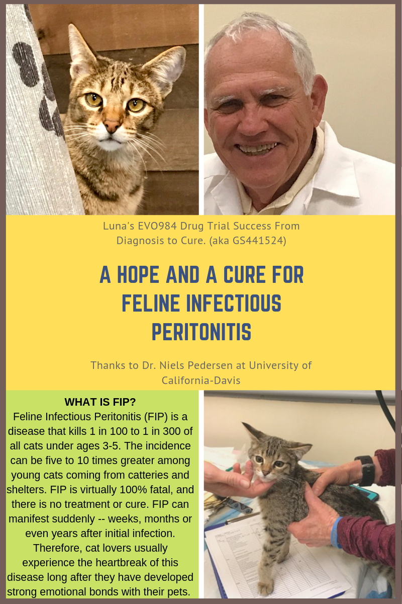
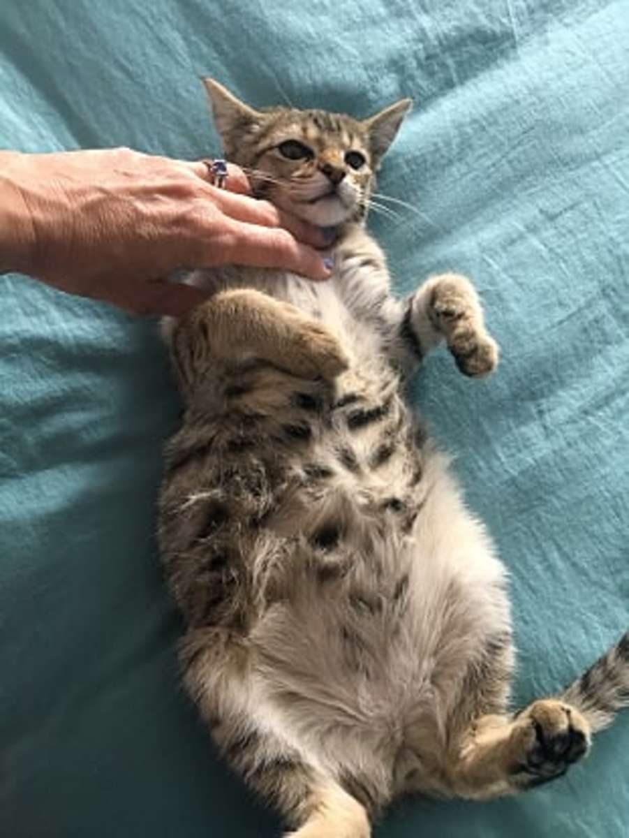
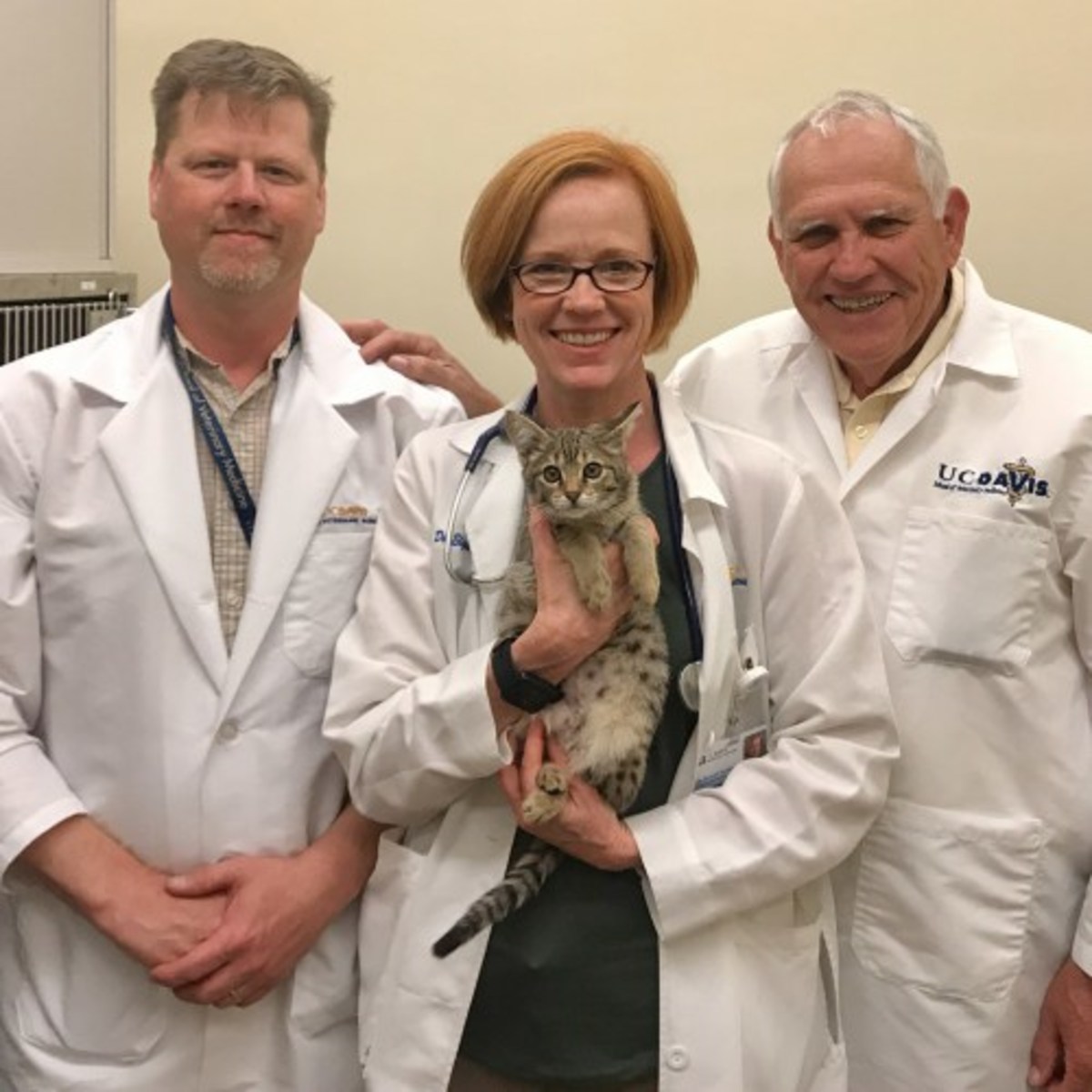
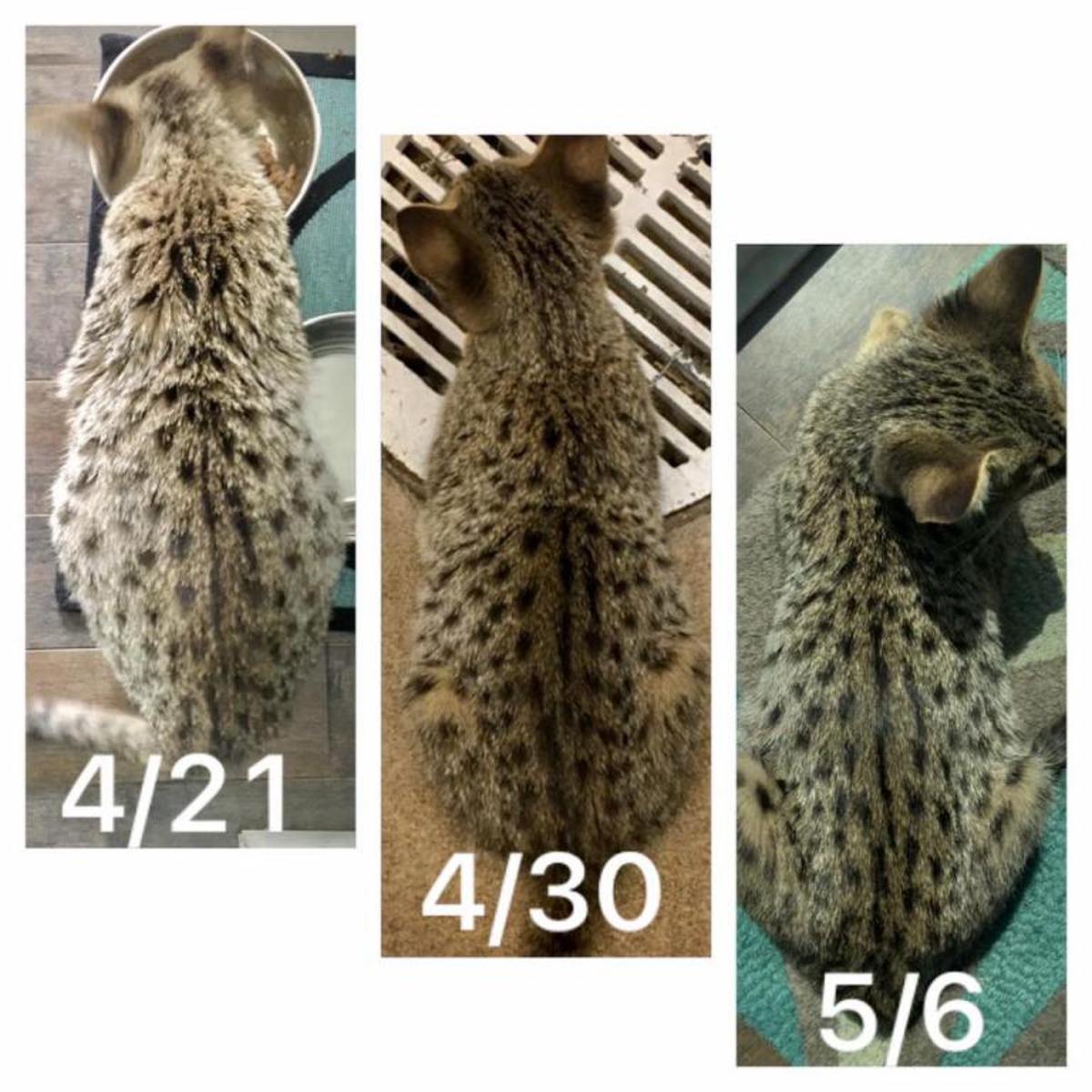
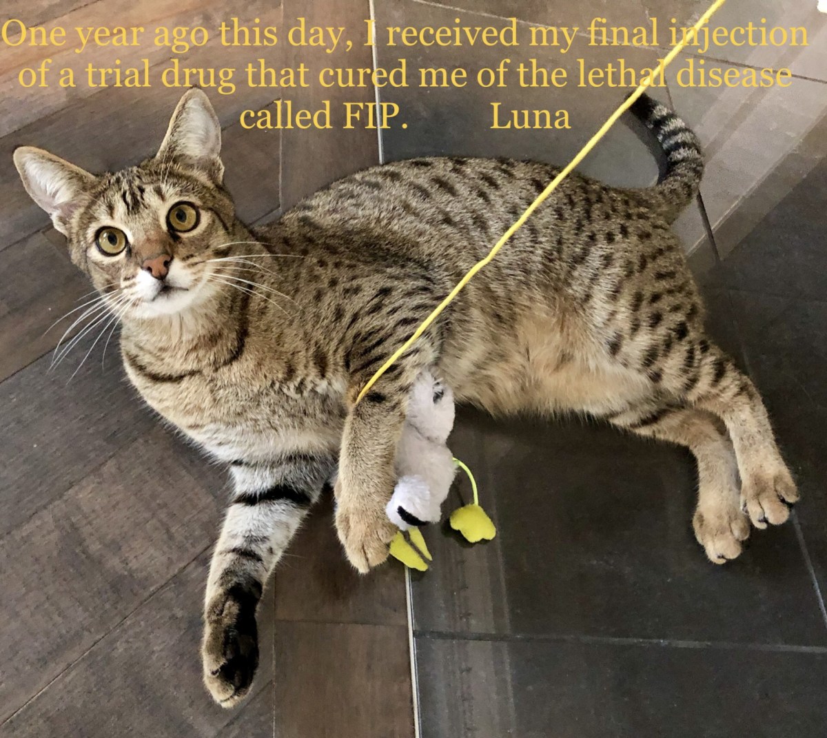
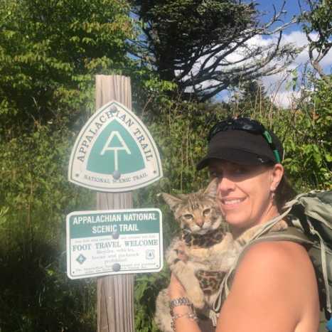
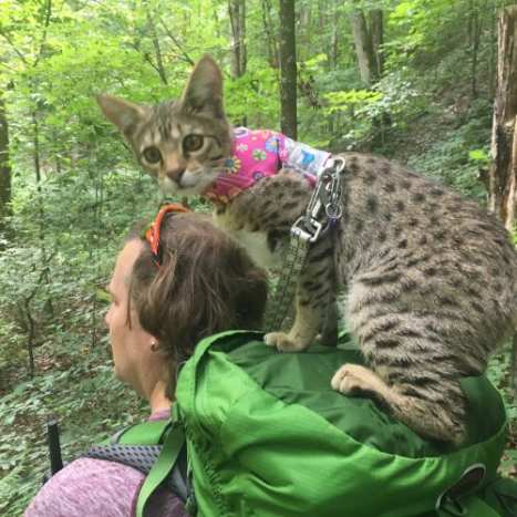


 EN
 SK